

The DOI matches its Crossref record; 72 of the 73 references it cites were found in a registry or an index.
Cytotoxic CD4+ T cells in cancer: Expanding the immune effector toolbox
Open accessAbstract
Cytotoxic T cells are important effectors of anti-tumor immunity. While tumor killing is ascribed to CD8 + T cell function, pre-clinical and clinical studies have identified intra-tumoral CD4 + T cells that possess cytotoxic programs and can directly kill cancer cells. Cytotoxic CD4 + T cells are found in other disease settings including infection and autoimmunity. Here, we review the phenotypic and functional characteristics of cytotoxic CD4 + T cells in non-cancer and cancer contexts. We conduct a comparative examination of cytolytic mechanisms of cytotoxic CD4 + T cells across disease states and synthesize features that define these cells independent of context. We discuss regulatory mechanisms driving ontogeny and effector function and evidence for the clinical relevance of cytotoxic CD4 + T cells in cancer. In this context, we highlight important gaps in understanding in the biology of cytotoxic CD4 + T cells as well as the potential use of these cells in immunotherapies for specific cancers.
Overview
The claims and statistics of this paper haven’t been checked yet.
Run the check — free, no account needed. It reads the open-access text and checks the sources it cites.
Integrity signals
Registry
Cites
Statistics
Figures
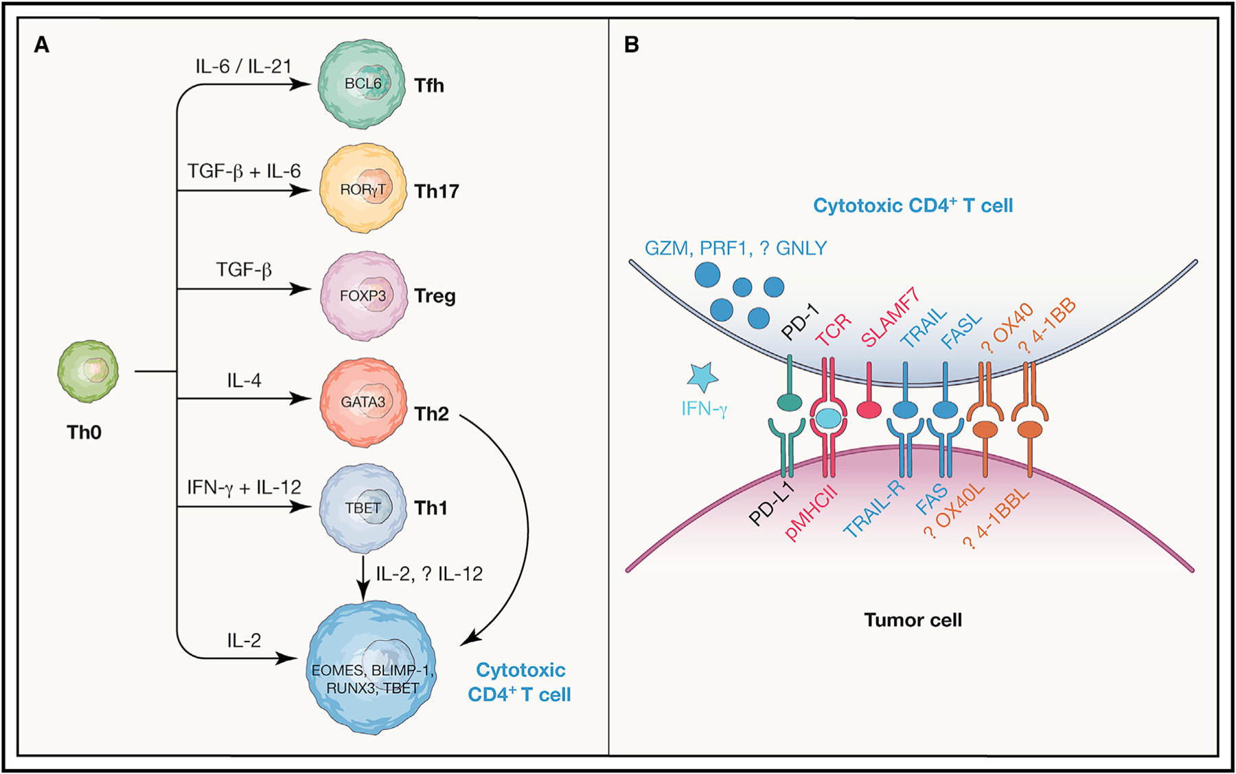
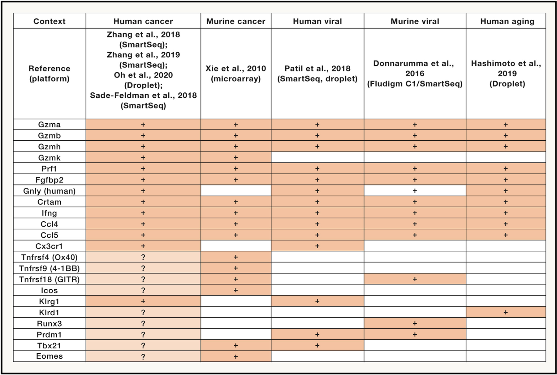
Verification
Claims the paper makes
The claims and statistics of this paper haven’t been checked yet.
Run the check — free, no account needed. It reads the open-access text and checks the sources it cites.
Claims other papers make about this one
Each is checked against this paper’s own text: does it say what the citing paper says it does?
The source backs part of the claim, not all of it — or backs it for a different population, condition or comparison.
In the citing paper
“we observed a significant increase in the total CD4+ T cells expressing IFN-γ (figure 4H) and granzyme B (GZMB; figure 4J),60–65 NK cells (figure 4N), and the amount of GZMB expression in CD8+ T cells (figure 4L) in the post-cryoablated RMS 76-9STINGwt TME as compared with RMS 76-9STING-/- TME in wild-type mice, as was Foxp3+ CD4+ cells (figure 4M).”
From Functional tumor cell-intrinsic STING, not host STING, drives local and systemic antitumor immunity and therapy efficacy following cryoablation (2023) · cites this paper as [64]
Experimental — accuracy not yet measured
Self-check
Percentages, sums and reported differences re-computed from the paper’s own numbers.
The self-check needs the paper’s full text.
Citations
Cites
An immunogenic personal neoantigen vaccine for patients with melanoma
Ott, P. A., Hu, Z., Keskin, D. B., Shukla, S. A., Sun, J., Bozym, D. J., Zhang, W., Luoma, A., Giobbie-Hurder, A., Peter, L., et al. (2017) · Nature · doi:10.1038/nature22991 · Cited by 2,936 · cited 1×
“Interestingly, in a complementary approach where high-risk resected stage III or stage IV melanoma patients were immunized with pools of long peptides containing predicted neoantigens prioritized based solely on HLA-A or HLA-B predicted binding affinity, neoantigen-specific CD4+ T cell responses were seen in addition…”
Puram, S. V., Tirosh, I., Parikh, A. S., Patel, A. P., Yizhak, K., Gillespie, S., Rodman, C., Luo, C. L., Mroz, E. A., Emerick, K. S., et al. (2017) · Cell · doi:10.1016/j.cell.2017.10.044 · Cited by 2,557
Personalized RNA mutanome vaccines mobilize poly-specific therapeutic immunity against cancer
Sahin, U., Derhovanessian, E., Miller, M., Kloke, B.-P., Simon, P., Löwer, M., Bukur, V., Tadmor, A. D., Luxemburger, U., Schrörs, B., et al. (2017) · Nature · doi:10.1038/nature23003 · Cited by 2,488
Defining T Cell States Associated with Response to Checkpoint Immunotherapy in Melanoma
Sade-Feldman, M., Yizhak, K., Bjorgaard, S. L., Ray, J. P., de Boer, C. G., Jenkins, R. W., Lieb, D. J., Chen, J. H., Frederick, D. T., Barzily-Rokni, M., et al. (2018) · Cell · doi:10.1016/j.cell.2018.10.038 · Cited by 2,053
Single-Cell Map of Diverse Immune Phenotypes in the Breast Tumor Microenvironment
Azizi, E., Carr, A. J., Plitas, G., Cornish, A. E., Konopacki, C., Prabhakaran, S., Nainys, J., Wu, K., Kiseliovas, V., Setty, M., et al. (2018) · Cell · doi:10.1016/j.cell.2018.05.060 · Cited by 1,976
Cancer Immunotherapy Based on Mutation-Specific CD4+ T Cells in a Patient with Epithelial Cancer
Tran, E., Turcotte, S., Gros, A., Robbins, P. F., Lu, Y., Dudley, M. E., Wunderlich, J. R., Somerville, R. P. T., Hogan, K., Hinrichs, C. S., et al. (2014) · Science · doi:10.1126/science.1251102 · Cited by 1,737
Global characterization of T cells in non-small-cell lung cancer by single-cell sequencing
Guo, X., Zhang, Y., Zheng, L., Zheng, C., Song, J., Zhang, Q., Kang, B., Liu, Z., Jin, L., Xing, R., et al. (2018) · Nature Medicine · doi:10.1038/s41591-018-0045-3 · Cited by 1,651
Landscape and Dynamics of Single Immune Cells in Hepatocellular Carcinoma
Zhang, Q., He, Y., Luo, N., Patel, S. J., Han, Y., Gao, R., Modak, M., Carotta, S., Haslinger, C., Kind, D., et al. (2019) · Cell · doi:10.1016/j.cell.2019.10.003 · Cited by 1,624
Lineage tracking reveals dynamic relationships of T cells in colorectal cancer
Zhang, L., Yu, X., Zheng, L., Zhang, Y., Li, Y., Fang, Q., Gao, R., Kang, B., Zhang, Q., Huang, J. Y., et al. (2018) · Nature · doi:10.1038/s41586-018-0694-x · Cited by 1,505
Treatment of Metastatic Melanoma with Autologous CD4+ T Cells against NY-ESO-1
Hunder, N. N. H., Wallen, H. D., Cao, J., Hendricks, D. W., Reilly, J. Z., Rodmyre, R. M., Jungbluth, A. A., Gnjatic, S., Thompson, J. A., & Yee, C. (2008) · New England Journal of Medicine · doi:10.1056/nejmoa0800251 · Cited by 904
Quezada, S. A., Simpson, T. R., Peggs, K. S., Merghoub, T., Vider, J., Fan, X., Blasberg, R., Yagita, H., Muranski, P., Antony, P. A., et al. (2010) · Journal of Experimental Medicine · doi:10.1084/jem.20091918 · Cited by 898
MHC-II neoantigens shape tumour immunity and response to immunotherapy
Alspach, E., Lussier, D. M., Miceli, A. P., Kizhvatov, I., DuPage, M., Luoma, A. M., Meng, W., Lichti, C. F., Esaulova, E., Vomund, A. N., et al. (2019) · Nature · doi:10.1038/s41586-019-1671-8 · Cited by 846 · cited 1×
“Furthermore, a pre-clinical study with poorly immunogenic sarcoma lines where either MHC class I- or MHC class II-restricted neoantigens were expressed demonstrated that recognition of MHC class II-dependent neoantigens on tumor cells by CD4+ T cells was necessary for tumor rejection in response to checkpoint inhibiti…”
Zhang, Y., Chen, H., Mo, H., Hu, X., Gao, R., Zhao, Y., Liu, B., Niu, L., Sun, X., Yu, X., et al. (2021) · Cancer Cell · doi:10.1016/j.ccell.2021.09.010 · Cited by 783
B cell maturation antigen–specific CAR T cells are clinically active in multiple myeloma
Cohen, A. D., Garfall, A. L., Stadtmauer, E. A., Melenhorst, J. J., Lacey, S. F., Lancaster, E., Vogl, D. T., Weiss, B. M., Dengel, K., Nelson, A., et al. (2019) · Journal of Clinical Investigation · doi:10.1172/jci126397 · Cited by 778
Intratumoral CD4+ T Cells Mediate Anti-tumor Cytotoxicity in Human Bladder Cancer
Oh, D. Y., Kwek, S. S., Raju, S. S., Li, T., McCarthy, E., Chow, E., Aran, D., Ilano, A., Pai, C.-C. S., Rancan, C., et al. (2020) · Cell · doi:10.1016/j.cell.2020.05.017 · Cited by 734 · cited 21×
“As far as the efficacy of MHC class II-dependent killing, of note, although the repertoire of naturally occurring cytotoxic CD4+ T cells in bladder cancer is noted to be restricted compared with other CD4+ T cell subsets (Oh et al., 2020), even within multimer-selected cytotoxic CD4+ T cell lines, the repertoire is po…”
InfluentialSelf-citationMogilenko, D. A., Shpynov, O., Andhey, P. S., Arthur, L., Swain, A., Esaulova, E., Brioschi, S., Shchukina, I., Kerndl, M., Bambouskova, M., et al. (2021) · Immunity · doi:10.1016/j.immuni.2020.11.005 · Cited by 683
Zhou, Y., Yang, D., Yang, Q., Lv, X., Huang, W., Zhou, Z., Wang, Y., Zhang, Z., Yuan, T., Ding, X., et al. (2020) · Nature Communications · doi:10.1038/s41467-020-20059-6 · Cited by 672
Johnson, D. B., Estrada, M. V., Salgado, R., Sanchez, V. M., Doxie, D. B., Opalenik, S. R., Vilgelm, A. E., Feld, E., Johnson, A. S., Greenplate, A. R., et al. (2016) · Nature Communications · doi:10.1038/ncomms10582 · Cited by 581 · cited 1×
“In fact, melanoma-specific MHC class II expression by tumor cells (and not antigen-presenting cells) correlated with both clinical response and overall survival in two separate cohorts of anti-PD-1-treated patients (Johnson et al., 2016).”
Rodig, S. J., Gusenleitner, D., Jackson, D. G., Gjini, E., Giobbie-Hurder, A., Jin, C., Chang, H., Lovitch, S. B., Horak, C., Weber, J. S., et al. (2018) · Science Translational Medicine · doi:10.1126/scitranslmed.aar3342 · Cited by 579 · cited 1×
“A separate examination of two independent phase II clinical trials of checkpoint inhibition in melanoma similarly found that anti-PD-1 responses associated with increased MHC class II expression and IFN-γ signaling prior to treatment (Rodig et al., 2018).”
Liakou, C. I., Kamat, A., Tang, D. N., Chen, H., Sun, J., Troncoso, P., Logothetis, C., & Sharma, P. (2008) · Proceedings of the National Academy of Sciences · doi:10.1073/pnas.0806075105 · Cited by 549
When worlds collide: Th17 and Treg cells in cancer and autoimmunity
Knochelmann, H. M., Dwyer, C. J., Bailey, S. R., Amaya, S. M., Elston, D. M., Mazza-McCrann, J. M., & Paulos, C. M. (2018) · Cellular & Molecular Immunology · doi:10.1038/s41423-018-0004-4 · Cited by 535
Runx3 and T-box proteins cooperate to establish the transcriptional program of effector CTLs
Cruz‐Guilloty, F., Pipkin, M. E., Djuretic, I. M., Levanon, D., Lotem, J., Lichtenheld, M. G., Groner, Y., & Rao, A. (2009) · The Journal of Experimental Medicine · doi:10.1084/jem.20081242 · Cited by 518 · cited 11×
“For a mixed population of GZMB+ and GZMK+ CD4+ in melanoma, higher RUNX3 and PRDM1 (BLIMP-1) expression was predominant over lowlevel TBX21/EOMES expression (Cachot et al., 2021), and the PRDM1 overexpression is supported by similar findings from GZMB+ CD4+ T cells in murine adenoviral infection (Donnarumma et al., 20…”
InfluentialHollern, D. P., Xu, N., Thennavan, A., Glodowski, C. R., García‐Recio, S., Mott, K. R., He, X., Garay, J. P., Carey-Ewend, K., Marron, D. L., et al. (2019) · Cell · doi:10.1016/j.cell.2019.10.028 · Cited by 480 · cited 1×
“, 2018), there is evidence to the contrary that Tfh cells can mediate response to checkpoint inhibition in genetically engineered mouse models (Hollern et al., 2019).”
Naive tumor-specific CD4+ T cells differentiated in vivo eradicate established melanoma
Xie, Y., Akpınarlı, A., Maris, C. H., Hipkiss, E. L., Lane, M. V., Kwon, E. M., Muranski, P. J., Restifo, N. P., & Antony, P. A. (2010) · The Journal of Experimental Medicine · doi:10.1084/jem.20091921 · Cited by 473
CD4 CTL, a cytotoxic subset of CD4(+) T cells, their differentiation and function
Takeuchi, A., & Saito, T. (2017) · Frontiers in Immunology · doi:10.3389/fimmu.2017.00194 · Cited by 431 · cited 1×
“Much of the current understanding on the functionality of cytotoxic CD4+ T cells comes from studies on anti-viral immunity (reviewed in Cheroutre and Husain, 2013; Marshall and Swain, 2011; Takeuchi and Saito, 2017).”
Mucida, D., Husain, M. M., Muroi, S., van Wijk, F., Shinnakasu, R., Naoe, Y., Reis, B. S., Huang, Y., Lambolez, F., Docherty, M., et al. (2013) · Nature Immunology · doi:10.1038/ni.2523 · Cited by 376
van Leeuwen, E. M. M., Remmerswaal, E. B. M., Vossen, M. T. M., Rowshani, A. T., Dillen, P. M. E. W., van Lier, R. A. W., & ten Berge, I. J. M. (2004) · The Journal of Immunology · doi:10.4049/jimmunol.173.3.1834 · Cited by 369
ICOS-based chimeric antigen receptors program bipolar TH17/TH1 cells
Guedan, S., Chen, X., Madar, A., Carpenito, C., McGettigan, S. E., Frigault, M. J., Lee, J., Posey, A. D., Scholler, J., Scholler, N., et al. (2014) · Blood · doi:10.1182/blood-2013-10-535245 · Cited by 359 · cited 1×
“Th17 CD4+ cells can either promote tumorigenesis or protect against established tumors depending on the context (reviewed in Vitiello and Miller, 2020), although several lines of evidence indicate that Th17 CD4+ T cells can eradicate tumors including when modified as CAR-T cells (Nelson et al., 2020; Guedan et al., 20…”
Single-cell transcriptomics reveals expansion of cytotoxic CD4 T cells in supercentenarians
Hashimoto, K., Kouno, T., Ikawa, T., Hayatsu, N., Miyajima, Y., Yabukami, H., Terooatea, T., Sasaki, T., Suzuki, T., Valentine, M., et al. (2019) · Proceedings of the National Academy of Sciences · doi:10.1073/pnas.1907883116 · Cited by 343
Tumor-specific cytolytic CD4 T cells mediate immunity against human cancer
Cachot, A., Bilous, M., Liu, Y., Li, X., Saillard, M., Cenerenti, M., Rockinger, G. A., Wyss, T., Guillaume, P., Schmidt, J., et al. (2021) · Science Advances · doi:10.1126/sciadv.abe3348 · Cited by 340
Lu, Y., Parker, L. L., Lu, T., Zheng, Z., Toomey, M. A., White, D. E., Yao, X., Li, Y. F., Robbins, P. F., Feldman, S. A., et al. (2017) · Journal of Clinical Oncology · doi:10.1200/jco.2017.74.5463 · Cited by 288 · cited 1×
“Cell therapy with retrovirally transduced CD4+ T cells expressing a TCR specific-for-another-cancertestis antigen, MAGE-A3, presented by HLA-DPB1*0401 also led to partial and complete response across several tumor types (Lu et al., 2017).”
Reis, B. S., Rogoz, A., Costa‐Pinto, F. A., Taniuchi, I., & Mucida, D. (2013) · Nature Immunology · doi:10.1038/ni.2518 · Cited by 284
HIV-Specific Cytolytic CD4 T Cell Responses During Acute HIV Infection Predict Disease Outcome
Soghoian, D. Z., Jessen, H. K., Flanders, M., Sierra-Davidson, K., Cutler, S., Pertel, T., Ranasinghe, S., Lindqvist, M., Davis, I., Lane, K., et al. (2012) · Science Translational Medicine · doi:10.1126/scitranslmed.3003165 · Cited by 277
Precursors of human CD4 + cytotoxic T lymphocytes identified by single-cell transcriptome analysis
Patil, V. S., Madrigal, A., Schmiedel, B. J., Clarke, J., O’Rourke, P., de Silva, A. D., Harris, E., Peters, B., Seumois, G., Weiskopf, D., et al. (2018) · Science Immunology · doi:10.1126/sciimmunol.aan8664 · Cited by 262 · cited 11×
“While all populations expressed GZMA and other granule-associated proteins such as GNLY and NKG7, distinct states co-expressed either GZMB or GZMK, and PRF1 was more predominantly coexpressed with GZMB.”
InfluentialBrown, D. M., Lee, S., García-Hernández, M. D. L. L., & Swain, S. L. (2012) · Journal of Virology · doi:10.1128/jvi.07172-11 · Cited by 245 · cited 3×
“The recognition of virally derived peptides by cytotoxic CD4+ T cells can lead to target cell killing (Jellison et al., 2005; Soghoian et al., 2012; Brown et al., 2012).”
InfluentialŚledzińska, A., Vila de Mucha, M., Bergerhoff, K., Hotblack, A., Demane, D. F., Ghorani, E., Akarca, A. U., Marzolini, M. A. V., Solomon, I., Vargas, F. A., et al. (2020) · Immunity · doi:10.1016/j.immuni.2019.12.007 · Cited by 228
Memory CD4+ T cells protect against influenza through multiple synergizing mechanisms
McKinstry, K. K., Strutt, T. M., Kuang, Y., Brown, D. M., Sell, S., Dutton, R. W., & Swain, S. L. (2012) · Journal of Clinical Investigation · doi:10.1172/jci63689 · Cited by 223
CRTAM determines the CD4+ cytotoxic T lymphocyte lineage
Takeuchi, A., Badr, M. E. S. G., Miyauchi, K., Ishihara, C., Onishi, R. M., Guo, Z., Sasaki, Y., Ike, H., Takumi, A., Tsuji, N. M., et al. (2015) · The Journal of Experimental Medicine · doi:10.1084/jem.20150519 · Cited by 220 · cited 3×
“Notably, in murine models, CD4+ CD8α+ cells expressing GZM, PRF1, interferon (IFN)-γ, and separately cytotoxic CD4+ T cells expressing the class I-restricted T cell-associated molecule (CRTAM), locate within the intraepithelial lymphocyte (IEL) compartment of the intestine (Takeuchi et al., 2016; Mucida et al., 2013;…”
Cytotoxic CD4+ T lymphocytes may induce endothelial cell apoptosis in systemic sclerosis
Maehara, T., Kaneko, N., Perugino, C. A., Mattoo, H., Kers, J., Allard‐Chamard, H., Mahajan, V. S., Liu, H., Murphy, S. J. H., Ghebremichael, M. S., et al. (2020) · Journal of Clinical Investigation · doi:10.1172/jci131700 · Cited by 202 · cited 1×
“Inflamed skin lesions from systemic sclerosis patients are also infiltrated by GZMA+ CD4+ T cells, which can be seen directly encountering human leukocyte antigen (HLA)-DR+ endothelial cells undergoing apoptosis, suggesting a direct cytotoxic role in end-organ tissue damage (Maehara et al., 2020).”
Biology and clinical relevance of granulysin
Krensky, A. M., & Clayberger, C. (2009) · Tissue Antigens · doi:10.1111/j.1399-0039.2008.01218.x · Cited by 200
Cytotoxic CD4 T Cells in Antiviral Immunity
Marshall, N. B., & Swain, S. L. (2011) · BioMed Research International · doi:10.1155/2011/954602 · Cited by 193
Martens, A., Wistuba-Hamprecht, K., Yuan, J., Postow, M. A., Wong, P., Capone, M., Madonna, G., Khammari, A., Schilling, B., Sucker, A., et al. (2016) · Clinical Cancer Research · doi:10.1158/1078-0432.ccr-16-0249 · Cited by 180 · cited 1×
“However, while increased ALC early after the first dose and CD8+ T cell percentages later on (between 8 and 14 weeks of treatment) associated with improved survival and clinical response in melanoma patients, increased CD4+ T cell percentages at 8–14 weeks also correlated with these positive clinical outcomes (Martens…”
Hirschhorn-Cymerman, D., Budhu, S., Kitano, S., Liu, C., Zhao, F., Zhong, H., Lesokhin, A. M., Avogadri-Connors, F., Yuan, J. D., Li, Y., et al. (2012) · The Journal of Experimental Medicine · doi:10.1084/jem.20120532 · Cited by 165
Qui, H. Z., Hagymasi, A. T., Bandyopadhyay, S., Rose, M. S., Ramanarasimhaiah, R., Ménoret, A., Mittler, R. S., Gordon, S. M., Reiner, S. L., Vella, A. T., et al. (2011) · The Journal of Immunology · doi:10.4049/jimmunol.1101244 · Cited by 165
Cytotoxic CD4 + T cells in viral hepatitis
Aslan, N., Yurdaydın, C., Wiegand, J., Greten, T. F., Ciner, A., Meyer, M., Heiken, H., Kuhlmann, B., Kaiser, T., Bozkaya, H., et al. (2006) · Journal of Viral Hepatitis · doi:10.1111/j.1365-2893.2006.00723.x · Cited by 153
Cutting Edge: MHC Class II-Restricted Killing In Vivo during Viral Infection
Jellison, E. R., Kim, S.-K., & Welsh, R. M. (2005) · The Journal of Immunology · doi:10.4049/jimmunol.174.2.614 · Cited by 152
Targeting the interleukin-17 immune axis for cancer immunotherapy
Vitiello, G. A., & Miller, G. (2020) · Journal of Experimental Medicine · doi:10.1084/jem.20190456 · Cited by 151
Control of Epstein-Barr virus infection in vitro by T helper cells specific for virion glycoproteins
Adhikary, D., Behrends, U., Moosmann, A., Witter, K., Bornkamm, G. W., & Mautner, J. (2006) · The Journal of Experimental Medicine · doi:10.1084/jem.20051287 · Cited by 145
Zappasodi, R., Budhu, S., Hellmann, M. D., Postow, M. A., Senbabaoglu, Y., Manne, S., Gasmi, B., Liu, C., Zhong, H., Li, Y., et al. (2018) · Cancer Cell · doi:10.1016/j.ccell.2018.05.009 · Cited by 142
Kitano, S., Tsuji, T., Liu, C., Hirschhorn-Cymerman, D., Kyi, C., Mu, Z., Allison, J. P., Gnjatic, S., Yuan, J. D., & Wolchok, J. D. (2013) · Cancer Immunology Research · doi:10.1158/2326-6066.cir-13-0068 · Cited by 136
Eomesodermin-expressing T-helper cells are essential for chronic neuroinflammation
Raveney, B. J. E., Oki, S., Hohjoh, H., Nakamura, M., Sato, W., Murata, M., & Yamamura, T. (2015) · Nature Communications · doi:10.1038/ncomms9437 · Cited by 134 · cited 1×
“In particular, EOMES appears to be important for the generation of pathogenic cytotoxic CD4+ T cells in experimental autoimmune encephalitis (Raveney et al., 2015); also, reduced EOMES expression impairs upregulation of GZMB after in vitro activation despite TBET levels being unaffected (Hirschhorn-Cymerman et al.”
Transcription Factor T-bet Regulates Intraepithelial Lymphocyte Functional Maturation
Reis, B. S., van Konijnenburg, D. P. H., Grivennikov, S. I., & Mucida, D. (2014) · Immunity · doi:10.1016/j.immuni.2014.06.017 · Cited by 129 · cited 1×
“Notably, in murine models, CD4+ CD8α+ cells expressing GZM, PRF1, interferon (IFN)-γ, and separately cytotoxic CD4+ T cells expressing the class I-restricted T cell-associated molecule (CRTAM), locate within the intraepithelial lymphocyte (IEL) compartment of the intestine (Takeuchi et al., 2016; Mucida et al., 2013;…”
Long, H. M., Haigh, T. A., Gudgeon, N. H., Leen, A. M., Tsang, C.-W., Brooks, J. M., Landais, E., Houssaint, E., Lee, S. P., Rickinson, A. B., et al. (2005) · Journal of Virology · doi:10.1128/jvi.79.8.4896-4907.2005 · Cited by 129 · cited 1×
“In regard to their activation requirements, it is clear that in viremic states, cytotoxic CD4+ T cells recognize cognate viral antigens in a major histocompatibility complex class II (MHC class II)-restricted manner, which mirrors the MHC restriction of non-cytotoxic CD4+ T cells (Jellison et al., 2005; Soghoian et al…”
Brown, D. M., Kamperschroer, C., Dilzer, A. M., Roberts, D. M., & Swain, S. L. (2009) · Cellular Immunology · doi:10.1016/j.cellimm.2009.03.002 · Cited by 128
Zappasodi, R., Budhu, S., Hellmann, M. D., Postow, M. A., Şenbabaoğlu, Y., Manne, S., Gasmi, B., Liu, C., Zhong, H., Li, Y., et al. (2018) · Cancer Cell · doi:10.1016/j.ccell.2018.09.007 · Cited by 124
CD4 CTL: Living up to the challenge
Cheroutre, H., & Husain, M. M. (2013) · Seminars in Immunology · doi:10.1016/j.smim.2013.10.022 · Cited by 119
Medley, Q. G., Kedersha, N., O'Brien, S., Tian, Q., Schlossman, S. F., Streuli, M., & Anderson, P. (1996) · Proceedings of the National Academy of Sciences · doi:10.1073/pnas.93.2.685 · Cited by 117 · cited 4×
“Additional granule constituents include the protein NKG7 (Medley et al., 1996) that is commonly seen in cytotoxic effector cells, including cytotoxic CD4+ T cells, whose function has been elusive.”
InfluentialDella‐Torre, E., Cassione, E. B., Sciorati, C., Ruggiero, E., Lanzillotta, M., Bonfiglio, S., Mattoo, H., Perugino, C. A., Bozzolo, E. P., Rovati, L., et al. (2018) · Arthritis & Rheumatology · doi:10.1002/art.40469 · Cited by 116 · cited 1×
“In addition, in immunoglobulin G4 (IgG4)-related disease, the infiltration of affected organs by cytotoxic CD4+ T cells is reduced by glucocorticoids; furthermore, dominant T cell receptor (TCR) β clonotypes in the circulation are shared with tissue-infiltrating cytotoxic CD4+ T cells (Della-Torre et al., 2018).”
Expression and function of NKG2D in CD4 + T cells specific for human cytomegalovirus
Sáez‐Borderías, A., Gumá, M., Angulo, A., Bellosillo, B., Pende, D., & López‐Botet, M. (2006) · European Journal of Immunology · doi:10.1002/eji.200636682 · Cited by 107
van Leeuwen, E. M. M., Remmerswaal, E. B. M., Heemskerk, M. H. M., ten Berge, I. J. M., & van Lier, R. A. W. (2006) · Blood · doi:10.1182/blood-2006-03-006809 · Cited by 106
Endogenous human cytomegalovirus gB is presented efficiently by MHC class II molecules to CD4 + CTL
Hegde, N. R., Dunn, C., Lewinsohn, D. M., Jarvis, M. A., Nelson, J. A., & Johnson, D. C. (2005) · The Journal of Experimental Medicine · doi:10.1084/jem.20050162 · Cited by 82 · cited 1×
“Beyond professional antigen-presenting cells, cytotoxic CD4+ T cells can also target other infected cells such as epithelial cells and B cells, which express MHC class II in humans (Hegde et al., 2005; Jellison et al., 2005; van Leeuwen et al., 2006).”
The transcriptional control of the perforin locus
Pipkin, M. E., Rao, A., & Lichtenheld, M. G. (2010) · Immunological Reviews · doi:10.1111/j.0105-2896.2010.00905.x · Cited by 76 · cited 1×
“For instance, anti-viral cytotoxic CD4+ T cells appear to require stimulation to increase and maintain expression of perforin, unlike the constitutive post-thymic expression of this cytolytic molecule in CD8+ T cells (Niiya et al., 2005; Pipkin et al., 2010).”
Landais, E., Saulquin, X., Scotet, E., Trautmann, L., Peyrat, M., Yates, J. L., Kwok, W. W., Bonneville, M., & Houssaint, E. (2003) · Blood · doi:10.1182/blood-2003-03-0930 · Cited by 75
Kwek, S. S., Lewis, J., Zhang, L., WEINBERG, V. K., Greaney, S. K., Harzstark, A. L., Lin, A., Ryan, C. J., Small, E. J., & Fong, L. H. (2015) · Cancer Immunology Research · doi:10.1158/2326-6066.cir-14-0227 · Cited by 62
Self-citationKhanna, R. (1997) · International Immunology · doi:10.1093/intimm/9.10.1537 · Cited by 60
Donnarumma, T., Young, G. R., Merkenschlager, J., Eksmond, U., Bongard, N., Nutt, S. L., Boyer, C., Dittmer, U., Le‐Trilling, V. T. K., Trilling, M., et al. (2016) · Cell Reports · doi:10.1016/j.celrep.2016.10.013 · Cited by 59
Identification of Neoantigen-Reactive Tumor-Infiltrating Lymphocytes in Primary Bladder Cancer
Leko, V., McDuffie, L. A., Zheng, Z., Gartner, J. J., Prickett, T. D., Apolo, A. B., Agarwal, P. K., Rosenberg, S. A., & Lu, Y. (2019) · The Journal of Immunology · doi:10.4049/jimmunol.1801022 · Cited by 49 · cited 1×
“Neoantigen-directed CD4+ adoptive T cell therapy, where reactive T cells are selected from tumor-infiltrating lymphocytes, also clearly has clinical activity, for instance, in cholangiocarcinoma with a clone directed against a mutation in ERBB2-interacting protein and in urothelial carcinoma with a clone directed agai…”
Identification of human CD4 + T cell populations with distinct antitumor activity
Nelson, M. H., Knochelmann, H. M., Bailey, S. R., Huff, L. W., Bowers, J. S., Majchrzak, K., Wyatt, M. M., Rubinstein, M. P., Mehrotra, S., Nishimura, M. I., et al. (2020) · Science Advances · doi:10.1126/sciadv.aba7443 · Cited by 49
CD4+ T cells induce rejection of urothelial tumors after immune checkpoint blockade
Sato, Y., Bolzenius, J. K., Eteleeb, A. M., Su, X., Maher, C. A., Sehn, J. K., & Arora, V. K. (2018) · JCI Insight · doi:10.1172/jci.insight.121062 · Cited by 48
Yang, D., Tian, Z., Zhang, M., Yang, W.-B., Tang, J., Wu, Y., & Ni, B. (2017) · Scientific Reports · doi:10.1038/s41598-017-01379-y · Cited by 46
Differential regulation of perforin expression in human CD4+ and CD8+ cytotoxic T lymphocytes
Niiya, H., Sakai, I., Jin, L., Azuma, T., Uchida, N., Yakushijin, Y., Hato, T., Fujita, S., & Yasukawa, M. (2005) · Experimental Hematology · doi:10.1016/j.exphem.2005.04.004 · Cited by 18 · cited 1×
“For instance, anti-viral cytotoxic CD4+ T cells appear to require stimulation to increase and maintain expression of perforin, unlike the constitutive post-thymic expression of this cytolytic molecule in CD8+ T cells (Niiya et al., 2005; Pipkin et al., 2010).”
Cited by
Sorted by citations
The cancer-immunity cycle: Indication, genotype, and immunotype
Mellman, I., Chen, D. S., Powles, T. B., & Turley, S. J. (2023) · Immunity · doi:10.1016/j.immuni.2023.09.011 · Cited by 1,018
Dynamics and specificities of T cells in cancer immunotherapy
Oliveira, G., & Wu, C. J. (2023) · Nature reviews. Cancer · doi:10.1038/s41568-023-00560-y · Cited by 574
Speiser, D. E., Chijioke, O., Schaeuble, K., & Münz, C. (2023) · Nature Cancer · doi:10.1038/s43018-023-00521-2 · Cited by 511
Xu, Z., Chen, Y., Ma, L., Chen, Y., Liu, J., Guo, Y., Yu, T., Zhang, L., Zhu, L., & Shu, Y. (2022) · Molecular Therapy · doi:10.1016/j.ymthe.2022.01.046 · Cited by 337
“Th cells can be further divided into Th1, Th17, and Tregs.(177)”
Nanomaterials for bone metastasis
Hao, X., Jiang, B., Wu, J., Xiang, D., Xiong, Z., Li, C., Li, Z., He, S., Tu, C., & Li, Z. (2024) · Journal of Controlled Release · doi:10.1016/j.jconrel.2024.07.067 · Cited by 240
Metadata
- Authors
- Oh, D. Y., & Fong, L.
- Affiliations
- University of California, San Francisco (US)
- Corresponding author
- Fong, Lawrence
- Venue
- Immunity
- Publisher
- Elsevier BV
- Published
- 2021-12
- Volume
- 54
- Issue
- 12
- Pages
- 2701-2711
- ISSN
- 1074-7613 (ISSN-L)
- PMC
- PMC8809482
- OpenAlex
- W4200244347
- Cited by
- 467
- References
- 73
- Citations by year
- 2026: 81 · 2025: 127 · 2024: 116 · 2023: 101 · 2022: 41 · 2021: 1
- FWCI
- 6.86
- Type
- journal article
- Language
- en
- Open access
- hybrid
- doi.org
- Licence
- Locations
- Immunity (publishedVersion, cc-by)
- PubMed (publishedVersion)
- PubMed Central (acceptedVersion, cc-by)
- eScholarship (California Digital Library) (submittedVersion, cc-by)
- Funders
- NCI
- National Institutes of Health (K08AI139375)
- Prostate Cancer Foundation (R35CA253175)
- Bavarian Nordic
- Genentech
- Parker Institute for Cancer Immunotherapy
- AbbVie
- Amgen
- Merck
- Merck KGaA
- Topics
- Life Sciences › Immunology and Microbiology › Immunology › T-cell and B-cell Immunology
- Health Sciences › Medicine › Oncology › Cancer Immunotherapy and Biomarkers
- Life Sciences › Immunology and Microbiology › Immunology › Immunotherapy and Immune Responses
- Keywords
- cytotoxic CD4+ T cells, antitumor immunity, cytotoxicity mechanism, cancer immunotherapy, infection, autoimmunity
- MeSH terms
- Animals, Cytotoxicity, Immunologic, Humans, Neoplasms, T-Lymphocytes, Cytotoxic, CD4-Positive T-Lymphocytes, Immunotherapy, Adoptive
About these checks
Registry, retraction, Cites and statistics results are computed, not judged, with one exception: a printed reference that no index lists is read once by a model before it counts as a citation error. Claims are extracted by a model and kept only when their quote appears verbatim in the text; each cited source is then checked, with a model’s help, for whether it says what the claim relies on. Those verdicts can be wrong — read the claim’s quote and the cited source before relying on one.
Statistics and arithmetic are recomputed, not judged.
Confidence bands are shown only for fields calibrated on our annotated test set. None is calibrated yet, so no band is shown.
What counts as a claim ›How we measure confidence ›How we check this ›
Are you an author, or do you see a problem with this page? Request a review
