

The DOI matches its Crossref record; 54 of the 55 references it cites were found in a registry or an index.
SARS-CoV-2 infects cells after viral entry via clathrin-mediated endocytosis
Open accessPublished version of a preprint
Abstract
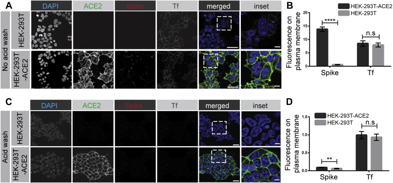
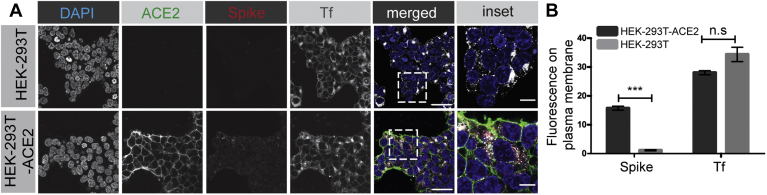
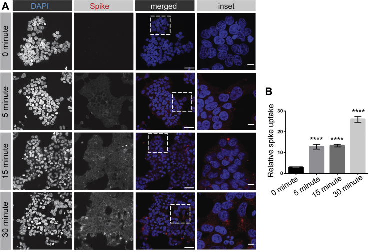
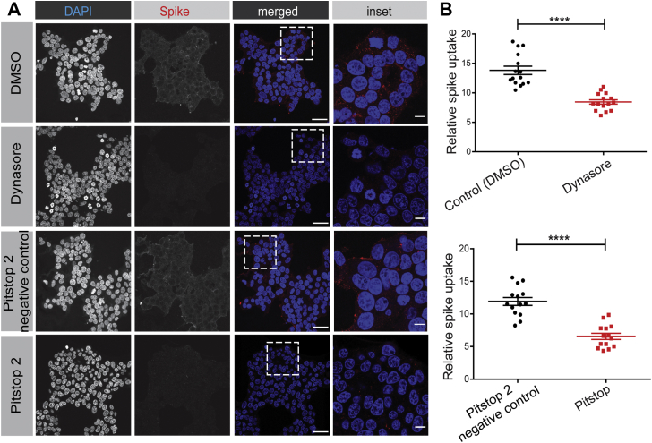
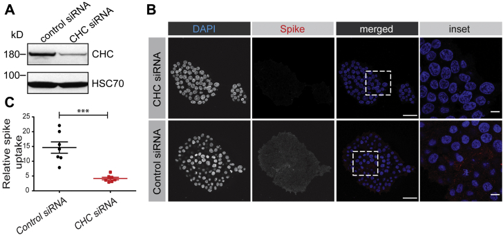
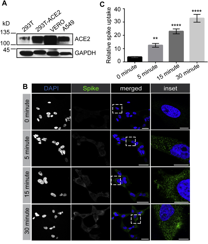
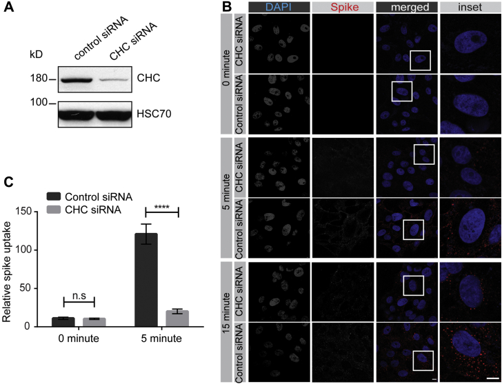
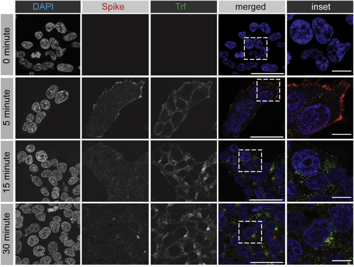
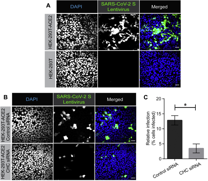
Severe acute respiratory syndrome coronavirus 2 (SARS-CoV-2) is the causative agent of COVID-19, so understanding its biology and infection mechanisms is critical to facing this major medical challenge. SARS-CoV-2 is known to use its spike glycoprotein to interact with the cell surface as a first step in the infection process. As for other coronaviruses, it is likely that SARS-CoV-2 next undergoes endocytosis, but whether or not this is required for infectivity and the precise endocytic mechanism used are unknown. Using purified spike glycoprotein and lentivirus pseudotyped with spike glycoprotein, a common model of SARS-CoV-2 infectivity, we now demonstrate that after engagement with the plasma membrane, SARS-CoV-2 undergoes rapid, clathrin-mediated endocytosis. This suggests that transfer of viral RNA to the cell cytosol occurs from the lumen of the endosomal system. Importantly, we further demonstrate that knockdown of clathrin heavy chain, which blocks clathrin-mediated endocytosis, reduces viral infectivity. These discoveries reveal that SARS-CoV-2 uses clathrin-mediated endocytosis to gain access into cells and suggests that this process is a key aspect of virus infectivity.
Overview
The claims and statistics of this paper haven’t been checked yet.
Run the check — free, no account needed. It reads the open-access text and checks the sources it cites.
Integrity signals
Registry
Cites
Statistics
Figures
Verification
Claims the paper makes
The claims and statistics of this paper haven’t been checked yet.
Run the check — free, no account needed. It reads the open-access text and checks the sources it cites.
Claims other papers make about this one
Each is checked against this paper’s own text: does it say what the citing paper says it does?
The source backs part of the claim, not all of it — or backs it for a different population, condition or comparison.
In the citing paper
“Interfering expression of clathrin heavy chain in HEK293 (stably expressing ACE2), limits SARS-CoV-2 viral infectivity [66].”
From Insights of Endocytosis Signaling in Health and Disease (2023) · cites this paper as [66]
Experimental — accuracy not yet measured
Self-check
Percentages, sums and reported differences re-computed from the paper’s own numbers.
The self-check needs the paper’s full text.
Citations
Cites
Clinical features of patients infected with 2019 novel coronavirus in Wuhan, China
Huang, C., Wang, Y., Li, X., Ren, L., Zhao, J., Hu, Y., Zhang, L., Fan, G., Xu, J., Gu, X., et al. (2020) · The Lancet · doi:10.1016/s0140-6736(20)30183-5 · Cited by 48,884
A novel coronavirus from patients with pneumonia in China, 2019
Zhu, N., Zhang, D., Wang, W., Li, X., Yang, B., Song, J., Zhao, X., Huang, B., Shi, W., Lu, R., et al. (2020) · New England Journal of Medicine · doi:10.1056/nejmoa2001017 · Cited by 28,244
A pneumonia outbreak associated with a new coronavirus of probable bat origin
Zhou, P., Yang, X.-L., Wang, X.-G., Hu, B., Zhang, L., Zhang, W., Si, H.-R., Zhu, Y., Li, B., Huang, C.-L., et al. (2020) · Nature · doi:10.1038/s41586-020-2012-7 · Cited by 21,969
Hoffmann, M., Kleine-Weber, H., Schroeder, S., Krüger, N., Herrler, T., Erichsen, S., Schiergens, T. S., Herrler, G., Wu, N.-H., Nitsche, A., et al. (2020) · Cell · doi:10.1016/j.cell.2020.02.052 · Cited by 20,286
Cryo-EM structure of the 2019-nCoV spike in the prefusion conformation
Wrapp, D., Wang, N., Corbett, K. S., Goldsmith, J. A., Hsieh, C.-L., Abiona, O., Graham, B. S., & McLellan, J. S. (2020) · Science · doi:10.1126/science.abb2507 · Cited by 8,897
Isolation of a Novel Coronavirus from a Man with Pneumonia in Saudi Arabia
Zaki, A. M., van Boheemen, S., Bestebroer, T. M., Osterhaus, A. D. M. E., & Fouchier, R. A. M. (2012) · New England Journal of Medicine · doi:10.1056/nejmoa1211721 · Cited by 5,677
Hamming, I., Timens, W., Bulthuis, M., Lely, A., Navis, G., & van Goor, H. (2004) · The Journal of Pathology · doi:10.1002/path.1570 · Cited by 5,676
Identification of a novel coronavirus in patients with severe acute respiratory syndrome
Drosten, C., Günther, S., Preiser, W., van der Werf, S., Brodt, H.-R., Becker, S., Rabenau, H., Panning, M., Kolesnikova, L., Fouchier, R. A. M., et al. (2003) · New England Journal of Medicine · doi:10.1056/nejmoa030747 · Cited by 4,574
A Novel Coronavirus Associated with Severe Acute Respiratory Syndrome
Tg, K., Erdman, D. D., Cs, G., Zaki, Peret, T. C. T., Emery, S. L., Tong, S., Urbani, C., Comer, J. A., Lim, W., et al. (2003) · New England Journal of Medicine · doi:10.1056/nejmoa030781 · Cited by 4,353
Letko, M., Marzi, A., & Munster, V. (2020) · Nature Microbiology · doi:10.1038/s41564-020-0688-y · Cited by 3,109
Cited by
Sorted by citations
Mechanisms of SARS-CoV-2 entry into cells
Jackson, C. B., Farzan, M., Chen, B., & Choe, H. (2022) · Nature Reviews Molecular Cell Biology · doi:10.1038/s41580-021-00418-x · Cited by 3,022
Innate immunity: the first line of defense against SARS-CoV-2
Diamond, M., & Kanneganti, T. (2022) · Nature Immunology · doi:10.1038/s41590-021-01091-0 · Cited by 639
SARS-CoV-2 Omicron variant: recent progress and future perspectives
Fan, Y., Li, X., Zhang, L., Wan, S., Zhang, L., & Zhou, F. (2022) · Signal Transduction and Targeted Therapy · doi:10.1038/s41392-022-00997-x · Cited by 599
Broadly neutralizing antibodies to SARS-CoV-2 and other human coronaviruses
Chen, Y., Zhao, X., Zhou, H., Zhu, H., Jiang, S., & Wang, P. (2022) · Nature reviews. Immunology · doi:10.1038/s41577-022-00784-3 · Cited by 389
SARS-CoV-2 replication in airway epithelia requires motile cilia and microvillar reprogramming
Wu, C.-T., Lidsky, P. V., Xiao, Y., Cheng, R., Lee, I. T., Nakayama, T., Jiang, S., He, W., Demeter, J., Garcia-Knight, M., et al. (2022) · Cell · doi:10.1016/j.cell.2022.11.030 · Cited by 204
Distinctive Roles of Furin and TMPRSS2 in SARS-CoV-2 Infectivity
Essalmani, R., Jain, J., Susan‐Resiga, D., Andréo, U., Evagelidis, A., Derbali, R. M., Huynh, D. N., Dallaire, F., Laporte, M., Delpal, A., et al. (2022) · Journal of Virology · doi:10.1128/jvi.00128-22 · Cited by 194
The role of high cholesterol in SARS-CoV-2 infectivity
Wang, H., Yuan, Z., Pavel, M. A., Jablonski, S. M., Jablonski, J. A., Hobson, R. J., Valente, S. T., Reddy, C. B., & Hansen, S. B. (2023) · Journal of Biological Chemistry · doi:10.1016/j.jbc.2023.104763 · Cited by 178
Peng, R., Wu, L.-A., Wang, Q., Qi, J., & Gao, G. F. (2021) · Trends in Biochemical Sciences · doi:10.1016/j.tibs.2021.06.001 · Cited by 168
Shirbhate, E., Pandey, J., Patel, V. K., Kamal, M., Jawaid, T., Gorain, B., Kesharwani, P., & Rajak, H. (2021) · Pharmacological Reports · doi:10.1007/s43440-021-00303-6 · Cited by 164
Endocytosis of abiotic nanomaterials and nanobiovectors: Inhibition of membrane trafficking
Makvandi, P., Chen, M., Sartorius, R., Zarrabi, A., Ashrafizadeh, M., Moghaddam, F. D., Ma, J., Mattoli, V., & Tay, F. R. (2021) · Nano Today · doi:10.1016/j.nantod.2021.101279 · Cited by 131
Cholesterol-Rich Lipid Rafts as Platforms for SARS-CoV-2 Entry
Palacios-Rápalo, S. N., Jesús‐González, L. A. D., Cordero‐Rivera, C. D., Farfán-Morales, C. N., Osuna‐Ramos, J. F., Martínez-Mier, G., Quistián-Galván, J., Muñoz-Pérez, A., Bernal-Dolores, V., del Ángel, R. M., et al. (2021) · Frontiers in Immunology · doi:10.3389/fimmu.2021.796855 · Cited by 125
mRNA vaccines for COVID-19 and diverse diseases
Hussain, A., Yang, H., Zhang, M., Liu, Q., Alotaibi, G., Irfan, M. F., He, H., Chang, J. K., Liang, X., Weng, Y., et al. (2022) · Journal of Controlled Release · doi:10.1016/j.jconrel.2022.03.032 · Cited by 121
A review on possible mechanistic insights of Nitazoxanide for repurposing in COVID-19
Lokhande, A. S., & Devarajan, P. V. (2020) · European Journal of Pharmacology · doi:10.1016/j.ejphar.2020.173748 · Cited by 109
SARS‐CoV‐2–host proteome interactions for antiviral drug discovery
Liu, X., Huuskonen, S., Laitinen, T., Redchuk, T. A., Bogacheva, M. S., Salokas, K., Pöhner, I., Öhman, T., Tonduru, A. K., Hassinen, A., et al. (2021) · Molecular Systems Biology · doi:10.15252/msb.202110396 · Cited by 104
COVID‐19: Current knowledge in clinical features, immunological responses, and vaccine development
Singh, R., Kang, A., Luo, X., Jeyanathan, M., Gillgrass, A. E., Afkhami, S., & Xing, Z. (2021) · The FASEB Journal · doi:10.1096/fj.202002662r · Cited by 101
Tan, Q., He, L., Meng, X., Wang, W., Pan, H., Yin, W., Zhu, T., Huang, X., & Shan, H. (2021) · Journal of Nanobiotechnology · doi:10.1186/s12951-021-00926-0 · Cited by 93
ACE2-Independent Alternative Receptors for SARS-CoV-2
Lim, S., Zhang, M., & Chang, T. L.-Y. (2022) · Viruses · doi:10.3390/v14112535 · Cited by 90
Rodrigues, L. A., Cunha, R. B., Vassilevskaia, T. D., Viveiros, M., & Cunha, C. V. (2022) · Molecules · doi:10.3390/molecules27092723 · Cited by 91
The role of lipid rafts in vesicle formation
Sapoń, K., Mańka, R., Janas, T., & Janas, T. (2023) · Journal of Cell Science · doi:10.1242/jcs.260887 · Cited by 95
Extracellular Vimentin as a Target Against SARS‐CoV‐2 Host Cell Invasion
Suprewicz, Ł., Swoger, M., Gupta, S., Piktel, E., Byfield, F. J., Iwamoto, D. V., Germann, D., Reszeć, J., Marcińczyk, N., Carroll, R. J., et al. (2021) · Small · doi:10.1002/smll.202105640 · Cited by 82
Pesce, E., Manfrini, N., Cordiglieri, C., Santi, S., Bandera, A., Gobbini, A., Gruarin, P., Favalli, A., Bombaci, M., Cuomo, A., et al. (2022) · Frontiers in Immunology · doi:10.3389/fimmu.2021.785941 · Cited by 85
Chen, P., Wu, M., He, Y., Jiang, B., & He, M. (2023) · Signal Transduction and Targeted Therapy · doi:10.1038/s41392-023-01510-8 · Cited by 89
Prabhakara, C., Godbole, R. K., Sil, P., Jahnavi, S., Gulzar, S.-E.-J., van Zanten, T. S., Sheth, D., Subhash, N., Chandra, A., Shivaraj, A., et al. (2021) · PLoS Pathogens · doi:10.1371/journal.ppat.1009706 · Cited by 82
Johansson, A., Mohamed, M. S., Moulin, T. C., & Schiöth, H. B. (2021) · Journal of Neuroimmunology · doi:10.1016/j.jneuroim.2021.577658 · Cited by 80
Can COVID-19 pandemic boost the epidemic of neurodegenerative diseases?
Verkhratsky, A., Li, Q., Melino, S., Melino, G., & Shi, Y. (2020) · Biology Direct · doi:10.1186/s13062-020-00282-3 · Cited by 81
Zhou, H., Wan, F., Jian, Y., Guo, F., Zhang, M., Shi, S., Yang, L., Li, S., Liu, Y., & Ding, W. (2023) · International Journal of Biological Macromolecules · doi:10.1016/j.ijbiomac.2023.127021 · Cited by 77
Coronavirus entry: how we arrived at SARS-CoV-2
Whittaker, G. R., Daniel, S., & Millet, J. K. (2021) · Current Opinion in Virology · doi:10.1016/j.coviro.2021.02.006 · Cited by 67
Xiang, Q., Li, L., Wu, J., Tian, M., & Fu, Y. (2022) · Microbiological Research · doi:10.1016/j.micres.2022.126993 · Cited by 80
Integrin activation is an essential component of SARS-CoV-2 infection
Simons, P. C., Rinaldi, D. A., Bondu, V., Kell, A. M., Bradfute, S. B., Lidke, D. S., & Buranda, T. (2021) · Scientific Reports · doi:10.1038/s41598-021-99893-7 · Cited by 69
Spike protein mediated membrane fusion during SARS‐CoV‐2 infection
Li, X., Yuan, H., Li, X., & Wang, H. (2022) · Journal of Medical Virology · doi:10.1002/jmv.28212 · Cited by 76
Endothelial Dysfunction in COVID-19: Potential Mechanisms and Possible Therapeutic Options
Pelle, M. C., Zaffina, I., Lucà, S., Forte, V., Trapanese, V., Melina, M., Giofrè, F., & Arturi, F. (2022) · Life · doi:10.3390/life12101605 · Cited by 72
Antiviral cyclic peptides targeting the main protease of SARS-CoV-2
Johansen‐Leete, J., Ullrich, S., Fry, S. E., Frkic, R. L., Bedding, M. J., Aggarwal, A., Ashhurst, A. S., Ekanayake, K. B., Mahawaththa, M. C., Sasi, V. M., et al. (2022) · Chemical Science · doi:10.1039/d1sc06750h · Cited by 67
Neurotropic Viruses, Astrocytes, and COVID-19
Verdev, P. T., Potokar, M., Kolenc, M., Korva, M., Avšič‐Županc, T., Zorec, R., & Jorgačevski, J. (2021) · Frontiers in Cellular Neuroscience · doi:10.3389/fncel.2021.662578 · Cited by 72
Choosing a cellular model to study SARS-CoV-2
de Souza, G. A. P., Bideau, M. L., Boschi, C., Wurtz, N., Colson, P., Aherfi, S., Devaux, C. A., & Scola, B. L. (2022) · Frontiers in Cellular and Infection Microbiology · doi:10.3389/fcimb.2022.1003608 · Cited by 63
Insights of Endocytosis Signaling in Health and Disease
Pathak, C., Vaidya, F. U., Waghela, B. N., Jaiswara, P. K., Gupta, V. K., Kumar, A., Rajendran, B. K., & Ranjan, K. (2023) · International Journal of Molecular Sciences · doi:10.3390/ijms24032971 · Cited by 59
“Interfering expression of clathrin heavy chain in HEK293 (stably expressing ACE2), limits SARS-CoV-2 viral infectivity [66].”
BackgroundEssaidi-Laziosi, M., Alvarez, C., Puhach, O., Sattonnet‐Roche, P., Torriani, G., Tapparel, C., Kaiser, L., & Eckerle, I. (2021) · Emerging Microbes & Infections · doi:10.1080/22221751.2021.2021806 · Cited by 58
The molecular mechanism of SARS-CoV-2 evading host antiviral innate immunity
Gu, W., Gan, H., Ma, Y., Xu, L., Cheng, Z. J., Li, B., Zhang, X., Jiang, W., Sun, J., Sun, B., et al. (2022) · Virology Journal · doi:10.1186/s12985-022-01783-5 · Cited by 64
Host cell-intrinsic innate immune recognition of SARS-CoV-2
Madden, E. A., & Diamond, M. S. (2021) · Current Opinion in Virology · doi:10.1016/j.coviro.2021.11.002 · Cited by 60
Virus interactions with the actin cytoskeleton—what we know and do not know about SARS-CoV-2
Kloc, M., Uosef, A., Wosik, J., Kubiak, J. Z., & Ghobrial, R. M. (2022) · Archives of Virology · doi:10.1007/s00705-022-05366-1 · Cited by 64
Rapid macropinocytic transfer of α-synuclein to lysosomes
Bayati, A., Banks, E., Han, C., Luo, W., Reintsch, W. E., Zorca, C. E., Shlaifer, I., Del Cid Pellitero, E., Vanderperre, B., McBride, H. M., et al. (2022) · Cell Reports · doi:10.1016/j.celrep.2022.111102 · Cited by 57
Self-citationProgrammed cell death: the pathways to severe COVID-19?
Bader, S. M., Cooney, J. P., Pellegrini, M., & Doerflinger, M. (2022) · Biochemical Journal · doi:10.1042/bcj20210602 · Cited by 55
Khezri, M. R., Varzandeh, R., & Ghasemnejad‐Berenji, M. (2022) · Cellular & Molecular Biology Letters · doi:10.1186/s11658-022-00308-w · Cited by 53
Rodrigues, R., & Costa‐de‐Oliveira, S. (2021) · Microorganisms · doi:10.3390/microorganisms9081692 · Cited by 56
Speciale, A., Muscarà, C., Molonia, M. S., Cimino, F., Saija, A., & Giofrè, S. V. (2021) · Phytotherapy Research · doi:10.1002/ptr.7107 · Cited by 51
Protective neutralizing epitopes in SARS‐CoV‐2
Liu, H., & Wilson, I. A. (2022) · Immunological Reviews · doi:10.1111/imr.13084 · Cited by 49
ACE2 Nascence, trafficking, and SARS-CoV-2 pathogenesis: the saga continues
Badawi, S., & Ali, B. R. (2021) · Human Genomics · doi:10.1186/s40246-021-00304-9 · Cited by 49
SARS-CoV-2 Cellular Entry Is Independent of the ACE2 Cytoplasmic Domain Signaling
Thankamani, K., Joseph, J., Das, V. R. A., Nair, N., Charulekha, P., Roji, M. D., & Raj, V. S. (2021) · Cells · doi:10.3390/cells10071814 · Cited by 51
Hydroxychloroquine blocks SARS-CoV-2 entry into the endocytic pathway in mammalian cell culture
Yuan, Z., Pavel, M. A., Wang, H., Kwachukwu, J. C., Mediouni, S., Jablonski, J. A., Nettles, K. W., Reddy, C. B., Valente, S. T., & Hansen, S. B. (2022) · Communications Biology · doi:10.1038/s42003-022-03841-8 · Cited by 69
Pharmacological features, health benefits and clinical implications of honokiol
Khatoon, F., Ali, S., Kumar, V., Elasbali, A. M., Alhassan, H. H., Alharethi, S. H., Islam, A., & Hassan, M. I. (2023) · Journal of Biomolecular Structure and Dynamics · doi:10.1080/07391102.2022.2120541 · Cited by 50
Insight into the role of clathrin‐mediated endocytosis inhibitors in SARS‐CoV‐2 infection
Alkafaas, S. S., Abdallah, A. M., Ghosh, S. B., Loutfy, S. A., Elkafas, S. S., Fattah, N. F. A., & Hessien, M. (2022) · Reviews in Medical Virology · doi:10.1002/rmv.2403 · Cited by 53
Knyazev, E. N., Nersisyan, S. A., & Tonevitsky, A. (2021) · Frontiers in Immunology · doi:10.3389/fimmu.2021.636966 · Cited by 47
Israeli, M., Finkel, Y., Yahalom-Ronen, Y., Paran, N., Chitlaru, T., Israeli, O., Cohen-Gihon, I., Aftalion, M., Falach, R., Rotem, S., et al. (2022) · Nature Communications · doi:10.1038/s41467-022-29896-z · Cited by 46
Kreutzberger, A. J. B., Sanyal, A., Ojha, R., Pyle, J. D., Vapalahti, O., Balistreri, G., & Kirchhausen, T. L. (2021) · Journal of Virology · doi:10.1128/jvi.00975-21 · Cited by 44
Zhang, Y., Liang, R., Wang, S., Ye, Z., Wang, T.-Y., Chen, M., Liu, J., Lei, N., Yang, Y., Yang, Y., et al. (2022) · Journal of Biological Chemistry · doi:10.1016/j.jbc.2022.102511 · Cited by 47
Nejat, R., Torshizi, M. F., & Najafi, D. J. (2023) · Vaccines · doi:10.3390/vaccines11020204 · Cited by 44
SNX27 suppresses SARS-CoV-2 infection by inhibiting viral lysosome/late endosome entry
Yang, B., Jia, Y., Meng, Y., Xue, Y., Liu, K., Li, Y., Liu, S., Li, X., Cui, K., Shang, L., et al. (2022) · Proceedings of the National Academy of Sciences · doi:10.1073/pnas.2117576119 · Cited by 42
Urata, R., Ikeda, K., Yamazaki, E., Ueno, D., Katayama, A., Shin‐Ya, M., Ohgitani, E., Mazda, O., & Matoba, S. (2022) · Scientific Reports · doi:10.1038/s41598-022-15976-z · Cited by 42
Jung, H. E., & Lee, H. K. (2021) · Viruses · doi:10.3390/v13112132 · Cited by 43
The AI-Assisted Identification and Clinical Efficacy of Baricitinib in the Treatment of COVID-19
Richardson, P. J., Robinson, B. W. S., Smith, D. P., & Stebbing, J. (2022) · Vaccines · doi:10.3390/vaccines10060951 · Cited by 44
Brennan, C., Nadella, S., Zhao, X., Dima, R. J., Jordan–Martin, N., Demestichas, B. R., Kleeman, S. O., Ferrer, M., Gablenz, E., Mourikis, N., et al. (2022) · Gut · doi:10.1136/gutjnl-2022-326952 · Cited by 42
The rapid adaptation of SARS-CoV-2–rise of the variants: transmission and resistance
Soh, S. M., Kim, Y., Kim, C., Jang, U. S., & Lee, H.-R. (2021) · The Journal of Microbiology · doi:10.1007/s12275-021-1348-5 · Cited by 39
Karim, M., Saul, S., Ghita, L., Sahoo, M. K., Ye, C., Bhalla, N., Lo, C., Jin, J., Park, J., Martínez-Gualda, B., et al. (2022) · Antiviral Research · doi:10.1016/j.antiviral.2022.105367 · Cited by 40
FCHO controls AP2’s initiating role in endocytosis through a PtdIns(4,5)P 2 -dependent switch
Zaccai, N. R., Kadlecová, Z., Dickson, V. K., Korobchevskaya, K., Kamenický, J., Kovtun, O., Umasankar, P. K., Wrobel, A. G., Kaufman, J. G. G., Gray, S. R., et al. (2022) · Science Advances · doi:10.1126/sciadv.abn2018 · Cited by 36
SARS-CoV-2 immunity in animal models
Chen, Z., Yuan, Y., Hu, Q., Zhu, A., Chen, F., Li, S., Guan, X., Lv, C., Tang, T., He, Y., et al. (2024) · Cellular and Molecular Immunology · doi:10.1038/s41423-023-01122-w · Cited by 39
Azithromycin: Immunomodulatory and antiviral properties for SARS-CoV-2 infection
Khezri, M. R., Zolbanin, N. M., Ghasemnejad‐Berenji, M., & Jafari, R. (2021) · European Journal of Pharmacology · doi:10.1016/j.ejphar.2021.174191 · Cited by 40
Baral, P. K., Yin, J., & James, M. N. G. (2021) · International Journal of Biological Macromolecules · doi:10.1016/j.ijbiomac.2021.07.013 · Cited by 36
Wang, J., Yang, G., Wang, X., Wen, Z., Shuai, L., Luo, J. D., Wang, C., Sun, Z., Liu, R., Ge, J., et al. (2021) · Cell Discovery · doi:10.1038/s41421-021-00357-z · Cited by 39
Common, low-frequency, rare, and ultra-rare coding variants contribute to COVID-19 severity
Fallerini, C., Picchiotti, N., Baldassarri, M., Zguro, K., Daga, S., Fava, F., Benetti, E., Amitrano, S., Bruttini, M., Palmieri, M., et al. (2021) · Human Genetics · doi:10.1007/s00439-021-02397-7 · Cited by 37
Viral modulation of lipid rafts and their potential as putative antiviral targets
Gee, Y. J., Sea, Y. L., & Lal, S. K. (2022) · Reviews in Medical Virology · doi:10.1002/rmv.2413 · Cited by 36
Evolutionary remodelling of N‐terminal domain loops fine‐tunes SARS‐CoV‐2 spike
Cantoni, D. M., Murray, M. J., Kalemera, M. D., Dicken, S. J., Stejskal, L., Brown, G., Lytras, S., Coey, J. D., McKenna, J. P., Bridgett, S., et al. (2022) · EMBO Reports · doi:10.15252/embr.202154322 · Cited by 37
Durairajan, S. S. K., Singh, A. K., Saravanan, U. B., Namachivayam, M., Radhakrishnan, M., Huang, J., Dhodapkar, R., & Zhang, H. (2023) · Viruses · doi:10.3390/v15061231 · Cited by 37
ACE2-EGFR-MAPK signaling contributes to SARS-CoV-2 infection
Engler, M., Albers, D., von Maltitz, P., Groß, R. M., Münch, J., & Cirstea, I. C. (2023) · Life Science Alliance · doi:10.26508/lsa.202201880 · Cited by 36
Akhtar, S., & Zuhair, F. (2025) · International Journal of Nanomedicine · doi:10.2147/ijn.s500978 · Cited by 39
Zhao, R., Shi, Q., Han, Z., Fan, Z., Ai, H., Chen, L., Li, L., Liu, T., Sun, J., & Liu, S. (2021) · Journal of Virology · doi:10.1128/jvi.02288-20 · Cited by 35
The Molecular Basis of COVID-19 Pathogenesis, Conventional and Nanomedicine Therapy
Kouhpayeh, S., Shariati, L., Boshtam, M., Rahimmanesh, I., Mirian, M., Esmaeili, Y., Najaflu, M., Khanahmad, N., Zeinalian, M., Trovato, M., et al. (2021) · International Journal of Molecular Sciences · doi:10.3390/ijms22115438 · Cited by 35
Dicken, S. J., Murray, M. J., Thorne, L., Reuschl, A., Forrest, C., Ganeshalingham, M., Muir, L., Kalemera, M. D., Palor, M., McCoy, L. E., et al. (2021) · bioRxiv (Cold Spring Harbor Laboratory) · doi:10.1101/2021.03.22.436468 · Cited by 30
Wallach, T. E., Raden, M., Hinkelmann, L., Brehm, M., Rabsch, D., Weidling, H., Krüger, C., Kettenmann, H., Backofen, R., & Lehnardt, S. (2023) · Frontiers in Immunology · doi:10.3389/fimmu.2022.1066456 · Cited by 33
Patil, P., Shetty, P. K., Kuriakose, N., Gollapalli, P., Shetty, S., Bhandary, R. R., Vishwanatha, J. K., & Ghate, S. D. (2021) · International Journal of Molecular Sciences · doi:10.3390/ijms222011028 · Cited by 33
Menéndez, J. C. (2022) · Molecules · doi:10.3390/molecules27030658 · Cited by 33
Microplastics interact with SARS-CoV-2 and facilitate host cell infection
Zhang, G., Cao, G., Luo, R., Song, Q., Zeng, Y., Liu, K., Qu, J. T., Lin, X., Liu, F., Wang, G., et al. (2022) · Environmental Science Nano · doi:10.1039/d2en00019a · Cited by 33
Yuan, H., Chen, P., Wan, C., Li, Y., & Liu, B. (2022) · TrAC Trends in Analytical Chemistry · doi:10.1016/j.trac.2022.116814 · Cited by 35
Degradative Effect of Nattokinase on Spike Protein of SARS-CoV-2
Tanikawa, T., Kiba, Y., Yu, J., Hsu, K., Chen, S., Ishii, A., Yokogawa, T., Suzuki, R., Inoue, Y., & Kitamura, M. (2022) · Molecules · doi:10.3390/molecules27175405 · Cited by 32
Ugalde, A. P., Bretones, G., Rodríguez, D., Quesada, V., Llorente, F. R., Fernández‐Delgado, R., Jiménez‐Clavero, M. Á., Vázquez, J., Calvo, E., Tamargo‐Gómez, I., et al. (2022) · The EMBO Journal · doi:10.15252/embj.2022110727 · Cited by 29
Matveeva, O. V., Nechipurenko, Y. D., Lagutkin, D., Yegorov, Y. E., & Kzhyshkowska, J. G. (2022) · Frontiers in Immunology · doi:10.3389/fimmu.2022.1050478 · Cited by 32
Cocherie, T., Zafilaza, K., Leducq, V., Marot, S., Cálvez, V., Marcelin, A., & Todesco, È. (2022) · Microorganisms · doi:10.3390/microorganisms11010030 · Cited by 33
COVID-19 in the Initiation and Progression of Atherosclerosis
Chidambaram, V., Kumar, A., Sadaf, M. I., Lu, E., Alaref, S. J., Tarun, T., Galiatsatos, P., Gulati, M., Blumenthal, R. S., Leucker, T. M., et al. (2024) · JACC Advances · doi:10.1016/j.jacadv.2024.101107 · Cited by 33
Serine Protease Inhibitors Restrict Host Susceptibility to SARS-CoV-2 Infections
Rosendal, E., Mihai, I. S., Becker, M., Das, D., Frängsmyr, L., Persson, B. D., Rankin, G. D., Gröning, R., Trygg, J., Forsell, M. N. E., et al. (2022) · mBio · doi:10.1128/mbio.00892-22 · Cited by 30
Endomembrane remodeling in SARS-CoV-2 infection
Chen, D., Zhao, Y. G., & Zhang, H. (2022) · Cell Insight · doi:10.1016/j.cellin.2022.100031 · Cited by 33
Goblet Cell Hyperplasia Increases SARS-CoV-2 Infection in Chronic Obstructive Pulmonary Disease
Osan, J. K., Talukdar, S. N., Feldmann, F., DeMontigny, B. A., Jerome, K., Bailey, K. L., Feldmann, H., & Mehedi, M. (2022) · Microbiology Spectrum · doi:10.1128/spectrum.00459-22 · Cited by 38
Antioxidant/anti‐inflammatory effect of Mg2+ in coronavirus disease 2019 (COVID‐19)
Arancibia‐Hernández, Y. L., Aranda‐Rivera, A. K., Cruz‐Gregorio, A., & Pedraza‐Chaverrí, J. (2022) · Reviews in Medical Virology · doi:10.1002/rmv.2348 · Cited by 30
Icho, S., Rujas, E., Muthuraman, K., Tam, J., Liang, H., Landreth, S. L., Liao, M., Falzarano, D., Julien, J., & Melnyk, R. A. (2022) · Antimicrobial Agents and Chemotherapy · doi:10.1128/aac.00439-22 · Cited by 30
SIM imaging resolves endocytosis of SARS-CoV-2 spike RBD in living cells
Miao, L., Yan, C., Chen, Y., Zhou, W., Zhou, X., Qiao, Q., & Xu, Z. (2023) · Cell chemical biology · doi:10.1016/j.chembiol.2023.02.001 · Cited by 29
Characterization of SARS-CoV-2 Omicron BA.2.75 clinical isolates
Uraki, R., Iida, S., Halfmann, P., Yamayoshi, S., Hirata, Y., Iwatsuki‐Horimoto, K., Kiso, M., Ito, M., Furusawa, Y., Ueki, H., et al. (2023) · Nature Communications · doi:10.1038/s41467-023-37059-x · Cited by 30
Khezri, M. R., Mohammadipanah, S., & Ghasemnejad‐Berenji, M. (2023) · Phytotherapy Research · doi:10.1002/ptr.8040 · Cited by 32
D’Ávila, H., Lima, C. N. R., Rampinelli, P. G., Mateus, L. C. O., Silva, R. V. D. S., Corrêa, J. R., & de Almeida, P. E. (2024) · International Journal of Molecular Sciences · doi:10.3390/ijms25010640 · Cited by 29
Structural understanding of SARS-CoV-2 virus entry to host cells
Le, K. P. U., Kannappan, S., Kim, T., Lee, J. H., Lee, H.-R., & Kim, K. K. (2023) · Frontiers in Molecular Biosciences · doi:10.3389/fmolb.2023.1288686 · Cited by 34
Studying SARS-CoV-2 with Fluorescence Microscopy
Putlyaeva, L. V., & Lukyanov, K. A. (2021) · International Journal of Molecular Sciences · doi:10.3390/ijms22126558 · Cited by 27
Inhibitory effect of honokiol on furin-like activity and SARS-CoV-2 infection
Tanikawa, T., Hayashi, T., Suzuki, R., Kitamura, M., & Inoue, Y. (2021) · Journal of Traditional and Complementary Medicine · doi:10.1016/j.jtcme.2021.09.005 · Cited by 28
Citicoline and COVID-19-Related Cognitive and Other Neurologic Complications
Turana, Y., Nathaniel, M., Shen, R., Ali, S., & Aparasu, R. R. (2021) · Brain Sciences · doi:10.3390/brainsci12010059 · Cited by 33
SARS-CoV-2 Infection Induces HMGB1 Secretion Through Post-Translational Modification and PANoptosis
Kwak, M. S., Choi, S., Kim, J., Lee, H., Park, I. H., Oh, J., Ngoc, D. T., Cho, N., Nam, K. T., & Shin, J. (2023) · Immune Network · doi:10.4110/in.2023.23.e26 · Cited by 34
Metadata
- Authors
- Bayati, A., Kumar, R., Francis, V., & McPherson, P. S.
- Affiliations
- Montreal Neurological Institute and Hospital (CA)
- Corresponding author
- McPherson, Peter S.
- Venue
- Journal of Biological Chemistry
- Publisher
- Elsevier BV
- Published
- 2021-01
- Volume
- 296
- Pages
- 100306
- ISSN
- 0021-9258 (ISSN-L)
- PMC
- PMC7816624
- OpenAlex
- W3121790907
- Cited by
- 499
- References
- 55
- Citations by year
- 2026: 28 · 2025: 53 · 2024: 87 · 2023: 106 · 2022: 150 · 2021: 70
- FWCI
- 32.78
- Type
- journal article
- Language
- en
- Open access
- gold
- doi.org
- Licence
- Locations
- Journal of Biological Chemistry (publishedVersion, cc-by)
- bioRxiv (Cold Spring Harbor Laboratory) (acceptedVersion)
- PubMed Central (publishedVersion, cc-by)
- PubMed (publishedVersion)
- Funders
- Natural Sciences and Engineering Research Council of Canada
- Topics
- Health Sciences › Medicine › Infectious Diseases › SARS-CoV-2 and COVID-19 Research
- Life Sciences › Biochemistry, Genetics and Molecular Biology › Cell Biology › Cellular transport and secretion
- Health Sciences › Medicine › Infectious Diseases › SARS-CoV-2 detection and testing
- Keywords
- SARS-CoV-2, clathrin-mediated endocytosis, viral entry, spike protein, clathrin heavy chain, endosomal escape, COVID-19 pandemic, infection mechanism
- MeSH terms
- A549 Cells, Angiotensin-Converting Enzyme 2, SARS-CoV-2, Animals, Chlorocebus aethiops, Endocytosis, Gene Expression Regulation, Genetic Vectors, Humans, Hydrazones, Protein Binding, Endosomes, Sulfonamides, Vero Cells, Signal Transduction, Lentivirus, Clathrin Heavy Chains, RNA, Small Interfering, Virus Internalization, Thiazolidines, Host-Pathogen Interactions, HEK293 Cells, Spike Glycoprotein, Coronavirus
About these checks
Registry, retraction, Cites and statistics results are computed, not judged, with one exception: a printed reference that no index lists is read once by a model before it counts as a citation error. Claims are extracted by a model and kept only when their quote appears verbatim in the text; each cited source is then checked, with a model’s help, for whether it says what the claim relies on. Those verdicts can be wrong — read the claim’s quote and the cited source before relying on one.
Statistics and arithmetic are recomputed, not judged.
Confidence bands are shown only for fields calibrated on our annotated test set. None is calibrated yet, so no band is shown.
What counts as a claim ›How we measure confidence ›How we check this ›
Are you an author, or do you see a problem with this page? Request a review
