Cited by 110Pancreatic and Hepatic Oncology ResearchPancreatitis Pathology and TreatmentPancreatic function and diabetes

The DOI matches its Crossref record; 83 of the 83 references it cites were found in a registry or an index.

Differential Contribution of Pancreatic Fibroblast Subsets to the Pancreatic Cancer Stroma

Open access
Garcia, Paloma E., Adoumie, Maeva, Kim, Esther C., Zhang, Yaqing, Scales, Michael K., El-Tawil, Yara S., Shaikh, Amara Z., Wen, Hui-Ju, Bednar, Filip, Allen, Ben L., Wellik, Deneen M., Crawford, Howard C., Pasca di Magliano, Marina·2020·Cellular and Molecular Gastroenterology and Hepatology

Abstract

Background & Aims Although the healthy pancreas consists mostly of epithelial cells, pancreatic cancer and the precursor lesions known as pancreatic intraepithelial neoplasia, are characterized by an extensive accumulation of fibroinflammatory stroma that includes a substantial and heterogeneous fibroblast population. The cellular origin of fibroblasts within the stroma has not been determined. Here, we show that the Gli1 and Hoxb6 markers label distinct fibroblast populations in the healthy mouse pancreas. We then set out to determine whether these distinct fibroblast populations expanded during carcinogenesis. Methods We developed genetically engineered models using a dual-recombinase approach that allowed us to induce pancreatic cancer formation through codon-optimized Flp recombinase-driven epithelial recombination of Kirsten rat sarcoma viral oncogene homolog while labeling Gli1 + or Hoxb6 + fibroblasts in an inducible manner. By using these models, we lineage-traced these 2 fibroblast populations during the process of carcinogenesis. Results Although in the healthy pancreas Gli1 + fibroblasts and Hoxb6 + fibroblasts are present in similar numbers, they contribute differently to the stroma in carcinogenesis. Namely, Gli1 + fibroblasts expand dramatically, whereas Hoxb6 + cells do not. Conclusions Fibroblasts present in the healthy pancreas expand during carcinogenesis, but with a different prevalence for different subtypes. Here, we compared Gli1 + and Hoxb6 + fibroblasts and found only Gli1 + expanded to contribute to the stroma during pancreatic carcinogenesis.

Overview

The claims and statistics of this paper haven’t been checked yet.

Run the check — free, no account needed. It reads the open-access text and checks the sources it cites.

A few free checks a day · papers already verified open instantly · results are public

Integrity signals

Registry

Crossref · 10.1016/j.jcmgh.2020.05.004

Cites

83 of 83 references verified0 citation errors · 0 not checkable

Statistics

Figures

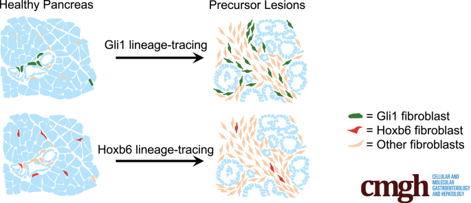
Graphical abstract
Graphical abstract CC BY-NC-ND · PubMed Central
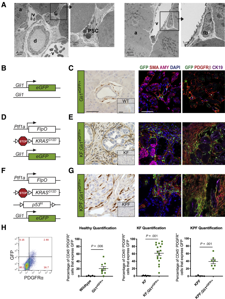
Figure 1 Gli1+fibroblasts are present in the healthy and neoplastic pancreas. (A) TEM images of a healthy mouse pancreas. Black squares and arrows indicate sections that are magnified. (B) Genetic scheme for the knock-in Gli1EGFP/+ reporter mouse. (C) Immunohistochemical staining for GFP and IF staining of GFP (green), αSMA (red), and amylase (pink); and GFP (green), PDGFRβ (red), and CK19 (pink) in healthy Gli1EGFP/+ mice and wild-type (inset) mice. Scale bar: 50 um. (D) Genetic scheme for the Ptf1aFlpO/+;KrasFRT-stop-FRT-G12D/+;Gli1EGFP/+ (KF;Gli1EGFP/+) mouse. (E) IHC staining for GFP and the IF staining panels in KF;Gli1EGFP/+ and KF-negative control (inset) mice after 3 weeks of cerulein-induced acute pancreatitis. (F) Genetic scheme for the KF;Trp53FRT-stop-FRT/+,Gli1EGFP/+ (KPF;Gli1EGFP/+) mouse. (G) IHC staining for GFP and the IF staining panels in KPF;Gli1EGFP/+ and KPF-negative control (inset) mice after evidence of disease burden. (H) Representative flow cytometry plot of GFP against PDGFR⍺, gated on 4′,6-diamidino-2-phenylindole (DAPI)- CD45- cells, and quantification of the percentage of GFP+ PDGFR⍺+ cells at the different disease stages (n ≥ 5). All data are expressed as means ± SEM. a, acinar cell; AMY, amylase; CK19, cytokeratin 19; d, duct; EGFP, enhanced green fluorescent protein; fb, fibroblast; PSC, pancreatic stellate cell; v, blood vessel.
Figure 1 Gli1+fibroblasts are present in the healthy and neoplastic pancreas. (A) TEM images of a healthy mouse pancreas. Black squares and arrows indicate sections that are magnified. (B) Genetic scheme for the knock-in Gli1EGFP/+ reporter mouse. (C) Immunohistochemical staining for GFP and IF staining of GFP (green), αSMA (red), and amylase (pink); and GFP (green), PDGFRβ (red), and CK19 (pink) in healthy Gli1EGFP/+ mice and wild-type (inset) mice. Scale bar: 50 um. (D) Genetic scheme for the Ptf1aFlpO/+;KrasFRT-stop-FRT-G12D/+;Gli1EGFP/+ (KF;Gli1EGFP/+) mouse. (E) IHC staining for GFP and the IF staining panels in KF;Gli1EGFP/+ and KF-negative control (inset) mice after 3 weeks of cerulein-induced acute pancreatitis. (F) Genetic scheme for the KF;Trp53FRT-stop-FRT/+,Gli1EGFP/+ (KPF;Gli1EGFP/+) mouse. (G) IHC staining for GFP and the IF staining panels in KPF;Gli1EGFP/+ and KPF-negative control (inset) mice after evidence of disease burden. (H) Representative flow cytometry plot of GFP against PDGFR⍺, gated on 4′,6-diamidino-2-phenylindole (DAPI)- CD45- cells, and quantification of the percentage of GFP+ PDGFR⍺+ cells at the different disease stages (n ≥ 5). All data are expressed as means ± SEM. a, acinar cell; AMY, amylase; CK19, cytokeratin 19; d, duct; EGFP, enhanced green fluorescent protein; fb, fibroblast; PSC, pancreatic stellate cell; v, blood vessel.CC BY-NC-ND · PubMed Central
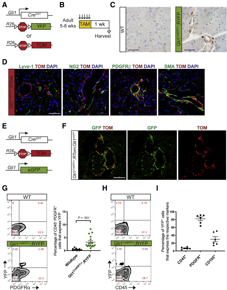
Figure 2 CreERT model labels healthy adult Gli1-expressing fibroblasts. (A) Genetic scheme for Gli1CreERT/+ crossed with either a Rosa26YFP/+ or a Rosa26tdTomato/+ reporter. (B) Experimental design for examining healthy adult mice expressing Gli1 in the pancreas. Adult mice ages 5–8 weeks were given tamoxifen gavages (4 mg/mouse/day) for 5 days. Tissue was examined 1 week after completing gavages. (C) IHC staining for YFP in healthy adult mice. Scale bar: 50 um. (D) IF staining of the following: Lyve-1 (green), Tomato (red), and DAPI (blue); NG2 (green), Tomato (red), and DAPI (blue); PDGFRβ (green), Tomato (red), and DAPI (blue); and αSMA (green), Tomato (red), and DAPI (blue) in Gli1CreERT/+;RTom samples. Scale bar: 50 um. (E) Genetic scheme for a Gli1CreER/+;Gli1EGFP/+,RTomato mouse model. (F) IF staining of GFP and Tomato in the Gli1CreERT/+;Gli1EGFP/+;RTom mouse. Scale bar: 50 um. (G) Representative YFP vs PDGFR⍺, flow cytometry plots of DAPI- cells in a healthy Gli1CreERT/+;RYFP mouse and wild-type control and the quantification (n ≥ 10). (H) Representative YFP vs CD45 flow cytometry plot. (I) Quantification of the percentage of YFP+ cells that expressed CD45, PDGFR⍺, or CD105, as determined by flow cytometry gating. All data are expressed as means ± SEM. TAM, tamoxifen; TOM, Tomato; WT, wild-type.
Figure 2 CreERT model labels healthy adult Gli1-expressing fibroblasts. (A) Genetic scheme for Gli1CreERT/+ crossed with either a Rosa26YFP/+ or a Rosa26tdTomato/+ reporter. (B) Experimental design for examining healthy adult mice expressing Gli1 in the pancreas. Adult mice ages 5–8 weeks were given tamoxifen gavages (4 mg/mouse/day) for 5 days. Tissue was examined 1 week after completing gavages. (C) IHC staining for YFP in healthy adult mice. Scale bar: 50 um. (D) IF staining of the following: Lyve-1 (green), Tomato (red), and DAPI (blue); NG2 (green), Tomato (red), and DAPI (blue); PDGFRβ (green), Tomato (red), and DAPI (blue); and αSMA (green), Tomato (red), and DAPI (blue) in Gli1CreERT/+;RTom samples. Scale bar: 50 um. (E) Genetic scheme for a Gli1CreER/+;Gli1EGFP/+,RTomato mouse model. (F) IF staining of GFP and Tomato in the Gli1CreERT/+;Gli1EGFP/+;RTom mouse. Scale bar: 50 um. (G) Representative YFP vs PDGFR⍺, flow cytometry plots of DAPI- cells in a healthy Gli1CreERT/+;RYFP mouse and wild-type control and the quantification (n ≥ 10). (H) Representative YFP vs CD45 flow cytometry plot. (I) Quantification of the percentage of YFP+ cells that expressed CD45, PDGFR⍺, or CD105, as determined by flow cytometry gating. All data are expressed as means ± SEM. TAM, tamoxifen; TOM, Tomato; WT, wild-type.CC BY-NC-ND · PubMed Central
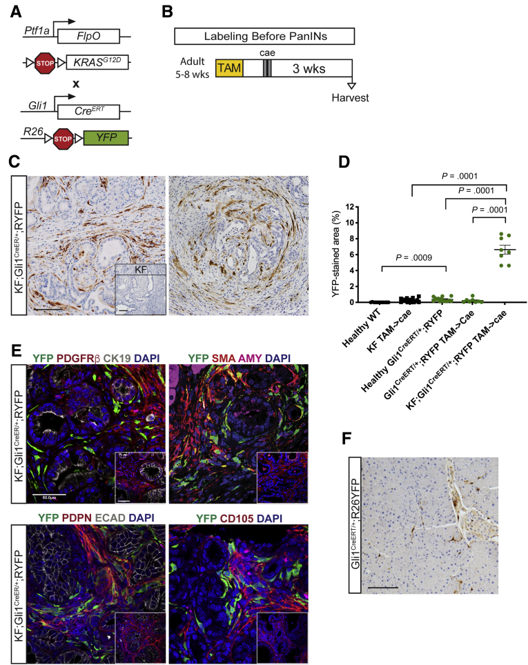
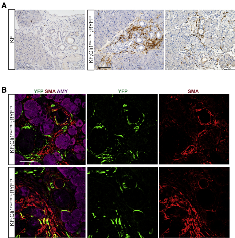
Figure 3 Gli1+fibroblasts lineage-traced before pancreatitis-induced PanIN lesion formation contribute to the stroma. (A) Genetic scheme for a KF;Gli1CreERT/+;RYFP mouse. (B) Experimental design for labeling healthy Gli1+ cells before PanIN generation. Adult mice 5–8 weeks old were given 5 tamoxifen gavages and rested for a week before 2 days of cerulein injections to induce pancreatitis. After 3 weeks, samples were collected. (C) IHC staining for YFP in KF;Gli1CreERT/+;RYFP and KF control tissue (inset) labeled before PanIN generation. Scale bar: 100 um. (D) Quantification of YFP+ staining area from IHC (n ≥ 8) (E) IF staining on KF;Gli1CreERT/+;RYFP and KF control (inset) samples of YFP (green), PDGFRβ (red), cytokeratin 19 (CK19) (white), and 4′,6-diamidino-2-phenylindole (DAPI) (blue); YFP (green), αSMA (red), amylase (pink), and DAPI (blue); YFP (green), podoplanin (red), E-cadherin (white), and DAPI (blue); and YFP (green), CD105 (red), and DAPI (blue). Scale bar: 50 um. (F) A representative YFP IHC image of a Gli1CreERT/+;RYFP mouse that followed the same labeling scheme. Scale bar: 100 um. AMY, amylase; ECAD, E-cadherin; TAM, tamoxifen; WT, wild-type.
Figure 3 Gli1+fibroblasts lineage-traced before pancreatitis-induced PanIN lesion formation contribute to the stroma. (A) Genetic scheme for a KF;Gli1CreERT/+;RYFP mouse. (B) Experimental design for labeling healthy Gli1+ cells before PanIN generation. Adult mice 5–8 weeks old were given 5 tamoxifen gavages and rested for a week before 2 days of cerulein injections to induce pancreatitis. After 3 weeks, samples were collected. (C) IHC staining for YFP in KF;Gli1CreERT/+;RYFP and KF control tissue (inset) labeled before PanIN generation. Scale bar: 100 um. (D) Quantification of YFP+ staining area from IHC (n ≥ 8) (E) IF staining on KF;Gli1CreERT/+;RYFP and KF control (inset) samples of YFP (green), PDGFRβ (red), cytokeratin 19 (CK19) (white), and 4′,6-diamidino-2-phenylindole (DAPI) (blue); YFP (green), αSMA (red), amylase (pink), and DAPI (blue); YFP (green), podoplanin (red), E-cadherin (white), and DAPI (blue); and YFP (green), CD105 (red), and DAPI (blue). Scale bar: 50 um. (F) A representative YFP IHC image of a Gli1CreERT/+;RYFP mouse that followed the same labeling scheme. Scale bar: 100 um. AMY, amylase; ECAD, E-cadherin; TAM, tamoxifen; WT, wild-type.CC BY-NC-ND · PubMed Central
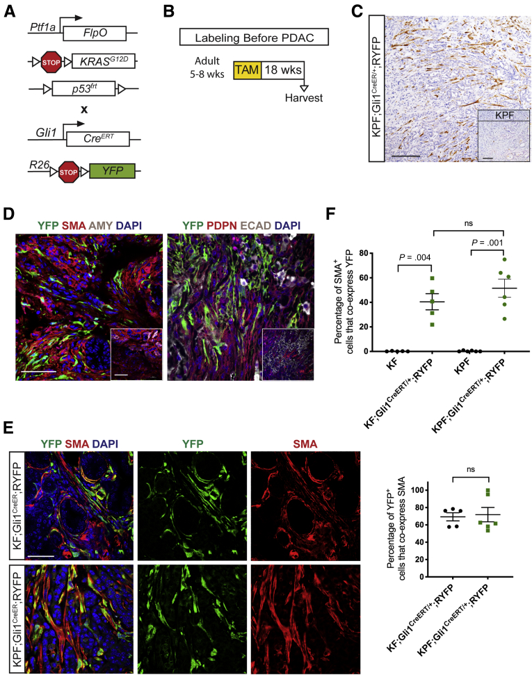
Figure 4 Gli1+fibroblasts contribute to the stroma when lineage-traced in a spontaneous pancreatic carcinogenesis model. (A) Genetic scheme for a KPF;Gli1CreERT/+;RYFP mouse. (B) Experimental design for labeling healthy Gli1+ cells before spontaneous carcinogenesis. Adult mice 5–8 weeks old were given 5 tamoxifen gavages, aged, and then harvested on evidence of disease burden. (C) IHC staining for YFP in KPF;Gli1CreERT/+;RYFP and KPF control (inset) mice. Scale bar: 100 um. (D) IF staining for YFP (green), αSMA (red), amylase (gray), and DAPI (blue); and YFP (green), podoplanin (red), E-cadherin (white), and DAPI (blue) in KPF;Gli1CreERT/+;RYFP and KPF control (inset) samples. Scale bar: 50 um. (E) Merged and single-channel IF images for YFP (green), αSMA (red), and DAPI (blue) in KF and KPF Gli1CreER;RYFP samples. Scale bar: 50 um. (F) IF quantification of the percentage of αSMA+ cells that co-expressed YFP in lineage-traced KF and KPF samples (n ≥ 5). (F) IF quantification of the percentage of YFP+ cells that co-expressed αSMA in lineage-traced KF and KPF samples (n ≥ 8). All data are expressed as means ± SEM. AMY, amylase; TAM, tamoxifen.
Figure 4 Gli1+fibroblasts contribute to the stroma when lineage-traced in a spontaneous pancreatic carcinogenesis model. (A) Genetic scheme for a KPF;Gli1CreERT/+;RYFP mouse. (B) Experimental design for labeling healthy Gli1+ cells before spontaneous carcinogenesis. Adult mice 5–8 weeks old were given 5 tamoxifen gavages, aged, and then harvested on evidence of disease burden. (C) IHC staining for YFP in KPF;Gli1CreERT/+;RYFP and KPF control (inset) mice. Scale bar: 100 um. (D) IF staining for YFP (green), αSMA (red), amylase (gray), and DAPI (blue); and YFP (green), podoplanin (red), E-cadherin (white), and DAPI (blue) in KPF;Gli1CreERT/+;RYFP and KPF control (inset) samples. Scale bar: 50 um. (E) Merged and single-channel IF images for YFP (green), αSMA (red), and DAPI (blue) in KF and KPF Gli1CreER;RYFP samples. Scale bar: 50 um. (F) IF quantification of the percentage of αSMA+ cells that co-expressed YFP in lineage-traced KF and KPF samples (n ≥ 5). (F) IF quantification of the percentage of YFP+ cells that co-expressed αSMA in lineage-traced KF and KPF samples (n ≥ 8). All data are expressed as means ± SEM. AMY, amylase; TAM, tamoxifen.CC BY-NC-ND · PubMed Central
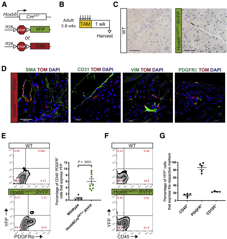
Figure 5 Hoxb6 labels a subset of mesenchymal cells in the healthy pancreas. (A) Genetic scheme for Hoxb6CreERT/+ crossed with either a RYFP reporter or RTomato reporter. (B) Experimental protocol for examining healthy adult mice expressing Hoxb6 in the pancreas. Adult mice aged 5–8 weeks were given tamoxifen gavages (4 mg/mouse/day) for 5 days. Tissue was examined 1 week after completing gavages. (C) IHC staining for YFP in healthy adult mice. Scale bar: 50 um. (D) IF staining of αSMA (green), Tomato (red), and 4′,6-diamidino-2-phenylindole (DAPI) (blue); CD31 (green), Tomato (red), and DAPI (blue); vimentin (green), Tomato (red), and DAPI (blue); and PDGFRβ (green), Tomato (red), and DAPI (blue) in healthy Hoxb6CreERT/+;RTom tissue. Scale bar: 50 um. (E) Representative YFP vs PDGFR⍺+ flow cytometry plots of DAPI- cells in a Hoxb6CreERT/+;RYFP mouse and wild-type control and the quantification (n ≥ 5). (F) Representative YFP vs CD45 flow cytometry plot. (G) Quantification of the percentage of YFP+ cells that expressed CD45, PDGFR⍺, or CD105 as determined by flow cytometry gating. All data are expressed as means ± SEM. TAM, tamoxifen; VIM, vimentin; WT, wild-type.
Figure 5 Hoxb6 labels a subset of mesenchymal cells in the healthy pancreas. (A) Genetic scheme for Hoxb6CreERT/+ crossed with either a RYFP reporter or RTomato reporter. (B) Experimental protocol for examining healthy adult mice expressing Hoxb6 in the pancreas. Adult mice aged 5–8 weeks were given tamoxifen gavages (4 mg/mouse/day) for 5 days. Tissue was examined 1 week after completing gavages. (C) IHC staining for YFP in healthy adult mice. Scale bar: 50 um. (D) IF staining of αSMA (green), Tomato (red), and 4′,6-diamidino-2-phenylindole (DAPI) (blue); CD31 (green), Tomato (red), and DAPI (blue); vimentin (green), Tomato (red), and DAPI (blue); and PDGFRβ (green), Tomato (red), and DAPI (blue) in healthy Hoxb6CreERT/+;RTom tissue. Scale bar: 50 um. (E) Representative YFP vs PDGFR⍺+ flow cytometry plots of DAPI- cells in a Hoxb6CreERT/+;RYFP mouse and wild-type control and the quantification (n ≥ 5). (F) Representative YFP vs CD45 flow cytometry plot. (G) Quantification of the percentage of YFP+ cells that expressed CD45, PDGFR⍺, or CD105 as determined by flow cytometry gating. All data are expressed as means ± SEM. TAM, tamoxifen; VIM, vimentin; WT, wild-type.CC BY-NC-ND · PubMed Central
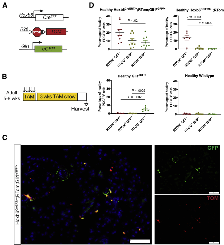
Figure 6 Hoxb6 and Gli1 are expressed in different populations. (A) Genetic scheme for a Hoxb6CreERT/+;RTom;Gli1EGFP/+ mouse model. (B) Experimental design for examining healthy Hoxb6CreERT/+;RTom;Gli1EGFP/+ mice. Adult mice aged 5–8 weeks were given tamoxifen gavages (4 mg/mouse/day) for 5 days. Mice then were placed on tamoxifen chow for 3 weeks. Tissue was examined 1 week after completing chow regimen. (C) IF staining of GFP and Tomato in the Hoxb6CreERT/+;RTom;Gli1EGFP/+ mouse. Scale bar: 50 um. (D) Flow cytometry quantification of the percentage of healthy CD45- PDGFR⍺+ cells that expressed Tomato, GFP, or both (n ≥ 6). All data are expressed as means ± SEM. RTOM, Rosa26LSL-tdTomato; TAM, tamoxifen.
Figure 6 Hoxb6 and Gli1 are expressed in different populations. (A) Genetic scheme for a Hoxb6CreERT/+;RTom;Gli1EGFP/+ mouse model. (B) Experimental design for examining healthy Hoxb6CreERT/+;RTom;Gli1EGFP/+ mice. Adult mice aged 5–8 weeks were given tamoxifen gavages (4 mg/mouse/day) for 5 days. Mice then were placed on tamoxifen chow for 3 weeks. Tissue was examined 1 week after completing chow regimen. (C) IF staining of GFP and Tomato in the Hoxb6CreERT/+;RTom;Gli1EGFP/+ mouse. Scale bar: 50 um. (D) Flow cytometry quantification of the percentage of healthy CD45- PDGFR⍺+ cells that expressed Tomato, GFP, or both (n ≥ 6). All data are expressed as means ± SEM. RTOM, Rosa26LSL-tdTomato; TAM, tamoxifen.CC BY-NC-ND · PubMed Central
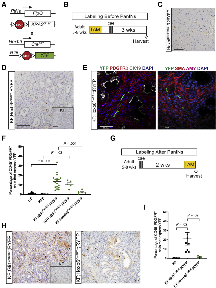
Figure 7 Hoxb6+fibroblasts do not contribute to the pancreatic stroma. (A) Genetic scheme for the KF;Hoxb6CreERT/+;RYFP model. (B) Experimental design for labeling healthy Hoxb6+ cells before PanIN generation. Adult mice 5–8 weeks old were given 5 tamoxifen gavages and rested a week before 2 days of cerulein injections to induce pancreatitis. After 3 weeks, mice were harvested. (C) A representative YFP IHC image of a Hoxb6CreERT/+;RYFP mouse following the protocol. Scale bar: 100 um. (D) IHC staining for YFP in KF;Hoxb6CreERT/+;RYFP and KF control (inset) mice labeled before PanIN generation. Scale bar: 100 um. (E) IF staining panels of YFP (green), PDGFRβ (red), cytokeratin 19 (CK19) (white), DAPI (blue); and YFP (green), αSMA (red), amylase (pink), and DAPI (blue) in KF;Hoxb6CreERT/+;RYFP mice labeled before PanIN generation. Scale bar: 50 um. (F) Flow cytometry quantification of the percentage of CD45- PDGFR+ cells that expressed YFP in lineage-traced Gli1 and Hoxb6 mice and their controls (n ≥ 5). (G) Experimental design for labeling after PanIN generation. Adult mice 5–8 weeks old were given cerulein injections, rested for 2 weeks, and then given 5 gavages of tamoxifen before tissue collection. (H) IHC staining for YFP in KF;Gli1CreERT/+;RYFP and KF;Hoxb6CreERT/+;RYFP mice labeled after PanIN generation. Inset shows KF control. Scale bar: 100 um. (C) Flow cytometry quantification of the percentage of CD45-PDGFR+ cells that express YFP in KF;Gli1CreERT/+;RYFP and KF;Hoxb6CreERT/+;RYFP mice labeled after PanIN generation (n ≥ 3). All data are expressed as means ± SEM. AMY, amylase.
Figure 7 Hoxb6+fibroblasts do not contribute to the pancreatic stroma. (A) Genetic scheme for the KF;Hoxb6CreERT/+;RYFP model. (B) Experimental design for labeling healthy Hoxb6+ cells before PanIN generation. Adult mice 5–8 weeks old were given 5 tamoxifen gavages and rested a week before 2 days of cerulein injections to induce pancreatitis. After 3 weeks, mice were harvested. (C) A representative YFP IHC image of a Hoxb6CreERT/+;RYFP mouse following the protocol. Scale bar: 100 um. (D) IHC staining for YFP in KF;Hoxb6CreERT/+;RYFP and KF control (inset) mice labeled before PanIN generation. Scale bar: 100 um. (E) IF staining panels of YFP (green), PDGFRβ (red), cytokeratin 19 (CK19) (white), DAPI (blue); and YFP (green), αSMA (red), amylase (pink), and DAPI (blue) in KF;Hoxb6CreERT/+;RYFP mice labeled before PanIN generation. Scale bar: 50 um. (F) Flow cytometry quantification of the percentage of CD45- PDGFR+ cells that expressed YFP in lineage-traced Gli1 and Hoxb6 mice and their controls (n ≥ 5). (G) Experimental design for labeling after PanIN generation. Adult mice 5–8 weeks old were given cerulein injections, rested for 2 weeks, and then given 5 gavages of tamoxifen before tissue collection. (H) IHC staining for YFP in KF;Gli1CreERT/+;RYFP and KF;Hoxb6CreERT/+;RYFP mice labeled after PanIN generation. Inset shows KF control. Scale bar: 100 um. (C) Flow cytometry quantification of the percentage of CD45-PDGFR+ cells that express YFP in KF;Gli1CreERT/+;RYFP and KF;Hoxb6CreERT/+;RYFP mice labeled after PanIN generation (n ≥ 3). All data are expressed as means ± SEM. AMY, amylase.CC BY-NC-ND · PubMed Central
Figure 8 Lineage-traced Gli1 fibroblasts are present around the earliest lesion development. (A) IHC staining for YFP in KF;Gli1CreERT/+;RYFP mice labeled before PanIN generation. Scale bar: 100 um. (B) IF merged and single-channel images for YFP (green), αSMA (red), and amylase (pink) in KF;Gli1CreERT/+;RYFP mice. Scale bar: 50 um. AMY, amylase.
Figure 8 Lineage-traced Gli1 fibroblasts are present around the earliest lesion development. (A) IHC staining for YFP in KF;Gli1CreERT/+;RYFP mice labeled before PanIN generation. Scale bar: 100 um. (B) IF merged and single-channel images for YFP (green), αSMA (red), and amylase (pink) in KF;Gli1CreERT/+;RYFP mice. Scale bar: 50 um. AMY, amylase.CC BY-NC-ND · PubMed Central

Verification

Claims the paper makes

The claims and statistics of this paper haven’t been checked yet.

Run the check — free, no account needed. It reads the open-access text and checks the sources it cites.

A few free checks a day · papers already verified open instantly · results are public

Claims other papers make about this one

Each is checked against this paper’s own text: does it say what the citing paper says it does?

Self-check

Percentages, sums and reported differences re-computed from the paper’s own numbers.

The self-check needs the paper’s full text.

Citations

Cites

Show all 83 references

Cited by

Sorted by citations

See all 110

Metadata

Authors
Garcia, P. E., Adoumie, M., Kim, E. C., Zhang, Y., Scales, M. K., El-Tawil, Y. S., Shaikh, A. Z., Wen, H.-J., Bednar, F., Allen, B. L., Wellik, D. M., Crawford, H. C., & Pasca di Magliano, M.
Affiliations
  • University of Michigan (US)
  • University of Wisconsin–Madison (US)
Venue
Cellular and Molecular Gastroenterology and Hepatology
Publisher
Elsevier BV
Published
2020
Volume
10
Issue
3
Pages
581-599
ISSN
2352-345X (ISSN-L)
OpenAlex
W3027333363
Cited by
110
References
83
Citations by year
2026: 16 · 2025: 16 · 2024: 25 · 2023: 18 · 2022: 16 · 2021: 14 · 2020: 5
FWCI
4.78
Type
journal article
Language
en
Open access
Locations
  • Cellular and Molecular Gastroenterology and Hepatology (publishedVersion, cc-by-nc-nd)
  • PubMed Central (publishedVersion, cc-by-nc-nd)
  • PubMed (publishedVersion)
  • DOAJ (DOAJ: Directory of Open Access Journals) (submittedVersion, cc-by-sa)
Funders
National Cancer Institute
Topics
  • Health Sciences › Medicine › Oncology › Pancreatic and Hepatic Oncology Research
  • Health Sciences › Medicine › Surgery › Pancreatitis Pathology and Treatment
  • Health Sciences › Medicine › Endocrinology, Diabetes and Metabolism › Pancreatic function and diabetes
Keywords
pancreatic cancer, pancreatic stroma, pancreatic fibroblasts, fibroblast heterogeneity, Gli1, HOXB6, pancreatic intraepithelial neoplasia, carcinogenesis
MeSH terms
Zinc Finger Protein GLI1, Animals, Disease Models, Animal, Fibroblasts, Humans, Mice, Transgenic, Pancreas, Pancreatic Neoplasms, Proto-Oncogene Proteins p21(ras), Homeodomain Proteins, Carcinoma, Pancreatic Ductal, Mice, Carcinogenesis

About these checks

Registry, retraction, Cites and statistics results are computed, not judged, with one exception: a printed reference that no index lists is read once by a model before it counts as a citation error. Claims are extracted by a model and kept only when their quote appears verbatim in the text; each cited source is then checked, with a model’s help, for whether it says what the claim relies on. Those verdicts can be wrong — read the claim’s quote and the cited source before relying on one.

Statistics and arithmetic are recomputed, not judged.

Confidence bands are shown only for fields calibrated on our annotated test set. None is calibrated yet, so no band is shown.

What counts as a claim ›How we measure confidence ›How we check this ›

Are you an author, or do you see a problem with this page? Request a review