

The DOI matches its Crossref record; 83 of the 83 references it cites were found in a registry or an index.
Differential Contribution of Pancreatic Fibroblast Subsets to the Pancreatic Cancer Stroma
Open accessAbstract
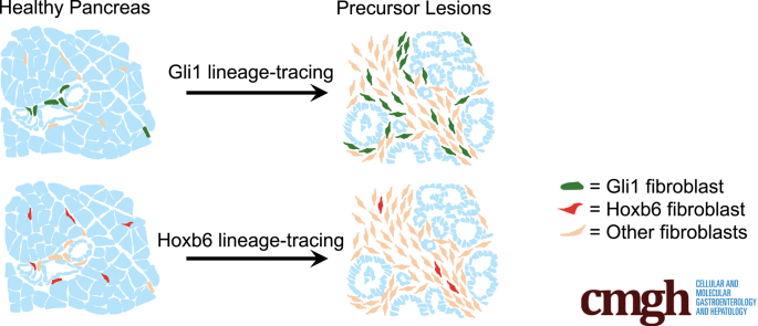
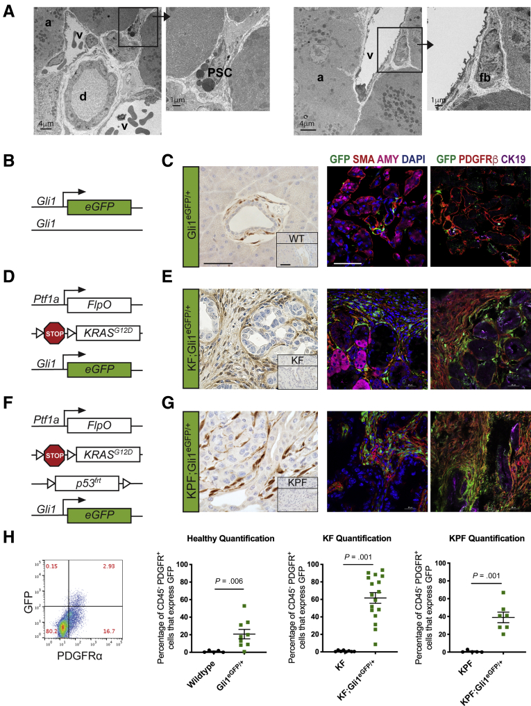
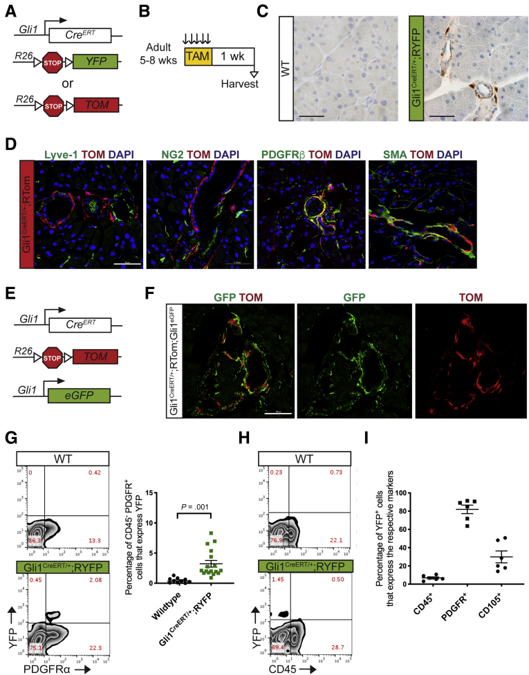
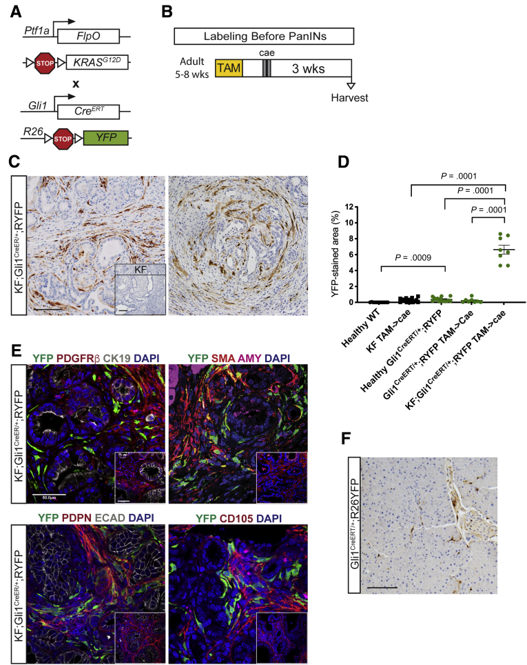
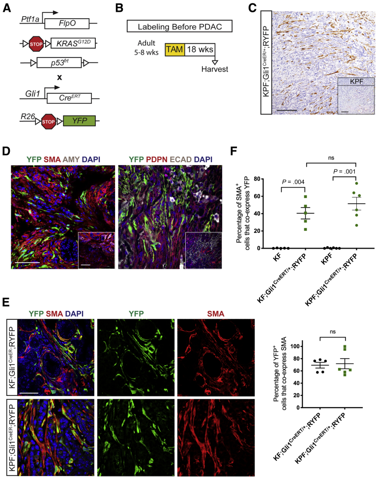
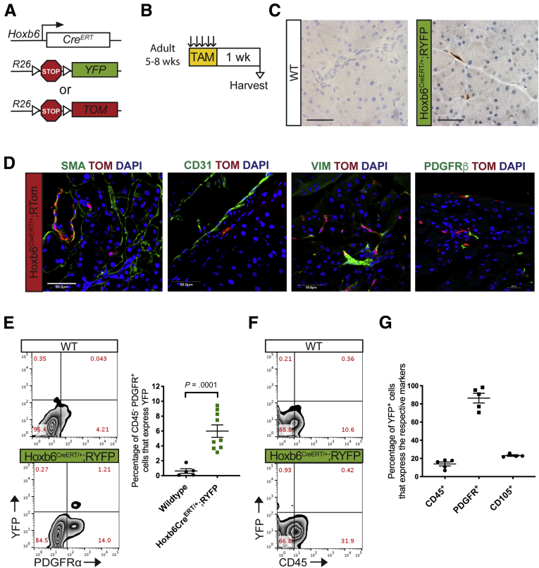
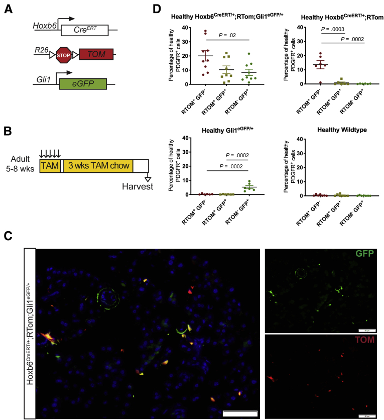
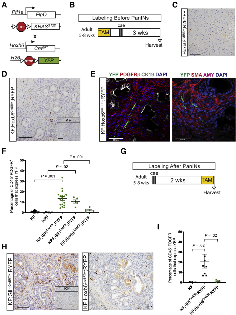
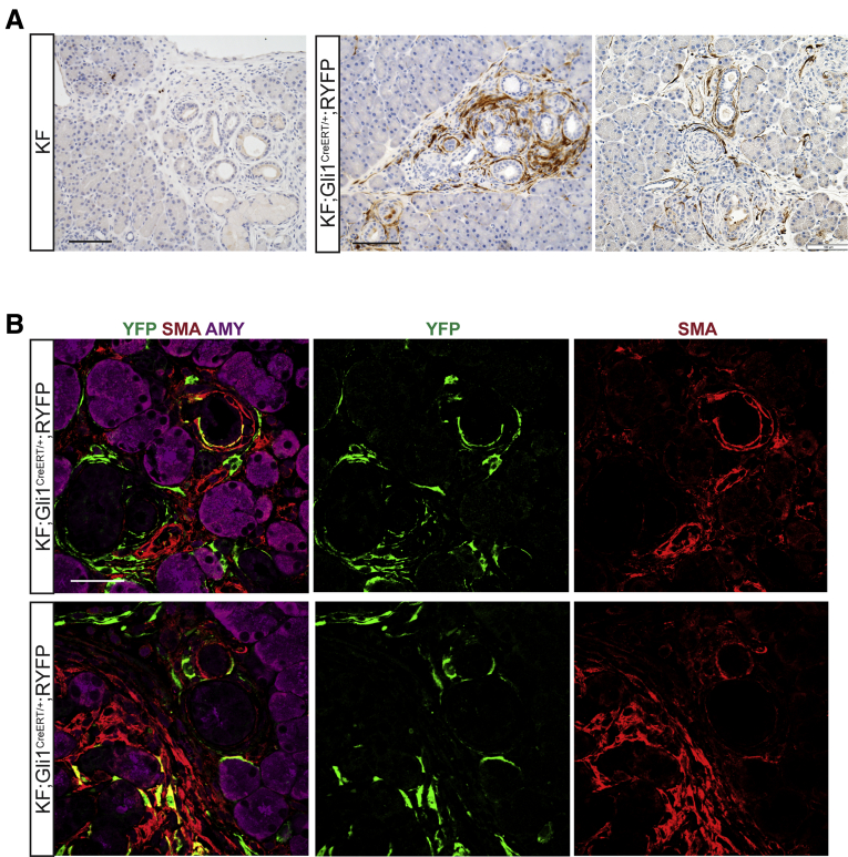
Background & Aims Although the healthy pancreas consists mostly of epithelial cells, pancreatic cancer and the precursor lesions known as pancreatic intraepithelial neoplasia, are characterized by an extensive accumulation of fibroinflammatory stroma that includes a substantial and heterogeneous fibroblast population. The cellular origin of fibroblasts within the stroma has not been determined. Here, we show that the Gli1 and Hoxb6 markers label distinct fibroblast populations in the healthy mouse pancreas. We then set out to determine whether these distinct fibroblast populations expanded during carcinogenesis. Methods We developed genetically engineered models using a dual-recombinase approach that allowed us to induce pancreatic cancer formation through codon-optimized Flp recombinase-driven epithelial recombination of Kirsten rat sarcoma viral oncogene homolog while labeling Gli1 + or Hoxb6 + fibroblasts in an inducible manner. By using these models, we lineage-traced these 2 fibroblast populations during the process of carcinogenesis. Results Although in the healthy pancreas Gli1 + fibroblasts and Hoxb6 + fibroblasts are present in similar numbers, they contribute differently to the stroma in carcinogenesis. Namely, Gli1 + fibroblasts expand dramatically, whereas Hoxb6 + cells do not. Conclusions Fibroblasts present in the healthy pancreas expand during carcinogenesis, but with a different prevalence for different subtypes. Here, we compared Gli1 + and Hoxb6 + fibroblasts and found only Gli1 + expanded to contribute to the stroma during pancreatic carcinogenesis.
Overview
The claims and statistics of this paper haven’t been checked yet.
Run the check — free, no account needed. It reads the open-access text and checks the sources it cites.
Integrity signals
Registry
Cites
Statistics
Figures
Verification
Claims the paper makes
The claims and statistics of this paper haven’t been checked yet.
Run the check — free, no account needed. It reads the open-access text and checks the sources it cites.
Claims other papers make about this one
Each is checked against this paper’s own text: does it say what the citing paper says it does?
In the citing paper
“recent lineage tracing studies targeting Fabp4+ stellate cells revealed that they give rise to a minority of CAFs142,143”
From Pancreatic cancer: Advances and challenges (2023) · cites this paper as [143]
Self-check
Percentages, sums and reported differences re-computed from the paper’s own numbers.
The self-check needs the paper’s full text.
Citations
Cites
Rahib, L., Smith, B. D., Aizenberg, R., Rosenzweig, A. B., Fleshman, J. M., & Matrisian, L. M. (2014) · Cancer Research · doi:10.1158/0008-5472.can-14-0155 · Cited by 7,094 · cited 1×
“ancreatic ductal adenocarcinoma (PDAC) is a deadly Pmalignancy that is projected to become the second leading cause of cancer-related death by 2030.(1) The 5-year”
BackgroundA framework for advancing our understanding of cancer-associated fibroblasts
Sahai, E., Astsaturov, I., Cukierman, E., DeNardo, D. G., Egeblad, M., Evans, R. M., Fearon, D., Greten, F. R., Hingorani, S. R., Hunter, T., et al. (2020) · Nature Reviews Cancer · doi:10.1038/s41568-019-0238-1 · Cited by 3,863
Olive, K. P., Jacobetz, M. A., Davidson, C. J., Gopinathan, A., McIntyre, D., Honess, D., Madhu, B., Goldgraben, M. A., Caldwell, M. E., Allard, D., et al. (2009) · Science · doi:10.1126/science.1171362 · Cited by 3,170
Özdemir, B. C., Pentcheva-Hoang, T., Carstens, J. L., Zheng, X., Wu, C.-C., Simpson, T. R., Laklai, H., Sugimoto, H., Kahlert, C., Novitskiy, S. V., et al. (2014) · Cancer Cell · doi:10.1016/j.ccr.2014.04.005 · Cited by 2,681 · cited 1×
“However, other results suggested that fibroblasts could restrain tumor formation and that depleting them might accelerate carcinogenesis.(13) This paradox might be explained by the observation that fibroblasts are not a homogenous population, but exist in subsets with different roles.”
ResultPericytes: Developmental, Physiological, and Pathological Perspectives, Problems, and Promises
Armulik, A., Genové, G., & Betsholtz, C. (2011) · Developmental Cell · doi:10.1016/j.devcel.2011.07.001 · Cited by 2,653 · cited 1×
“In the pancreas, themesenchyme surrounds and supports epithelium development(50) and differentiates to form cell types such as fibroblasts, pancreatic stellate cells, and pericytes.(51,52) Activated fibroblasts, also known as myofibroblasts, help regulate wound repair and recovery,(53,54) but in the context of certain…”
BackgroundHingorani, S. R., Wang, L., Multani, A. S., Combs, C., Deramaudt, T. B., Hruban, R. H., Rustgi, A. K., Chang, S., & Tuveson, D. A. (2005) · Cancer Cell · doi:10.1016/j.ccr.2005.04.023 · Cited by 2,609
Distinct populations of inflammatory fibroblasts and myofibroblasts in pancreatic cancer
Öhlund, D., Handly-Santana, A., Biffi, G., Elyada, E., Almeida, A. S., Ponz-Sarvise, M., Corbo, V., Oni, T. E., Hearn, S. A., Lee, E. J., et al. (2017) · Journal of Experimental Medicine · doi:10.1084/jem.20162024 · Cited by 2,590
Preinvasive and invasive ductal pancreatic cancer and its early detection in the mouse
Hingorani, S. R., Petricoin, E. F., Maitra, A., Rajapakse, V., King, C., Jacobetz, M. A., Ross, S., Conrads, T. P., Veenstra, T. D., Hitt, B. A., et al. (2003) · Cancer Cell · doi:10.1016/s1535-6108(03)00309-x · Cited by 2,471
Self-citationEpidemiology of Pancreatic Cancer: Global Trends, Etiology and Risk Factors
Rawla, P., Sunkara, T., & Gaduputi, V. (2019) · World Journal of Oncology · doi:10.14740/wjon1166 · Cited by 2,269 · cited 1×
“survival rate for PDAC is only 10%, which can be attributed to the aggressive and chemoresistant nature of the cancer, in addition to the frequently late diagnosis.(2) In PDAC, more”
BackgroundElyada, E., Bolisetty, M., Laise, P., Flynn, W. F., Courtois, E. T., Burkhart, R. A., Teinor, J. A., Belleau, P., Biffi, G., Lucito, M. S., et al. (2019) · Cancer Discovery · doi:10.1158/2159-8290.cd-19-0094 · Cited by 2,241 · cited 1×
“populations with potentially distinct functions and activation pathways.(14,15,20) Here, we sought to determine which fibroblasts in the”
Background
Cited by
Sorted by citations
Pancreatic cancer: Advances and challenges
Halbrook, C. J., Lyssiotis, C. A., Pasca di Magliano, M., & Maitra, A. (2023) · Cell · doi:10.1016/j.cell.2023.02.014 · Cited by 1,321
Clinical and therapeutic relevance of cancer-associated fibroblasts
Chen, Y., McAndrews, K. M., & Kalluri, R. (2021) · Nature Reviews Clinical Oncology · doi:10.1038/s41571-021-00546-5 · Cited by 1,230
Cancer-associated fibroblasts: from basic science to anticancer therapy
Yang, D., Liu, J., Qian, H., & Zhuang, Q. (2023) · Experimental & Molecular Medicine · doi:10.1038/s12276-023-01013-0 · Cited by 572
Tumor Microenvironment in Pancreatic Cancer Pathogenesis and Therapeutic Resistance
Sherman, M. H., & Beatty, G. L. (2022) · Annual Review of Pathology Mechanisms of Disease · doi:10.1146/annurev-pathmechdis-031621-024600 · Cited by 501
Activated fibroblasts in cancer: Perspectives and challenges
Caligiuri, G., & Tuveson, D. A. (2023) · Cancer Cell · doi:10.1016/j.ccell.2023.02.015 · Cited by 450
Han, L., Wu, Y., Fang, K., Sweeney, S., Roesner, U. K., Parrish, M., Patel, K., Walter, T., Piermattei, J., Trimboli, A., et al. (2023) · Nature Communications · doi:10.1038/s41467-022-34464-6 · Cited by 369
Cancer-associated fibroblasts and their influence on tumor immunity and immunotherapy
Barrett, R. L., & Puré, E. (2020) · eLife · doi:10.7554/elife.57243 · Cited by 357
“Further adding to the complexity is that fibroblasts in healthy tissue, from skin to pancreas, exhibit heterogeneity even before activation (Philippeos et al., 2018; Garcia et al., 2020).”
BackgroundInhibition of Hedgehog Signaling Alters Fibroblast Composition in Pancreatic Cancer
Steele, N. G., Biffi, G., Kemp, S. B., Zhang, Y., Drouillard, D., Syu, L.-J., Yuan, H., Oni, T. E., Brosnan, E., Elyada, E., et al. (2021) · Clinical Cancer Research · doi:10.1158/1078-0432.ccr-20-3715 · Cited by 289
“The healthy pancreas is home to distinct fibroblast populations (24, 25), which have differing potential to expand during carcinogenesis (26).”
BackgroundSelf-citationXiao, Z., Todd, L., Huang, L., Noguera-Ortega, E., Lu, Z., Huang, L., Kopp, M., Li, Y., Pattada, N., Zhong, W., et al. (2023) · Nature Communications · doi:10.1038/s41467-023-40850-5 · Cited by 272
Helms, E. J., Berry, M. W., Chaw, R. C., DuFort, C. C., Sun, D., Onate, M. K., Oon, C., Bhattacharyya, S., Sanford-Crane, H., Horton, W., et al. (2022) · Cancer Discovery · doi:10.1158/2159-8290.cd-21-0601 · Cited by 238
The Origin and Contribution of Cancer-Associated Fibroblasts in Colorectal Carcinogenesis
Kobayashi, H., Gieniec, K. A., Lannagan, T. R. M., Wang, T., Asai, N., Mizutani, Y., Iida, T., Ando, R., Thomas, E. M., Sakai, A., et al. (2021) · Gastroenterology · doi:10.1053/j.gastro.2021.11.037 · Cited by 214
“Interestingly, a recent paper found that Gli1þ pancreatic stellate cells could contribute to approximately half of ACTA2þ CAFs in a mouse model of pancreatic cancer.(33) Further research is required to determine the hierarchic or overlapping relationship between Lepr-lineage and Gli1-lineage CAFs in different tissues…”
BackgroundArginase 1 is a key driver of immune suppression in pancreatic cancer
Menjivar, R. E., Nwosu, Z. C., Du, W., Donahue, K. L., Hong, H. S., Espinoza, C. E., Brown, K. L., Velez-Delgado, A., Yan, W., Lima, F., et al. (2023) · eLife · doi:10.7554/elife.80721 · Cited by 189
“The dualrecombinase system integrates both FlippaseFRT (FlpFRT) and Cre- loxP recombination technologies to independently modify epithelial cells and myeloid cells (Garcia et al., 2020; Wen et al., 2019).”
BackgroundMethodInfluentialSelf-citationHarnessing metabolic dependencies in pancreatic cancers
Encarnación-Rosado, J., & Kimmelman, A. C. (2021) · Nature Reviews Gastroenterology & Hepatology · doi:10.1038/s41575-021-00431-7 · Cited by 190
Hox genes in development and beyond
Hubert, K. A., & Wellik, D. M. (2023) · Development · doi:10.1242/dev.192476 · Cited by 129
Self-citationExtrinsic KRAS signaling shapes the pancreatic microenvironment through fibroblast reprogramming
Velez-Delgado, A., Donahue, K. L., Brown, K. L., Du, W., Irizarry-Negron, V., Menjivar, R. E., Lasse Opsahl, E. L., Steele, N. G., The, S., Lazarus, J., et al. (2022) · Cellular and Molecular Gastroenterology and Hepatology · doi:10.1016/j.jcmgh.2022.02.016 · Cited by 91
“cells), but recent lineage tracing work by our group has shown that a substantial proportion of CAFs derive from perivascular fibroblasts present in the normal pancreas.(68) Accordingly, a lineage tracing study following pancreatic stellate cells in pancreatic cancer showed that they only contribute to a small subset…”
BackgroundSelf-citationQin, Q., Yu, R., Eriksson, J., Tsai, H., & Zhu, H. (2024) · Cancer Letters · doi:10.1016/j.canlet.2024.216859 · Cited by 102
Vaish, U., Jain, T., Are, A. C., & Dudeja, V. (2021) · International Journal of Molecular Sciences · doi:10.3390/ijms222413408 · Cited by 102
“further shed light on the heterogenous fibroblastic precursors of CAFs in PDAC when they performed lineage-tracing studies using Gli1 and Hoxb6, two mesenchymal markers that have not been associated with stellate cells [43].”
BackgroundLi, T., Hu, C., Huang, T., Zhou, Y., Tian, Q., Chen, H., He, R., Yuan, Y., Yong, J., Jiang, H., et al. (2025) · Cancer Research · doi:10.1158/0008-5472.can-24-3173 · Cited by 94
“47), CD105 + and CD105 � CAFs, LRRC15 + CAFs (48), Gli1 + CAFs, and Hoxb6 + CAFs (49), among others, and each sub-type has a distinct influence on tumor dynamics (46).”
Hypoxia promotes an inflammatory phenotype of fibroblasts in pancreatic cancer
Mello, A. M., Ngodup, T., Lee, Y., Donahue, K. L., Li, J., Rao, A. U. K., Carpenter, E. S., Crawford, H. C., di Magliano, M. P., & Lee, K. E. (2022) · Oncogenesis · doi:10.1038/s41389-022-00434-2 · Cited by 91
“However, recent reports suggest that besides PSCs, PDAC CAFs can arise from multiple cell types [33, 34].”
BackgroundSelf-citationTranscription factors in fibroblast plasticity and CAF heterogeneity
Melchionna, R., Trono, P., Carlo, A. D., Modugno, F. D., & Nisticò, P. (2023) · Journal of Experimental & Clinical Cancer Research · doi:10.1186/s13046-023-02934-4 · Cited by 92
“…in liver fibrosis [25] Normal fibroblast conversion into CAFs GLI1 Specific Gli1 + fibroblasts expansion in tumor stroma during carcinogenesis [26] TBX4 Lost during lung CAF activation Promotes fibroblast proliferation and collagen gel contraction capacity [27] CSL/p53 complex Lost during…”
BackgroundCancer‐associated fibroblasts in pancreatic cancer: new subtypes, new markers, new targets
Menezes, S., Okail, M. H., Jalil, S. M. A., Kocher, H. M., & Cameron, A. J. M. (2022) · The Journal of Pathology · doi:10.1002/path.5926 · Cited by 82
“Garcia et al reported distinct Gli1 and HoxB6 fibroblast lineages in the healthy mouse pancreas, which can both contribute significantly to KF (Ptf1aFlpO/+;KrasFSF-G12D/+) and KPF (Ptf1aFlpO/+;KrasFSF-G12D/+; Trp53FRT-STOP-FRT/+)-driven tumour CAFs, with Gli1+ cells dominating [68].”
BackgroundMasugi, Y. (2022) · Cancers · doi:10.3390/cancers14133293 · Cited by 90
“A recent study using a lineage-tracing platform has revealed differential contributions between two differentially located resident fibroblasts (GLI family zinc finger 1 (GLI1)+ fibroblasts and homeobox B6 (HOXB6)+ fibroblasts) to CAF generation during murine PDAC carcinogenesis [99].”
BackgroundMacrophages and fibroblasts as regulators of the immune response in pancreatic cancer
Shakiba, M., & Tuveson, D. A. (2025) · Nature Immunology · doi:10.1038/s41590-025-02134-6 · Cited by 81
“However, PSCs were later shown to constitute only a minority of CAFs, whereas other cell types, such as pericytes, adipocytes, endothelial cells and mesothelial cells, were shown to also contribute to the generation of CAFs in PDAC 39–42 (Fig.”
Zheng, S., Hu, C., Lin, Q., Li, T., Li, G., Tian, Q., Zhang, X., Huang, T., Ye, Y., He, R., et al. (2024) · Science Translational Medicine · doi:10.1126/scitranslmed.adi0178 · Cited by 64
“CAF subpopulations like myCAFs, iCAFs, and others influence tumor dynamics ( 7 , 29 – 33 ).”
BackgroundHerting, C. J., Karpovsky, I., & Lesinski, G. B. (2021) · Cancer and Metastasis Reviews · doi:10.1007/s10555-021-09988-w · Cited by 60
Sunami, Y., Häußler, J., & Kleeff, J. H. (2020) · Cancers · doi:10.3390/cancers12123770 · Cited by 66
“In a pancreatic cancer mouse model (Ptf1-Cre; frp-stop-frp-KrasG12D/+; Gli1CreERT), lineage-traced, GLI1-positive cells account for a little less than half of the total myofibroblast population [45].”
BackgroundInfluentialDonahue, K. L., Watkoske, H. R., Kadiyala, P., Du, W., Brown, K. L., Scales, M. K., Elhossiny, A. M., Espinoza, C. E., Opsahl, E. L. L., Griffith, B. D., et al. (2024) · Cancer Discovery · doi:10.1158/2159-8290.cd-24-0100 · Cited by 60
“Likewise, the mechanism through which normal fibroblasts and other cells of the healthy pancreas are converted to PAFs and CAFs also remains unclear.”
InfluentialSelf-citationThe fibroinflammatory response in cancer
Xiao, Z., & Puré, E. (2025) · Nature reviews. Cancer · doi:10.1038/s41568-025-00798-8 · Cited by 65
Yan, W., Menjivar, R. E., Bonilla, M. E., Steele, N. G., Kemp, S. B., Du, W., Donahue, K. L., Brown, K. L., Carpenter, E. S., Avritt, F. R., et al. (2023) · Cancer Immunology Research · doi:10.1158/2326-6066.cir-23-0037 · Cited by 52
Self-citationA bird’s eye view of fibroblast heterogeneity: A pan‐disease, pan‐cancer perspective
Pradhan, R., Krishnamurty, A. T., Fletcher, A. L., Turley, S. J., & Müller, S. (2021) · Immunological Reviews · doi:10.1111/imr.12990 · Cited by 51
Pancreatic Fibroblast Heterogeneity: From Development to Cancer
Garcia, P. E., Scales, M. K., Allen, B. L., & di Magliano, M. P. (2020) · Cells · doi:10.3390/cells9112464 · Cited by 48
“Gli1 Lineage Derived from Gli1+ fibroblasts in healthy pancreas Not fully understood Partially contributes to myCAFs [28]”
BackgroundSelf-citationA Quick Guide to CAF Subtypes in Pancreatic Cancer
Brichkina, A. I., Polo, P., Sharma, S. D., Visestamkul, N., & Lauth, M. (2023) · Cancers · doi:10.3390/cancers15092614 · Cited by 45
“However, one recent study used lineage-tracing experiments to delineate the origins of Gli1+ mesenchymal cells in murine PDAC [84].”
BackgroundCancer-Associated Fibroblasts: Lessons from Pancreatic Cancer
Sherman, M. H., & di Magliano, M. P. (2023) · Annual Review of Cancer Biology · doi:10.1146/annurev-cancerbio-061421-035400 · Cited by 39
Ganguly, K., Cox, J. L., Ghersi, D., Grandgenett, P. M., Hollingsworth, M. A., Jain, M., Sushil Kumar, K., & Batra, S. K. (2022) · Gastroenterology · doi:10.1053/j.gastro.2022.02.032 · Cited by 39
Murphy, K. J., Chambers, C. R., Herrmann, D., Timpson, P., & Pereira, B. (2021) · Cancers · doi:10.3390/cancers13143481 · Cited by 37
“The progenitors of these CAF subpopulations are yet to be fully elucidated, however Garcia et al. (2020) recently reported that some myCAFs could be derived from GLI1+ fibroblast progenitors [47].”
BackgroundCancer-Associated Fibroblast Diversity Shapes Tumor Metabolism in Pancreatic Cancer
Peiffer, R., Boumahd, Y., Gullo, C., Crake, R., Letellier, E., Bellahcène, A., & Peulen, O. (2022) · Cancers · doi:10.3390/cancers15010061 · Cited by 38
“In another study, healthy fibroblasts were discriminated, based on GLI1 and HOXB6 expression and upon lineage tracing; both populations only partially contributed to the PDAC CAF landscape [13].”
Combinatorial Gli activity directs immune infiltration and tumor growth in pancreatic cancer
Scales, M. K., Velez-Delgado, A., Steele, N. G., Schrader, H. E., Stabnick, A. M., Yan, W., Mercado-Soto, N. M., Nwosu, Z., Johnson, C. N., Zhang, Y.-Q., et al. (2022) · PLoS Genetics · doi:10.1371/journal.pgen.1010315 · Cited by 28
“To target Gli2 and Gli3 in fibroblasts in vivo, we crossed the Pdgfrα allele [49] into Kras;Ptf1a (KF) [50] mice.”
BackgroundMethodSelf-citationTumor Microenvironment in Pancreatic Intraepithelial Neoplasia
Opitz, F. V., Häberle, L., Daum, A., & Esposito, I. (2021) · Cancers · doi:10.3390/cancers13246188 · Cited by 35
“This fibroblast population upregulated the expression of Gli1 and was found in the progression to PDAC over PanIN lesions [61].”
BackgroundPancreatic Cancer–Associated Fibroblasts: Where Do We Go from Here?
Carpenter, E. S., Costa, D. B. V., Hasselluhn, M. C., Maitra, A., Olive, K. P., Cukierman, E., di Magliano, M. P., & Sherman, M. H. (2024) · Cancer Research · doi:10.1158/0008-5472.can-24-2860 · Cited by 28
Stromal FOXF2 suppresses prostate cancer progression and metastasis by enhancing antitumor immunity
Jia, D., Zhou, Z., Kwon, O., Zhang, L., Wei, X., Zhang, Y., Yi, M., Roudier, M. P., Regier, M. C., Dumpit, R. F., et al. (2022) · Nature Communications · doi:10.1038/s41467-022-34665-z · Cited by 30
Self-citationMacena, Y. D. O., Cezar, M. E. N., Lira, C. B. F., Oliveira, L. B. D. M. D., Almeida, T. N., Costa, A., Araujo, B. M. D. D., Junior, D. D. A., Dantas, H. M., Mélo, E. C. D., et al. (2024) · Cancer and Metastasis Reviews · doi:10.1007/s10555-024-10233-3 · Cited by 28
Fibroblast Subsets in Intestinal Homeostasis, Carcinogenesis, Tumor Progression, and Metastasis
Dang, H., Harryvan, T. J., & Hawinkels, L. J. A. C. (2021) · Cancers · doi:10.3390/cancers13020183 · Cited by 26
Giustarini, G., Teng, G., Pavesi, A., & Adriani, G. (2023) · Frontiers in Oncology · doi:10.3389/fonc.2023.1156769 · Cited by 29
Pereira, B., Ritchie, S., Chambers, C. R., Gordon, K. A., Magenau, A., Murphy, K. J., Nobis, M., Tyma, V. M., Liew, Y. F., Lucas, M. C., et al. (2024) · Science Advances · doi:10.1126/sciadv.adl1197 · Cited by 29
“Although vascular CAFs have not been explicitly identified as a separate CAF subpopulation in pancreatic cancer, Garcia et al. ( 13 ) recently identified that perivascular Gli1 + - resident fibroblasts are one of the main origins of CAFs in murine pancreatic tumors using lineage-tracing models.”
The plasticity of cancer-associated fibroblasts
Xia, Z., & Wever, O. D. (2025) · Trends in cancer · doi:10.1016/j.trecan.2025.04.012 · Cited by 27
Barrera, L. N., Ridley, P., Bermejo‐Rodríguez, C., Costello, E., & Pérez-Mancera, P. A. (2022) · Journal of Physiology and Biochemistry · doi:10.1007/s13105-022-00899-0 · Cited by 22
“Interestingly, Gli1+ fibroblasts express αSMA and may represent early precursors of the myCAF population [34].”
BackgroundCancer-Associated Fibroblast Heterogeneity in Malignancy with Focus on Oral Squamous Cell Carcinoma
Arebro, J., Lee, C.-M., Bennewith, K. L., & Garnis, C. (2024) · International Journal of Molecular Sciences · doi:10.3390/ijms25021300 · Cited by 25
“CAFs may originate from normal tissue fibroblasts, as reported by others [20], with lineage tracing studies in genetically engineered mouse models supporting this concept [21].”
MethodResultSaúde-Conde, R., Öztürk, A. A., Stosic, K., Senar, O. A., Navez, J., Bouchart, C., Arsenijevic, T., Flamen, P., & Laethem, J. V. (2024) · Biomedicines · doi:10.3390/biomedicines12030591 · Cited by 20
Challenges and Opportunities in Targeting the Complex Pancreatic Tumor Microenvironment
Finan, J. M., Guo, Y., Goodyear, S. M., & Brody, J. R. (2024) · JCO oncology advances. · doi:10.1200/oa-24-00050 · Cited by 21
Salas-Escabillas, D. J., Hoffman, M. T., Brender, S. M., Moore, J. S., Wen, H., Benitz, S., Davis, E. T., Long, D., Wombwell, A. M., Chianis, E. R. D., et al. (2024) · Developmental Cell · doi:10.1016/j.devcel.2024.12.003 · Cited by 20
Self-citationMolecular phenotypes and spatial archetypes: A new framework for cancer-associated fibroblasts
Liu, Y., Chen, X., Dai, Y., Jia, Y., Kieffer, Y., Xie, L., Zhou, Z., Tyler, L. A., Kim, A. C., Biffi, G., et al. (2026) · Cancer Cell · doi:10.1016/j.ccell.2026.06.001 · Cited by 19
An integrated study to decipher immunosuppressive cellular communication in the PDAC environment
Avşar, G., & Pir, P. (2023) · npj Systems Biology and Applications · doi:10.1038/s41540-023-00320-6 · Cited by 19
Lloyd, E. G., Jihad, M., Manansala, J. S., Li, W., Cheng, P. S. W., Teles, S. P., Mucciolo, G., Henríquez, J. A., Ashworth, S., Luo, W., et al. (2025) · Cancer Research · doi:10.1158/0008-5472.can-24-2330 · Cited by 18
Fibroblast heterogeneity in pancreatic ductal adenocarcinoma: Perspectives in immunotherapy
Luong, T., Golivi, Y., Nagaraju, G. P., & El‐Rayes, B. F. (2022) · Cytokine & Growth Factor Reviews · doi:10.1016/j.cytogfr.2022.09.001 · Cited by 18
Sun, L., Zheng, M., Gao, Y., Brigstock, D. R., & Gao, R. (2024) · European Journal of Pharmacology · doi:10.1016/j.ejphar.2024.176374 · Cited by 22
Organotypic Culture of Acinar Cells for the Study of Pancreatic Cancer Initiation
Carrer, A., & Paoli, C. (2020) · Cancers · doi:10.3390/cancers12092606 · Cited by 16
Targeting the Stromal Pro-Tumoral Hyaluronan-CD44 Pathway in Pancreatic Cancer
Koltai, T., Reshkin, S. J., Carvalho, T. M. A., & Cardone, R. A. (2021) · International Journal of Molecular Sciences · doi:10.3390/ijms22083953 · Cited by 15
“creas which expands during carcinogenesis with a different prevalence according to subtypes [22].”
BackgroundGraham, S., Dmitrieva, M., Costa, D. B. V., Francescone, R., Trujillo, M. A. M., Cukierman, E., & Wood, L. D. (2024) · Carcinogenesis · doi:10.1093/carcin/bgae064 · Cited by 15
Ando, R., Shiraki, Y., Miyai, Y., Shimizu, H., Furuhashi, K., Minatoguchi, S., Kato, K., Kato, A., Iida, T., Mizutani, Y., et al. (2023) · The Journal of Pathology · doi:10.1002/path.6211 · Cited by 13
Heterocellular crosstalk and architecture of the pancreatic tumour microenvironment
Arnold, F., Vecchio, A. D., Hussain, Z., & Sherman, M. H. (2026) · Nature reviews. Cancer · doi:10.1038/s41568-025-00905-9 · Cited by 15
Heterogeneity and plasticity of cancer-associated fibroblasts
Pramod, S., Lavon, H., Scherz‐Shouval, R., & Sherman, M. H. (2026) · Nature Cancer · doi:10.1038/s43018-026-01146-x · Cited by 14
Spatiotemporal signaling underlies progressive vascular rarefaction in myocardial infarction
Tung, L. W., Groppa, E., Soliman, H., Lin, B., Chang, C.-K., Cheung, C. W., Ritso, M., Guo, D., Rempel, L., Sinha, S., et al. (2023) · Nature Communications · doi:10.1038/s41467-023-44227-6 · Cited by 12
Heterogeneity in Pancreatic Cancer Fibroblasts—TGFβ as a Master Regulator?
Watt, D. M., & Morton, J. P. (2021) · Cancers · doi:10.3390/cancers13194984 · Cited by 12
Reyes, J., Priore, I. S. D., Chaikovsky, A. C., Pasnuri, N., Elhossiny, A. M., Park, J., Weiler, P., Krause, T., Moorman, A. R., Snopkowski, C., et al. (2026) · Cell · doi:10.1016/j.cell.2026.03.032 · Cited by 12
Hox genes and patterning the vertebrate body
Wellik, D. M. (2024) · Current topics in developmental biology/Current Topics in Developmental Biology · doi:10.1016/bs.ctdb.2024.02.011 · Cited by 11
Self-citationStromal fibrin shapes immune infiltration landscape of pancreatic ductal adenocarcinoma
Karim, M., Hasan, M. M., Kim, S. H., Azam, Z., Wahab, R., Islam, T., Alam, F., Kim, Y.-J., Bae, D., Roy, S., et al. (2025) · Biomaterials · doi:10.1016/j.biomaterials.2025.123280 · Cited by 11
Vera, R. E., Lamberti, M. J., Almada, L. L., Tolosa, E. J., Vrabel, A. M., Sigafoos, A. N., Toruner, M. D., Flores, L. F., Rivarola, V. A., Vittar, N. B. R., et al. (2023) · Biochemical Journal · doi:10.1042/bcj20220521 · Cited by 11
Kou, Z., Liu, C., Zhang, W., Sun, C., Liu, L., & Zhang, Q. (2024) · International Journal of Oncology · doi:10.3892/ijo.2024.5642 · Cited by 11
“Through lineage tracing, Garcia et al (76) found that resident Gli1+ fibroblasts, which are distinct from PSCs in healthy pancreas, may a source of myCAF populations, providing clues for this hypothesis.”
BackgroundCancer-associated fibroblasts mediate resistance to anti-EGFR therapies in cancer
Dai, S., Liu, Y., Liu, Z., Li, R., Luo, F., Li, Y., Dai, L., & Peng, X. (2024) · Pharmacological Research · doi:10.1016/j.phrs.2024.107304 · Cited by 10
Spatially organized cellular communities shape functional tissue architecture in the pancreas
Torres-Cano, A., Darrigrand, J.-F., Herrera-Oropeza, G. E., Goss, G., Willnow, D., Salowka, A., Ma, S., Chitnis, D., Rouault, M., Vigilante, A., et al. (2025) · Science Advances · doi:10.1126/sciadv.adx5791 · Cited by 11
“Notably, it has been shown that not all resident fibroblast populations expand upon injury and transition into cancer-associated fibroblasts within the pancreas ( 56 – 58 ).”
Cellular collusion: cracking the code of immunosuppression and chemo resistance in PDAC
Musiu, C., Lupo, F., Agostini, A., Lionetto, G., Bevere, M., Paiella, S., Carbone, C., Corbo, V., Ugel, S., & Sanctis, F. D. (2024) · Frontiers in Immunology · doi:10.3389/fimmu.2024.1341079 · Cited by 9
Cancer-associated fibroblasts in acute leukemia
Gu, L., Liao, P. H., & Liu, H. (2022) · Frontiers in Oncology · doi:10.3389/fonc.2022.1022979 · Cited by 8
McAndrews, K. M., Mahadevan, K. K., & Kalluri, R. (2024) · Cold Spring Harbor Perspectives in Medicine · doi:10.1101/cshperspect.a041411 · Cited by 8
Concerted BAG3 and SIRPα blockade impairs pancreatic tumor growth
Marco, M. D., Gauttier, V., Pengam, S., Mary, C., Ranieri, B., Basile, A. M., Festa, M., Falco, A., Reppucci, F., Cammarota, A. L., et al. (2022) · Cell Death Discovery · doi:10.1038/s41420-022-00817-9 · Cited by 7
CAFomics: convergence to translation for precision stroma approaches
McCabe, I. C., Peng, X. L., Kearney, J. F., & Yeh, J. J. (2024) · Carcinogenesis · doi:10.1093/carcin/bgae063 · Cited by 6
Seidel, T., Ohri, N., Glaß, M., Sunami, Y., Müller, L. P., & Kleeff, J. H. (2025) · Cancers · doi:10.3390/cancers17091541 · Cited by 7
“Currently, no unique markers distinguish pancreatic fibroblasts from fibroblasts in other organs [20].”
Bahng, I. (2026) · Frontiers in Immunology · doi:10.3389/fimmu.2026.1713123 · Cited by 6
Tracing the Origin of Fibroblasts in Pancreatic Cancer
Biffi, G. (2020) · Cellular and Molecular Gastroenterology and Hepatology · doi:10.1016/j.jcmgh.2020.06.008 · Cited by 6
Type 2 diabetes alters quiescent pancreatic stellate cells to tumor-prone state
Hara, Y., Mizukami, H., Yamada, T., Shimoyama, S., Yamazaki, K., Sasaki, T., Wang, Z., Kushibiki, H., Ryuzaki, M., Ogasawara, S., et al. (2025) · JCI Insight · doi:10.1172/jci.insight.187424 · Cited by 6
“Pdpn , Pdgfra , S100a4 , Fap , Cd34 , and Acta2 are known as PSC markers (14, 25, 26).”
Khouzam, R. A., Salman, S., Thiery, J., Zaarour, R. F., Vela, V., Limani, P., Janji, B., & Chouaı̈b, S. (2026) · International Journal of Molecular Sciences · doi:10.3390/ijms27093873 · Cited by 7
“In PDAC, resident quiescent pancreatic stellate cells (PSCs) are thought to be the primary source of CAFs [44].”
Zhang, G., Shu, Z., Yu, J. Q., Li, J., Yi, P., Wu, B., Deng, D., Yan, S., Li, Y., Ren, D., et al. (2024) · Frontiers in Immunology · doi:10.3389/fimmu.2024.1341209 · Cited by 5
Paracrine regulation of pancreatic cancer cell response to chemotherapy by GLI2–collagen I signaling
Vera, R. E., Fernadez-Barrena, M. G., Falero, J. M., Kwon, J., Garza, R. A., Sigafoos, A. N., Ross, M. D., Toruner, M. D., Toruner, M., Tolosa, E. J., et al. (2025) · Journal of Biological Chemistry · doi:10.1016/j.jbc.2025.110311 · Cited by 5
Lloyd, E. G., Henríquez, J. A., & Biffi, G. (2024) · Disease Models & Mechanisms · doi:10.1242/dmm.050624 · Cited by 4
“Malignant cell-dependent reprogramming and distinct cells of origin have been shown to contribute to CAF heterogeneity and function (Öhlund et al., 2017; Biffi et al., 2019; Biffi and Tuveson, 2020; Garcia et al., 2020; Helms et al., 2021; Hutton et al., 2021).”
Stromal KITL/SCF Maintains Pancreas Tissue Homeostasis and Restrains Tumor Progression
Oñate, M. K., Oon, C. H., Bhattacharyya, S., Low, V., Chen, C., Zhao, X., Arnold, F., Yan, Z., Pramod, S., Hang, Y., et al. (2025) · Cancer Discovery · doi:10.1158/2159-8290.cd-24-1079 · Cited by 4
“To further understand Kitl expression patterns in the pancreatic mesenchyme, we used standard density centrifugation to isolate PSCs from the gradient interface (30) and also isolated PDGFR α + pancreatic fibroblasts from the lower layers of the gradient (31).”
Handschin, C., Shalhoub, H., Mazet, A., Guyon, C., Dusserre, N., Boutet-Robinet, E. A., Oliveira, H., & Guillermet‐Guibert, J. (2025) · Biofabrication · doi:10.1088/1758-5090/adb51c · Cited by 5
Periostin-positive stellate cells associated with perineural invasion in pancreatic adenocarcinoma
Fraga, C. A. D. C., Santos, C. R. E. S., Lima, K. F. B., Rodrigues, G., Porto, C. C. F. N., de Menezes, M. N., da Silva, G. V. L., Simizo, A., Duarte, A. K. D. S. F., Baggio, J. A. D. O., et al. (2025) · Molecular and Cellular Endocrinology · doi:10.1016/j.mce.2025.112678 · Cited by 3
Netrin G1 Ligand is a new stromal immunomodulator that promotes pancreatic cancerPreprint
Costa, D. B. V., Francescone, R., Franco‐Barraza, J., Luong, T., Graves, M., de Aquino, A. M., Steele, N. G., Gardiner, J. C., dos Santos, S. A. A., Ogier, C., et al. (2024) · bioRxiv (Cold Spring Harbor Laboratory) · doi:10.1101/2024.05.15.594354 · Cited by 3
Wilms Tumor 1–Expressing Stromal Cells Promote Pancreatic Cancer Progression
Bischoff, A. C., Brown, K. L., Lasse-Opsahl, E., Watkoske, H. R., Espinoza, C. E., Okoye, J., Olivei, A., Green, L. M., Rai, R., The, S., et al. (2025) · Cancer Research · doi:10.1158/0008-5472.can-25-1014
“Human PDAC samples were acquired from patients referred for Whipple or distal pancreatectomy according to Institutional Review Board protocol HUM000025339.”
InfluentialSelf-citationCancer-Associated Fibroblasts: Heterogeneity and Bimodality in Oncogenesis
Lunina, N. A., Safina, D. R., & Kostrov, S. V. (2023) · Molecular Biology · doi:10.31857/s0026898423050105 · Cited by 2
Integrins in Cancer: Refocusing on the Tumor Microenvironment
Zeltz, C., Lü, N., Heljäsvaara, R., & Gullberg, D. (2022) · Biology of extracellular matrix · doi:10.1007/978-3-030-99708-3_11 · Cited by 3
Cancer-Associated Fibroblasts: Heterogeneity and Bimodality in Oncogenesis
Lunina, N. A., Safina, D. R., & Kostrov, S. V. (2023) · Molecular Biology · doi:10.1134/s0026893323050096 · Cited by 1
Bertonnier‐Brouty, L., Bsharat, S., Achanta, K., Andersson, J., Pranomphon, T., Singh, T., Kaprio, T., Hagström, J., Haglund, C. H., Seppänen, H. E., et al. (2025) · Molecular Biomedicine · doi:10.1186/s43556-025-00292-5 · Cited by 1
“…of the microRNA miR-2682-5p in a signaling cascade involving LINC01006-miR-2682-5p-HOXB8 that promotes pancreatic cancer proliferation and metastasis [47], while HOXB6 expression has been reported previously in mouse pancreatic fibroblasts which were not associated with the tumor stroma [23].”
Singh, R., Yousefian, N., Allen, W. M., Silva, A. D., Anaraki, C., Michels, A. K., Calderon, S. E., Loveless, I. M., Rastelli, D., George, A., et al. (2026) · Cellular and Molecular Gastroenterology and Hepatology · doi:10.1016/j.jcmgh.2026.101872 · Cited by 1
Li, W., Jihad, M., Lloyd, E. G., Zaccaria, M., Mucciolo, G., Nsubuga, G., Koonan-Lonappan, A., Henríquez, J. A., Cheng, P. S. W., Harish, S., et al. (2026) · Nature Cell Biology · doi:10.1038/s41556-026-02057-w · Cited by 1
Origin, activation and heterogeneity of fibroblasts associated with pancreas and breast cancers
Han, L., Seward, C., Leone, G. W., & Ostrowski, M. C. (2022) · Advances in cancer research · doi:10.1016/bs.acr.2022.03.003 · Cited by 1
HOXB6 and HOXB8 control immune-cancer cell interactions in pancreatic cancerPreprint
Bertonnier‐Brouty, L., Achanta, K., Andersson, J., Bsharat, S., Singh, T., Kaprio, T., Hagström, J., Haglund, C., Seppänen, H. E., Prasad, R. B., et al. (2024) · bioRxiv (Cold Spring Harbor Laboratory) · doi:10.1101/2024.09.06.611619 · Cited by 1
Zhang, B., & Shang, D. (2026) · Molecular Cancer · doi:10.1186/s12943-026-02656-z · Cited by 2
“Accumulating evidence indicates that fibroblasts comprise heterogeneous subpopulations with distinct transcriptional programs and functional roles, some of which may not originate directly from PSCs [5, 6].”
Tracing back to one of the origins of immune evasion in pancreatic cancer
Henríquez, J. A., & Biffi, G. (2022) · Cancer Cell · doi:10.1016/j.ccell.2022.05.002
O’Fee, K., Burley, A., & Wilkins, A. C. (2024) · Elsevier eBooks · doi:10.1016/b978-0-443-14064-8.00005-9
O’Fee, K., Burley, A., & Wilkins, A. C. (2024) · Elsevier eBooks · doi:10.1016/b978-0-443-14064-8.90005-5
Metadata
- Authors
- Garcia, P. E., Adoumie, M., Kim, E. C., Zhang, Y., Scales, M. K., El-Tawil, Y. S., Shaikh, A. Z., Wen, H.-J., Bednar, F., Allen, B. L., Wellik, D. M., Crawford, H. C., & Pasca di Magliano, M.
- Affiliations
- University of Michigan (US)
- University of Wisconsin–Madison (US)
- ORCID
- Venue
- Cellular and Molecular Gastroenterology and Hepatology
- Publisher
- Elsevier BV
- Published
- 2020
- Volume
- 10
- Issue
- 3
- Pages
- 581-599
- ISSN
- 2352-345X (ISSN-L)
- PMC
- PMC7399194
- OpenAlex
- W3027333363
- Cited by
- 110
- References
- 83
- Citations by year
- 2026: 16 · 2025: 16 · 2024: 25 · 2023: 18 · 2022: 16 · 2021: 14 · 2020: 5
- FWCI
- 4.78
- Type
- journal article
- Language
- en
- Open access
- gold
- doi.org
- Licence
- Locations
- Cellular and Molecular Gastroenterology and Hepatology (publishedVersion, cc-by-nc-nd)
- PubMed Central (publishedVersion, cc-by-nc-nd)
- PubMed (publishedVersion)
- DOAJ (DOAJ: Directory of Open Access Journals) (submittedVersion, cc-by-sa)
- Funders
- National Cancer Institute
- Topics
- Health Sciences › Medicine › Oncology › Pancreatic and Hepatic Oncology Research
- Health Sciences › Medicine › Surgery › Pancreatitis Pathology and Treatment
- Health Sciences › Medicine › Endocrinology, Diabetes and Metabolism › Pancreatic function and diabetes
- Keywords
- pancreatic cancer, pancreatic stroma, pancreatic fibroblasts, fibroblast heterogeneity, Gli1, HOXB6, pancreatic intraepithelial neoplasia, carcinogenesis
- MeSH terms
- Zinc Finger Protein GLI1, Animals, Disease Models, Animal, Fibroblasts, Humans, Mice, Transgenic, Pancreas, Pancreatic Neoplasms, Proto-Oncogene Proteins p21(ras), Homeodomain Proteins, Carcinoma, Pancreatic Ductal, Mice, Carcinogenesis
About these checks
Registry, retraction, Cites and statistics results are computed, not judged, with one exception: a printed reference that no index lists is read once by a model before it counts as a citation error. Claims are extracted by a model and kept only when their quote appears verbatim in the text; each cited source is then checked, with a model’s help, for whether it says what the claim relies on. Those verdicts can be wrong — read the claim’s quote and the cited source before relying on one.
Statistics and arithmetic are recomputed, not judged.
Confidence bands are shown only for fields calibrated on our annotated test set. None is calibrated yet, so no band is shown.
What counts as a claim ›How we measure confidence ›How we check this ›
Are you an author, or do you see a problem with this page? Request a review
