

The DOI matches its Crossref record; 83 of the 83 references it cites were found in a registry or an index.
Differential Contribution of Pancreatic Fibroblast Subsets to the Pancreatic Cancer Stroma
Open accessAbstract
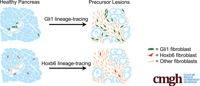
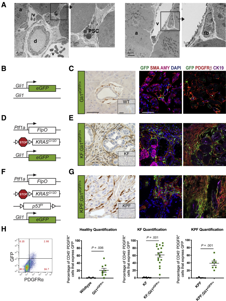
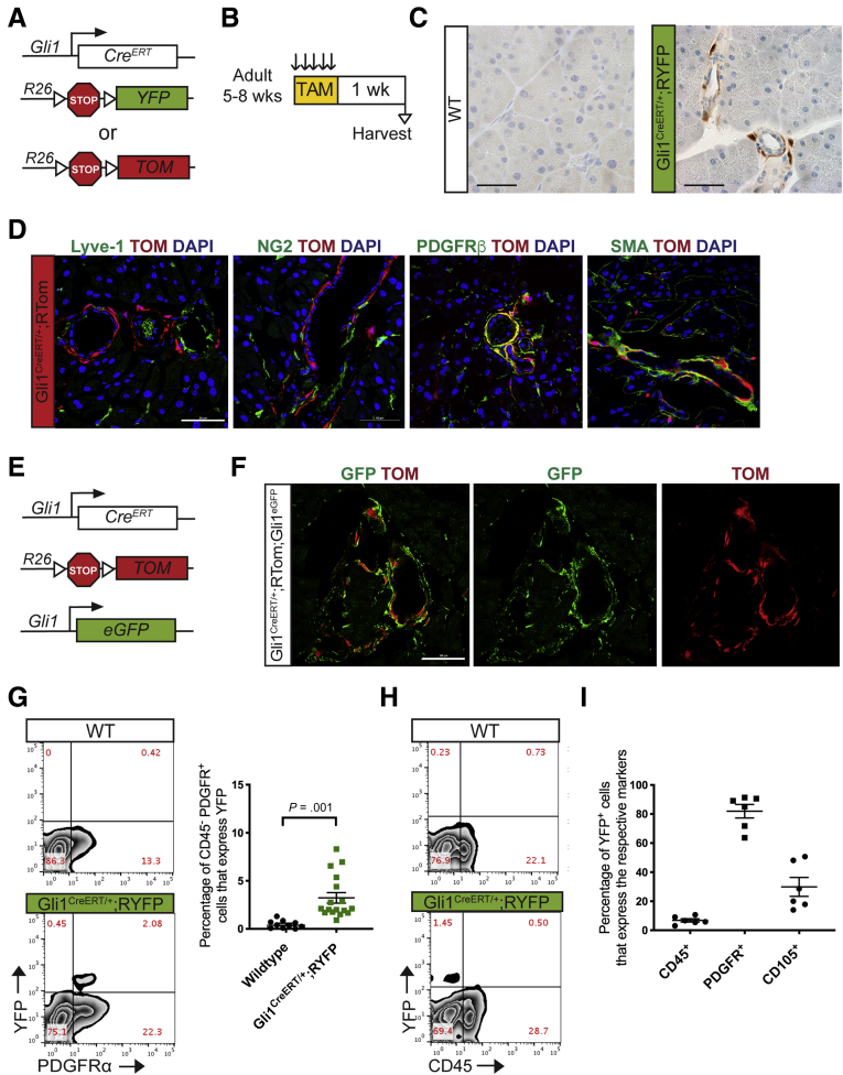
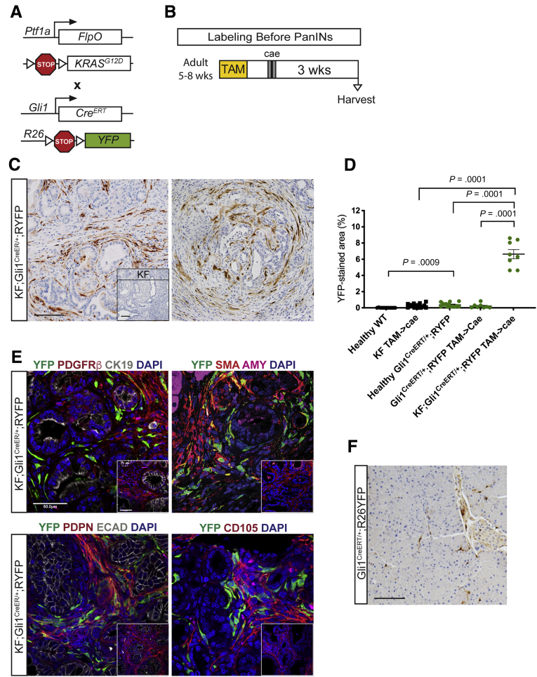
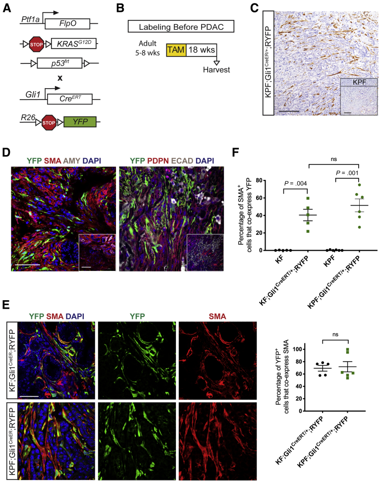
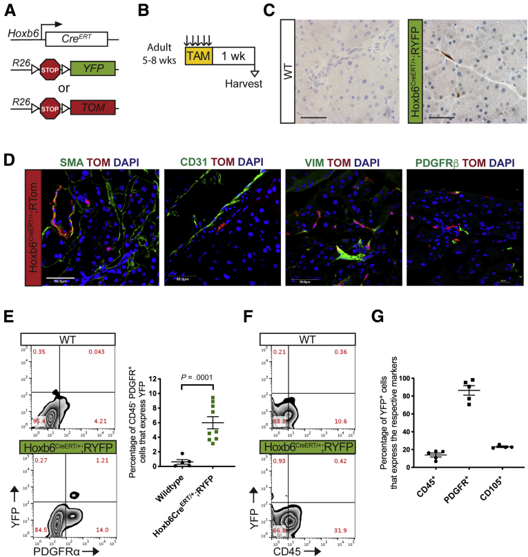
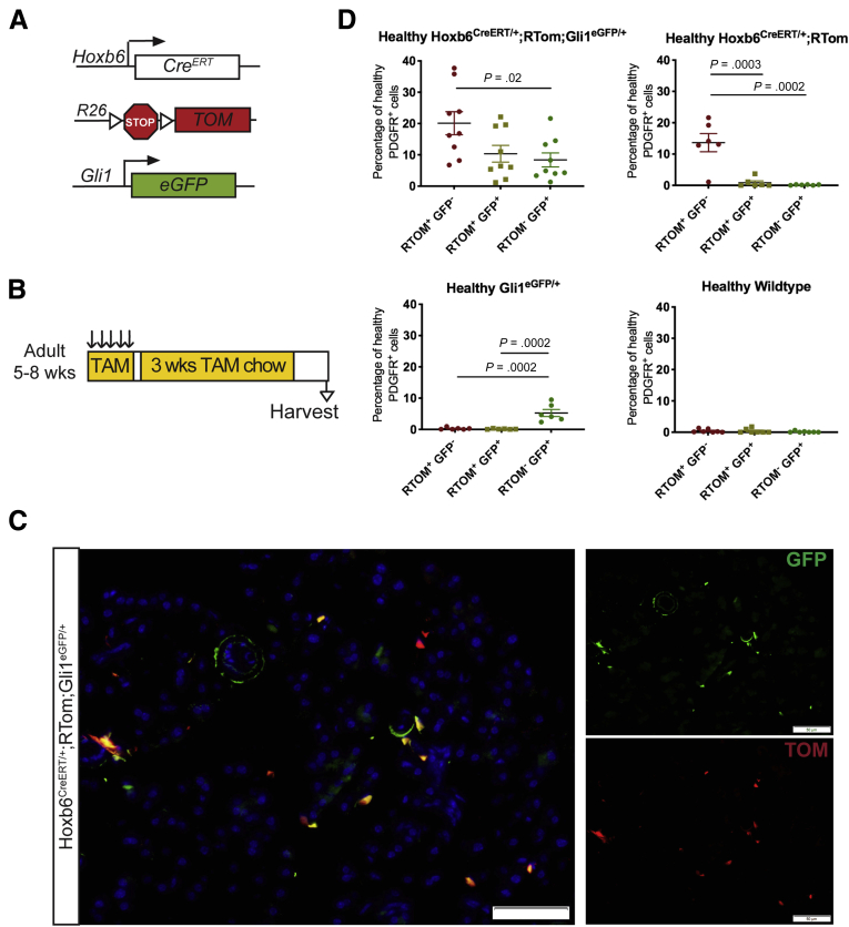
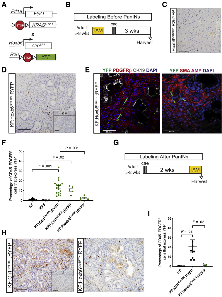
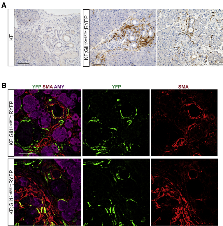
Background & Aims Although the healthy pancreas consists mostly of epithelial cells, pancreatic cancer and the precursor lesions known as pancreatic intraepithelial neoplasia, are characterized by an extensive accumulation of fibroinflammatory stroma that includes a substantial and heterogeneous fibroblast population. The cellular origin of fibroblasts within the stroma has not been determined. Here, we show that the Gli1 and Hoxb6 markers label distinct fibroblast populations in the healthy mouse pancreas. We then set out to determine whether these distinct fibroblast populations expanded during carcinogenesis. Methods We developed genetically engineered models using a dual-recombinase approach that allowed us to induce pancreatic cancer formation through codon-optimized Flp recombinase-driven epithelial recombination of Kirsten rat sarcoma viral oncogene homolog while labeling Gli1 + or Hoxb6 + fibroblasts in an inducible manner. By using these models, we lineage-traced these 2 fibroblast populations during the process of carcinogenesis. Results Although in the healthy pancreas Gli1 + fibroblasts and Hoxb6 + fibroblasts are present in similar numbers, they contribute differently to the stroma in carcinogenesis. Namely, Gli1 + fibroblasts expand dramatically, whereas Hoxb6 + cells do not. Conclusions Fibroblasts present in the healthy pancreas expand during carcinogenesis, but with a different prevalence for different subtypes. Here, we compared Gli1 + and Hoxb6 + fibroblasts and found only Gli1 + expanded to contribute to the stroma during pancreatic carcinogenesis.
Overview
The claims and statistics of this paper haven’t been checked yet.
Run the check — free, no account needed. It reads the open-access text and checks the sources it cites.
Integrity signals
Registry
Cites
Statistics
Figures
Verification
Claims the paper makes
The claims and statistics of this paper haven’t been checked yet.
Run the check — free, no account needed. It reads the open-access text and checks the sources it cites.
Claims other papers make about this one
Each is checked against this paper’s own text: does it say what the citing paper says it does?
In the citing paper
“recent lineage tracing studies targeting Fabp4+ stellate cells revealed that they give rise to a minority of CAFs142,143”
From Pancreatic cancer: Advances and challenges (2023) · cites this paper as [143]
Self-check
Percentages, sums and reported differences re-computed from the paper’s own numbers.
The self-check needs the paper’s full text.
Citations
Cites
Rahib, L., Smith, B. D., Aizenberg, R., Rosenzweig, A. B., Fleshman, J. M., & Matrisian, L. M. (2014) · Cancer Research · doi:10.1158/0008-5472.can-14-0155 · Cited by 7,094 · cited 1×
“ancreatic ductal adenocarcinoma (PDAC) is a deadly Pmalignancy that is projected to become the second leading cause of cancer-related death by 2030.(1) The 5-year”
BackgroundA framework for advancing our understanding of cancer-associated fibroblasts
Sahai, E., Astsaturov, I., Cukierman, E., DeNardo, D. G., Egeblad, M., Evans, R. M., Fearon, D., Greten, F. R., Hingorani, S. R., Hunter, T., et al. (2020) · Nature Reviews Cancer · doi:10.1038/s41568-019-0238-1 · Cited by 3,863
Olive, K. P., Jacobetz, M. A., Davidson, C. J., Gopinathan, A., McIntyre, D., Honess, D., Madhu, B., Goldgraben, M. A., Caldwell, M. E., Allard, D., et al. (2009) · Science · doi:10.1126/science.1171362 · Cited by 3,170
Özdemir, B. C., Pentcheva-Hoang, T., Carstens, J. L., Zheng, X., Wu, C.-C., Simpson, T. R., Laklai, H., Sugimoto, H., Kahlert, C., Novitskiy, S. V., et al. (2014) · Cancer Cell · doi:10.1016/j.ccr.2014.04.005 · Cited by 2,681 · cited 1×
“However, other results suggested that fibroblasts could restrain tumor formation and that depleting them might accelerate carcinogenesis.(13) This paradox might be explained by the observation that fibroblasts are not a homogenous population, but exist in subsets with different roles.”
ResultPericytes: Developmental, Physiological, and Pathological Perspectives, Problems, and Promises
Armulik, A., Genové, G., & Betsholtz, C. (2011) · Developmental Cell · doi:10.1016/j.devcel.2011.07.001 · Cited by 2,653 · cited 1×
“In the pancreas, themesenchyme surrounds and supports epithelium development(50) and differentiates to form cell types such as fibroblasts, pancreatic stellate cells, and pericytes.(51,52) Activated fibroblasts, also known as myofibroblasts, help regulate wound repair and recovery,(53,54) but in the context of certain…”
BackgroundHingorani, S. R., Wang, L., Multani, A. S., Combs, C., Deramaudt, T. B., Hruban, R. H., Rustgi, A. K., Chang, S., & Tuveson, D. A. (2005) · Cancer Cell · doi:10.1016/j.ccr.2005.04.023 · Cited by 2,609
Distinct populations of inflammatory fibroblasts and myofibroblasts in pancreatic cancer
Öhlund, D., Handly-Santana, A., Biffi, G., Elyada, E., Almeida, A. S., Ponz-Sarvise, M., Corbo, V., Oni, T. E., Hearn, S. A., Lee, E. J., et al. (2017) · Journal of Experimental Medicine · doi:10.1084/jem.20162024 · Cited by 2,590
Preinvasive and invasive ductal pancreatic cancer and its early detection in the mouse
Hingorani, S. R., Petricoin, E. F., Maitra, A., Rajapakse, V., King, C., Jacobetz, M. A., Ross, S., Conrads, T. P., Veenstra, T. D., Hitt, B. A., et al. (2003) · Cancer Cell · doi:10.1016/s1535-6108(03)00309-x · Cited by 2,471
Self-citationEpidemiology of Pancreatic Cancer: Global Trends, Etiology and Risk Factors
Rawla, P., Sunkara, T., & Gaduputi, V. (2019) · World Journal of Oncology · doi:10.14740/wjon1166 · Cited by 2,269 · cited 1×
“survival rate for PDAC is only 10%, which can be attributed to the aggressive and chemoresistant nature of the cancer, in addition to the frequently late diagnosis.(2) In PDAC, more”
BackgroundElyada, E., Bolisetty, M., Laise, P., Flynn, W. F., Courtois, E. T., Burkhart, R. A., Teinor, J. A., Belleau, P., Biffi, G., Lucito, M. S., et al. (2019) · Cancer Discovery · doi:10.1158/2159-8290.cd-19-0094 · Cited by 2,241 · cited 1×
“populations with potentially distinct functions and activation pathways.(14,15,20) Here, we sought to determine which fibroblasts in the”
BackgroundProvenzano, P. P., Cuevas, C., Chang, A. E., Goel, V. K., Von Hoff, D. D., & Hingorani, S. R. (2012) · Cancer Cell · doi:10.1016/j.ccr.2012.01.007 · Cited by 2,195
Feig, C., Jones, J. O., Kraman, M., Wells, R. J. B., Deonarine, A., Chan, D. S., Connell, C. M., Roberts, E. W., Zhao, Q., Caballero, O. L., et al. (2013) · Proceedings of the National Academy of Sciences · doi:10.1073/pnas.1320318110 · Cited by 2,100
Fibroblast Heterogeneity and Immunosuppressive Environment in Human Breast Cancer
Costa, A., Kieffer, Y., Scholer-Dahirel, A., Pelon, F., Bourachot, B., Cardon, M., Sirven, P., Magagna, I., Fuhrmann, L., Bernard, C., et al. (2018) · Cancer Cell · doi:10.1016/j.ccell.2018.01.011 · Cited by 1,705
Fate Tracing Reveals the Pericyte and Not Epithelial Origin of Myofibroblasts in Kidney Fibrosis
Humphreys, B. D., Lin, S., Kobayashi, A., Hudson, T. E., Nowlin, B. T., Bonventre, J. V., Valerius, M. T., McMahon, A. P., & Duffield, J. S. (2009) · American Journal Of Pathology · doi:10.2353/ajpath.2010.090517 · Cited by 1,474
Mederacke, I., Hsu, C. C., Troeger, J. S., Huebener, P., Mu, X., Dapito, D. H., Pradere, J.-P., & Schwabe, R. F. (2013) · Nature Communications · doi:10.1038/ncomms3823 · Cited by 1,443
Biffi, G., Oni, T. E., Spielman, B., Hao, Y., Elyada, E., Park, Y., Preall, J., & Tuveson, D. A. (2019) · Cancer Discovery · doi:10.1158/2159-8290.cd-18-0710 · Cited by 1,360 · cited 1×
“populations with potentially distinct functions and activation pathways.(14,15,20) Here, we sought to determine which fibroblasts in the”
BackgroundPeng, J., Sun, B.-F., Chen, C.-Y., Zhou, J.-Y., Chen, Y.-S., Chen, H., Liu, L., Huang, D., Jiang, J., Cui, G.-S., et al. (2019) · Cell Research · doi:10.1038/s41422-019-0195-y · Cited by 1,321
Distinct fibroblast lineages determine dermal architecture in skin development and repair
Driskell, R. R., Lichtenberger, B. M., Hoste, E., Kretzschmar, K., Simons, B. D., Charalambous, M., Ferron, S. R., Herault, Y., Pavlovic, G., Ferguson-Smith, A. C., et al. (2013) · Nature · doi:10.1038/nature12783 · Cited by 1,291
Su, S., Chen, J., Yao, H., Liu, J., Yu, S., Lao, L., Wang, M., Luo, M., Xing, Y., Chen, F., et al. (2018) · Cell · doi:10.1016/j.cell.2018.01.009 · Cited by 1,230 · cited 1×
“Currently, functionally distinct subclasses of CAFs have been defined in other cancers such as breast carcinoma,(55,56) colorectal carcinoma,(57) and lung adenocarcinoma.(58) In pancreatic cancer, a putative mesenchymal stem cell population has been identified inmouse and human samples,(16,17) and CAFs have been class…”
BackgroundGuerra, C., Schuhmacher, A. J., Cañamero, M., Grippo, P. J., Verdaguer, L., Pérez-Gallego, L., Dubus, P., Sandgren, E. P., & Barbacid, M. (2007) · Cancer Cell · doi:10.1016/j.ccr.2007.01.012 · Cited by 1,224
Sherman, M. H., Yu, R. T., Engle, D. D., Ding, N., Atkins, A. R., Tiriac, H., Collisson, E. A., Connor, F., Van Dyke, T., Kozlov, S., et al. (2014) · Cell · doi:10.1016/j.cell.2014.08.007 · Cited by 1,158
Cancer-Associated Stromal Fibroblasts Promote Pancreatic Tumor Progression
Hwang, R. F., Moore, T. T., Arumugam, T., Ramachandran, V., Amos, K. D., Rivera, A. M., Ji, B., Evans, D. B., & Logsdon, C. D. (2008) · Cancer Research · doi:10.1158/0008-5472.can-07-5714 · Cited by 1,117
Li, H., Courtois, E. T., Sengupta, D., Tan, Y., Chen, K. H., Goh, J. J. L., Kong, S. L., Chua, C., Hon, L. K., Tan, W. S., et al. (2017) · Nature Genetics · doi:10.1038/ng.3818 · Cited by 1,080
Fibroblasts and myofibroblasts in wound healing
Desmouliere, A., Darby, I. A., Laverdet, B., & Bonté, F. (2014) · Clinical, Cosmetic and Investigational Dermatology · doi:10.2147/ccid.s50046 · Cited by 1,039
Effect of friction on oxidative graphite intercalation and high-quality graphene formation
Seiler, S., Halbig, C. E., Grote, F., Rietsch, P., Börrnert, F., Kaiser, U. A., Meyer, B., & Eigler, S. (2018) · Nature Communications · doi:10.1038/s41467-018-03211-1 · Cited by 990
A paracrine requirement for hedgehog signalling in cancer
Yauch, R. L., Gould, S. E., Scales, S. J., Tang, T. T.-L., Tian, H., Ahn, C. P., Marshall, D. C. L., Fu, L., Januario, T., Kallop, D. Y., et al. (2008) · Nature · doi:10.1038/nature07275 · Cited by 961
Genetic lineage tracing defines myofibroblast origin and function in the injured heart
Kanisicak, O., Khalil, H., Ivey, M. J., Karch, J. M., Maliken, B. D., Correll, R. N., Brody, M. J., Lin, S. J., Aronow, B. J., Tallquist, M. D., et al. (2016) · Nature Communications · doi:10.1038/ncomms12260 · Cited by 926
Perivascular Gli1+ Progenitors Are Key Contributors to Injury-Induced Organ Fibrosis
Kramann, R., Schneider, R. K., DiRocco, D. P., Machado, F., Fleig, S., Bondzie, P. A., Henderson, J. M., Ebert, B. L., & Humphreys, B. D. (2015) · Cell Stem Cell · doi:10.1016/j.stem.2014.11.004 · Cited by 922 · cited 2×
“Approximately 40% of myofibroblasts in the kidney, liver, and lung, and 60% of myofibroblasts in the heart originated from tissue-resident Gli1þ progenitors after a fibrosisinducing injury.(32,33) In addition, Gli1 progenitors”
Periacinar stellate shaped cells in rat pancreas: identification, isolation, and culture
Apte, M. V., Haber, P., Applegate, T. L., Norton, I. D., McCaughan, G. W., Korsten, M. A., Pirola, R. C., & Wilson, J. S. (1998) · Gut · doi:10.1136/gut.43.1.128 · Cited by 920
A Pericyte Origin of Spinal Cord Scar Tissue
Göritz, C., Dias, D. O., Tomilin, N., Barbacid, M., Shupliakov, O., & Frisén, J. (2011) · Science · doi:10.1126/science.1203165 · Cited by 858
Dominguez, C. X., Müller, S., Keerthivasan, S., Koeppen, H., Hung, J., Gierke, S., Breart, B., Foreman, O., Bainbridge, T. W., Castiglioni, A., et al. (2020) · Cancer Discovery · doi:10.1158/2159-8290.cd-19-0644 · Cited by 857 · cited 5×
“tumor-associated myCAF or inflammatory-CAF populations than to themselves.(23) Understanding the complexity of fibroblasts in healthy organs and disease has been hampered by the limited availability of specific markers.”
BackgroundInfluentialBartoschek, M., Oskolkov, N., Bocci, M., Lövrot, J., Larsson, C., Sommarin, M., Madsen, C. D., Lindgren, D., Pekar, G., Karlsson, G., et al. (2018) · Nature Communications · doi:10.1038/s41467-018-07582-3 · Cited by 826
Oncogenic Kras is required for both the initiation and maintenance of pancreatic cancer in mice
Collins, M. A., Bednar, F., Zhang, Y., Brisset, J.-C., Galbán, S., Galbán, C. J., Rakshit, S., Flannagan, K. S., Adsay, N. V., & Pasca di Magliano, M. (2012) · Journal of Clinical Investigation · doi:10.1172/jci59227 · Cited by 770 · cited 1×
“Immunohistochemistry and Immunofluorescence All immunohistochemical and immunofluorescent stains were performed as previously described.(78) Primary antibodies used were as follows: YFP (1:200; Abcam, Cambridge, MA), PDGFRb (1:100; Abcam), cytokeratin 19 (1:50; Iowa Developmental Hybridoma Bank, Iowa City, IA), aSMA (…”
MethodSelf-citationGli Proteins in Development and Disease
Hui, C., & Angers, S. (2011) · Annual Review of Cell and Developmental Biology · doi:10.1146/annurev-cellbio-092910-154048 · Cited by 713
Notochord repression of endodermal Sonic hedgehog permits pancreas development
Hebrok, M., Kim, S. K., & Melton, D. A. (1998) · Genes & Development · doi:10.1101/gad.12.11.1705 · Cited by 628
A peek into cancer-associated fibroblasts: origins, functions and translational impact
LeBleu, V. S., & Kalluri, R. (2018) · Disease Models & Mechanisms · doi:10.1242/dmm.029447 · Cited by 557
Ahn, S., & Joyner, A. (2004) · Cell · doi:10.1016/j.cell.2004.07.023 · Cited by 545 · cited 1×
“The mice lack 1 functional allele of Gli1 but nevertheless are viable and fertile.(39) Because the natural fluorescence of EGFP does not persist after fixation and paraffin embedding, we used an anti–green fluorescent protein antibody to visualize the reporter expression in immunohistochemistry (IHC) or immunofluoresc…”
MethodZhao, H., Feng, J., Seidel, K., Shi, S., Klein, O. D., Sharpe, P. T., & Chai, Y. (2014) · Cell stem cell · doi:10.1016/j.stem.2013.12.013 · Cited by 464
Fibroblasts and myofibroblasts: Their source, function and role in disease
McAnulty, R. J. (2006) · The International Journal of Biochemistry & Cell Biology · doi:10.1016/j.biocel.2006.11.005 · Cited by 455
Sonic hedgehog directs specialised mesoderm differentiation in the intestine and pancreas
Apelqvist, Å., Ahlgren, U., & Edlund, H. (1997) · Current Biology · doi:10.1016/s0960-9822(06)00340-x · Cited by 445
Erkan, M., Michalski, C. W., Rieder, S., Reiser–Erkan, C., Abiatari, I., Kolb, A., Giese, N. A., Esposito, I., Frieß, H., & Kleeff, J. H. (2008) · Clinical Gastroenterology and Hepatology · doi:10.1016/j.cgh.2008.05.006 · Cited by 433 · cited 1×
“so than in other solid tumors, a high percentage of the tumor volume is occupied by nonmalignant cells that form the tumor stroma.(3) The cellular component of the stroma includes fibroblasts, infiltrating immune cells, vascular elements, and nerves.”
BackgroundMesenchymal stem cells in fibrotic disease
El Agha, E., Kramann, R., Schneider, R. K., Li, X., Seeger, W., Humphreys, B. D., & Bellusci, S. (2017) · Cell stem cell · doi:10.1016/j.stem.2017.07.011 · Cited by 430
Morris, J. P., Cano, D., Sekine, S., Wang, S. C., & Hebrok, M. (2010) · Journal of Clinical Investigation · doi:10.1172/jci40045 · Cited by 391 · cited 1×
“Louis, MO) as previously described.(47,77) To activate the CreERT transgene, mice received 4 mg tamoxifen in corn oil/3% ethanol (SigmaAldrich) per day via oral gavage for 5 days.”
MethodThe role of stromal cancer-associated fibroblasts in pancreatic cancer
Ahrens, D. V., Bhagat, T. D., Nagrath, D., Maitra, A., & Verma, A. K. (2017) · Journal of Hematology & Oncology · doi:10.1186/s13045-017-0448-5 · Cited by 389
Bernard, V., Semaan, A., Huang, J. H., Lucas, F. A. S., Mulu, F. C., Stephens, B. M., Guerrero, P. A., Huang, Y., Zhao, J., Kamyabi, N., et al. (2018) · Clinical Cancer Research · doi:10.1158/1078-0432.ccr-18-1955 · Cited by 387 · cited 4×
“However, no experimental validation supports the notion that stellate cells lead to CAFs.”
BackgroundInfluentialEl Agha, E., Moiseenko, A., Kheirollahi, V., De Langhe, S., Crnkovic, S., Kwapiszewska, G., Szibor, M., Kosanovic, D., Schwind, F., Schermuly, R. T., et al. (2017) · Cell Stem Cell · doi:10.1016/j.stem.2016.10.004 · Cited by 347
Stromal biology of pancreatic cancer
Chu, G. C., Kimmelman, A. C., Hezel, A. F., & DePinho, R. A. (2007) · Journal of Cellular Biochemistry · doi:10.1002/jcb.21209 · Cited by 330 · cited 1×
“In the pancreas, themesenchyme surrounds and supports epithelium development(50) and differentiates to form cell types such as fibroblasts, pancreatic stellate cells, and pericytes.(51,52) Activated fibroblasts, also known as myofibroblasts, help regulate wound repair and recovery,(53,54) but in the context of certain…”
BackgroundNeuzillet, C., Tijeras‐Raballand, A., Ragulan, C., Cros, J. F., Patil, Y., Martinet, M., Erkan, M., Kleeff, J. H., Wilson, J. S., Apte, M. V., et al. (2018) · The Journal of Pathology · doi:10.1002/path.5224 · Cited by 310
Kramann, R., Goettsch, C., Wongboonsin, J., Iwata, H., Schneider, R. K., Kuppe, C., Kaesler, N., Chang-Panesso, M., Machado, F. G., Gratwohl, S., et al. (2016) · Cell stem cell · doi:10.1016/j.stem.2016.08.001 · Cited by 308 · cited 1×
“Approximately 40% of myofibroblasts in the kidney, liver, and lung, and 60% of myofibroblasts in the heart originated from tissue-resident Gli1þ progenitors after a fibrosisinducing injury.(32,33) In addition, Gli1 progenitors”
BackgroundHosein, A. N., Huang, H., Wang, Z., Parmar, K., Du, W., Huang, J., Maitra, A., Olson, E., Verma, U., & Brekken, R. A. (2019) · JCI Insight · doi:10.1172/jci.insight.129212 · Cited by 302
Fibroblast Heterogeneity in the Pancreatic Tumor Microenvironment
Helms, E. J., Oñate, M. K., & Sherman, M. H. (2020) · Cancer Discovery · doi:10.1158/2159-8290.cd-19-1353 · Cited by 288
Schneider, R. K., Mullally, A., Dugourd, A. J. F., Peisker, F., Hoogenboezem, R. M., van Strien, P. M. H., Bindels, E. M., Heckl, D., Büsche, G., Fleck, D., et al. (2017) · Cell stem cell · doi:10.1016/j.stem.2017.03.008 · Cited by 281
Matsuo, Y., Ochi, N., Sawai, H., Yasuda, A., Takahashi, H., Funahashi, H., Takeyama, H., Tong, Z., & Guha, S. (2008) · International Journal of Cancer · doi:10.1002/ijc.24040 · Cited by 280
Zhang, Y., Yan, W., Collins, M. A., Bednar, F., Rakshit, S., Zetter, B. R., Stanger, B. Z., Chung, I., Rhim, A. D., & di Magliano, M. P. (2013) · Cancer Research · doi:10.1158/0008-5472.can-13-1558-t · Cited by 268
Self-citationK-ras mutations in pancreatic ductal proliferative lesions.
Klimstra, D. S., & Longnecker, D. S. (1994) · PubMed · Cited by 197
GM-CSF Mediates Mesenchymal–Epithelial Cross-talk in Pancreatic Cancer
Waghray, M., Yalamanchili, M., Dziubinski, M. L., Zeinali, M., Erkkinen, M., Yang, H., Schradle, K. A., Urs, S., di Magliano, M. P., Welling, T. H., et al. (2016) · Cancer Discovery · doi:10.1158/2159-8290.cd-15-0947 · Cited by 197
Evolving Concepts of Mitochondrial Dynamics
Dorn, G. W. (2018) · Annual Review of Physiology · doi:10.1146/annurev-physiol-020518-114358 · Cited by 197
Hedgehog Signaling Is Required for Effective Regeneration of Exocrine Pancreas
Fendrich, V., Esni, F., Garay, M. V. R., Feldmann, G., Habbe, N., Jensen, J. N., Dor, Y., Stoffers, D., Jensen, J., Leach, S. D., et al. (2008) · Gastroenterology · doi:10.1053/j.gastro.2008.04.011 · Cited by 191 · cited 1×
“However, some low level of Hedgehog signaling persists(64) to maintain homeostasis and assist in injury recovery.(32,65,66) Thus, we found that a subset of fibroblasts in the healthy pancreas consistently express Gli1 and do not have characteristics of stellate cells.”
BackgroundLineage dynamics of murine pancreatic development at single-cell resolution
Byrnes, L. E., Wong, D. M., Subramaniam, M., Meyer, N. P., Gilchrist, C. L., Knox, S. M., Tward, A. D., Ye, C., & Sneddon, J. B. (2018) · Nature Communications · doi:10.1038/s41467-018-06176-3 · Cited by 185 · cited 1×
“Even in the developing pancreas, distinct fibroblast populations were observed.(26) However, which type(s) of fibroblasts in the healthy pancreas gives rise to CAFs remains an open question.”
BackgroundMolecular Pathology of Pancreatic Cancer
Wood, L. D., Adsay, V. N., & Hruban, R. H. (2012) · Springer eBooks · doi:10.1007/978-1-4614-4900-3_2 · Cited by 182
Sonic Hedgehog Regulates Discrete Populations of Astrocytes in the Adult Mouse Forebrain
Garcia, A. D. R., Petrova, R., Eng, L., & Joyner, A. L. (2010) · Journal of Neuroscience · doi:10.1523/jneurosci.0830-10.2010 · Cited by 178
Metastatic pancreatic cancer is dependent on oncogenic Kras in mice
Collins, M. A., Brisset, J.-C., Zhang, Y., Bednar, F., Pierre, J., Heist, K. A., Galbán, C. J., Galbán, S., & di Magliano, M. P. (2012) · PLoS ONE · doi:10.1371/journal.pone.0049707 · Cited by 178 · cited 1×
“Louis, MO) as previously described.(47,77) To activate the CreERT transgene, mice received 4 mg tamoxifen in corn oil/3% ethanol (SigmaAldrich) per day via oral gavage for 5 days.”
MethodSelf-citationControl of Cell Identity in Pancreas Development and Regeneration
Stanger, B. Z., & Hebrok, M. (2013) · Gastroenterology · doi:10.1053/j.gastro.2013.01.074 · Cited by 158
Hedgehog signaling regulation of insulin production by pancreatic beta-cells.
Thomas, M. K., Rastalsky, N., Lee, J. H., & Habener, J. F. (2000) · Diabetes · doi:10.2337/diabetes.49.12.2039 · Cited by 153 · cited 1×
“However, some low level of Hedgehog signaling persists(64) to maintain homeostasis and assist in injury recovery.(32,65,66) Thus, we found that a subset of fibroblasts in the healthy pancreas consistently express Gli1 and do not have characteristics of stellate cells.”
BackgroundKoelewijn, L., Lancaster, T. M., Linden, D. E. J., Dima, D. C., Routley, B. C., Magazzini, L., Barawi, K., Brindley, L. M., Adams, R., Tansey, K. E., et al. (2019) · eLife · doi:10.7554/elife.36011 · Cited by 151
McCarroll, J. A., Phillips, P. A., Santucci, N., Pirola, R. C., Wilson, J. S., & Apte, M. V. (2005) · Gut · doi:10.1136/gut.2005.064543 · Cited by 147
Dangerous liaisons: Pancreatic stellate cells and pancreatic cancer cells
Apte, M. V., & Wilson, J. S. (2012) · Journal of Gastroenterology and Hepatology · doi:10.1111/j.1440-1746.2011.07000.x · Cited by 146 · cited 1×
“Results Gli1þ Fibroblasts Are Present in the Healthy and Neoplastic Pancreas Pancreatic stellate cells (PSCs) have long been presumed to be the predominant mesenchymal population in the healthy pancreas.(37) Because Gli1þ cells have not been described as stellate cells,(32) we sought to confirm the existence of differ…”
BackgroundTranscription factor TBX4 regulates myofibroblast accumulation and lung fibrosis
Xie, T., Liang, J., Liu, N., Huan, C., Zhang, Y., Liu, W., Kumar, M. E., Xiao, R., D'Armiento, J. M., METZGER, D., et al. (2016) · Journal of Clinical Investigation · doi:10.1172/jci85328 · Cited by 139
Self-citationThe Molecular Pathology of Pancreatic Cancer
Lemoine, N. R. (1993) · Springer eBooks · doi:10.1007/978-3-642-77437-9_63 · Cited by 124
Mathew, E., Brannon, A., Vecchio, A. D., Garcia, P. E., Penny, M. K., Kane, K. T., Vinta, A., Buckanovich, R. J., & di Magliano, M. P. (2016) · Neoplasia · doi:10.1016/j.neo.2016.01.005 · Cited by 122
Self-citationThe activity of Gli transcription factors is essential for Kras-induced pancreatic tumorigenesis
Rajurkar, M., DeJesus-Monge, W. E., Driscoll, D. R., Appleman, V. A., Huang, H., Cotton, J. L., Klimstra, D., Zhu, L., Simin, K., Xu, L., et al. (2012) · Proceedings of the National Academy of Sciences of the United States of America · doi:10.1073/pnas.1114168109 · Cited by 119 · cited 1×
“Gli1 has been found to be expressed in epithelial cells in a variable manner across tumor lines.(28,49) In this advanced disease progression model, we did not observe epithelial Gli1 expression.”
BackgroundCD4+ T lymphocyte ablation prevents pancreatic carcinogenesis in mice
Zhang, Y., Yan, W., Mathew, E., Bednar, F., Wan, S., Collins, M. A., Evans, R. A., Welling, T. H., Vonderheide, R. H., & di Magliano, M. P. (2014) · Cancer Immunology Research · doi:10.1158/2326-6066.cir-14-0016-t · Cited by 102
Self-citationHedgehog signaling in pancreas development and disease
Lau, J., Kawahira, H., & Hebrok, M. (2006) · Cellular and Molecular Life Sciences · doi:10.1007/s00018-005-5357-z · Cited by 95
DOSAGE-DEPENDENT REGULATION OF PANCREATIC CANCER GROWTH AND ANGIOGENESIS BY HEDGEHOG SIGNALING
Mathew, E., Zhang, Y.-Q., Holtz, A., Kane, K. T., Song, J. Y., Allen, B. L., & di Magliano, M. P. (2014) · Cell Reports · doi:10.1016/j.celrep.2014.09.010 · Cited by 92 · cited 1×
“We previously described a small population of Gli1þ fibroblasts in the healthy pancreas.(27,30) In a number of other organs, tissue-resident Gli1þ cells are mesenchymal progenitors that associate closely with the vasculature until activation into proliferative myofibroblasts by injury.”
BackgroundSelf-citationTranscriptional control of subtype switching ensures adaptation and growth of pancreatic cancer
Adams, C. R., Htwe, H. H., Marsh, T., Wang, A. L., Montoya, M., Subbaraj, L., Tward, A. D., Bardeesy, N. M., & Perera, R. M. (2019) · eLife · doi:10.7554/elife.45313 · Cited by 88 · cited 1×
“Gli1 has been found to be expressed in epithelial cells in a variable manner across tumor lines.(28,49) In this advanced disease progression model, we did not observe epithelial Gli1 expression.”
BackgroundCarrière, C., Young, A. L., Gunn, J. R., Longnecker, D. S., & Korc, M. (2011) · PLoS ONE · doi:10.1371/journal.pone.0027725 · Cited by 77
Chen, K., Chen, L., Hu, Y., Ding, N., Li, Y., McGuire, T. L., Lu, H., Kessler, J. A., & Kan, L. (2017) · Bone · doi:10.1016/j.bone.2017.06.014 · Cited by 76 · cited 1×
“All data are expressed as means ± S instances of endochondral heterotopic ossification and bone marrow fibrosis.(31,35) Gli1þ fibroblasts have not been characterized in the pancreas, and their contribution to tumor-”
MethodLau, J., & Hebrok, M. (2010) · Diabetes · doi:10.2337/db09-0914 · Cited by 59
Mathew, E., Collins, M. A., Fernández‐Barrena, M. G., Holtz, A. M., Yan, W., Hogan, J. O., Tata, Z., Allen, B. L., Fernández-Zapico, M. E., & di Magliano, M. P. (2014) · Journal of Biological Chemistry · doi:10.1074/jbc.m114.556563 · Cited by 52
Self-citationEpithelial-Stromal Interactions in Pancreatic Cancer
Zhang, Y., Crawford, H. C., & di Magliano, M. P. (2018) · Annual Review of Physiology · doi:10.1146/annurev-physiol-020518-114515 · Cited by 52 · cited 2×
“This fibroinflammatory environment develops during the early steps of tumorigenesis and supports tumor growth and metastasis, although the roles of the individual components are still not fully understood.(4) Fibroblasts are a prominent and active cellular component of the stroma; they contribute to the secretion of e…”
BackgroundSelf-citationMesenchymal Hox6 function is required for pancreatic endocrine cell differentiation
Larsen, B. M., Hrycaj, S., Newman, M., Li, Y., & Wellik, D. M. (2015) · Development · doi:10.1242/dev.126888 · Cited by 46 · cited 2×
“developing pancreas, and regulates the embryonic development of the organ.(36) To assess the expression of Hoxb6 in the adult pancreas, we followed a similar strategy to that described before for Gli1.”
BackgroundSelf-citationNguyen, M., Zhu, J., Nakamura, E., Bao, X., & Mackem, S. (2009) · Developmental Dynamics · doi:10.1002/dvdy.21846 · Cited by 41
Myeloid Cell-Derived HB-EGF Drives Tissue Recovery After Pancreatitis
Wen, H.-J., Gao, S., Wang, Y., Ray, M., Magnuson, M., Wright, C., di Magliano, M. P., Frankel, T., & Crawford, H. (2019) · Cellular and Molecular Gastroenterology and Hepatology · doi:10.1016/j.jcmgh.2019.05.006 · Cited by 30
Self-citation
Cited by
Sorted by citations
Pancreatic cancer: Advances and challenges
Halbrook, C. J., Lyssiotis, C. A., Pasca di Magliano, M., & Maitra, A. (2023) · Cell · doi:10.1016/j.cell.2023.02.014 · Cited by 1,321
Clinical and therapeutic relevance of cancer-associated fibroblasts
Chen, Y., McAndrews, K. M., & Kalluri, R. (2021) · Nature Reviews Clinical Oncology · doi:10.1038/s41571-021-00546-5 · Cited by 1,230
Cancer-associated fibroblasts: from basic science to anticancer therapy
Yang, D., Liu, J., Qian, H., & Zhuang, Q. (2023) · Experimental & Molecular Medicine · doi:10.1038/s12276-023-01013-0 · Cited by 572
Tumor Microenvironment in Pancreatic Cancer Pathogenesis and Therapeutic Resistance
Sherman, M. H., & Beatty, G. L. (2022) · Annual Review of Pathology Mechanisms of Disease · doi:10.1146/annurev-pathmechdis-031621-024600 · Cited by 501
Activated fibroblasts in cancer: Perspectives and challenges
Caligiuri, G., & Tuveson, D. A. (2023) · Cancer Cell · doi:10.1016/j.ccell.2023.02.015 · Cited by 450
Metadata
- Authors
- Garcia, P. E., Adoumie, M., Kim, E. C., Zhang, Y., Scales, M. K., El-Tawil, Y. S., Shaikh, A. Z., Wen, H.-J., Bednar, F., Allen, B. L., Wellik, D. M., Crawford, H. C., & Pasca di Magliano, M.
- Affiliations
- University of Michigan (US)
- University of Wisconsin–Madison (US)
- ORCID
- Venue
- Cellular and Molecular Gastroenterology and Hepatology
- Publisher
- Elsevier BV
- Published
- 2020
- Volume
- 10
- Issue
- 3
- Pages
- 581-599
- ISSN
- 2352-345X (ISSN-L)
- PMC
- PMC7399194
- OpenAlex
- W3027333363
- Cited by
- 110
- References
- 83
- Citations by year
- 2026: 16 · 2025: 16 · 2024: 25 · 2023: 18 · 2022: 16 · 2021: 14 · 2020: 5
- FWCI
- 4.78
- Type
- journal article
- Language
- en
- Open access
- gold
- doi.org
- Licence
- Locations
- Cellular and Molecular Gastroenterology and Hepatology (publishedVersion, cc-by-nc-nd)
- PubMed Central (publishedVersion, cc-by-nc-nd)
- PubMed (publishedVersion)
- DOAJ (DOAJ: Directory of Open Access Journals) (submittedVersion, cc-by-sa)
- Funders
- National Cancer Institute
- Topics
- Health Sciences › Medicine › Oncology › Pancreatic and Hepatic Oncology Research
- Health Sciences › Medicine › Surgery › Pancreatitis Pathology and Treatment
- Health Sciences › Medicine › Endocrinology, Diabetes and Metabolism › Pancreatic function and diabetes
- Keywords
- pancreatic cancer, pancreatic stroma, pancreatic fibroblasts, fibroblast heterogeneity, Gli1, HOXB6, pancreatic intraepithelial neoplasia, carcinogenesis
- MeSH terms
- Zinc Finger Protein GLI1, Animals, Disease Models, Animal, Fibroblasts, Humans, Mice, Transgenic, Pancreas, Pancreatic Neoplasms, Proto-Oncogene Proteins p21(ras), Homeodomain Proteins, Carcinoma, Pancreatic Ductal, Mice, Carcinogenesis
About these checks
Registry, retraction, Cites and statistics results are computed, not judged, with one exception: a printed reference that no index lists is read once by a model before it counts as a citation error. Claims are extracted by a model and kept only when their quote appears verbatim in the text; each cited source is then checked, with a model’s help, for whether it says what the claim relies on. Those verdicts can be wrong — read the claim’s quote and the cited source before relying on one.
Statistics and arithmetic are recomputed, not judged.
Confidence bands are shown only for fields calibrated on our annotated test set. None is calibrated yet, so no band is shown.
What counts as a claim ›How we measure confidence ›How we check this ›
Are you an author, or do you see a problem with this page? Request a review
