

The DOI matches its Crossref record; 137 of the 146 references it cites were found in a registry or an index.
The Liver Tumor Segmentation Benchmark (LiTS)
Open accessAbstract
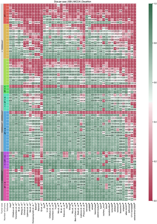
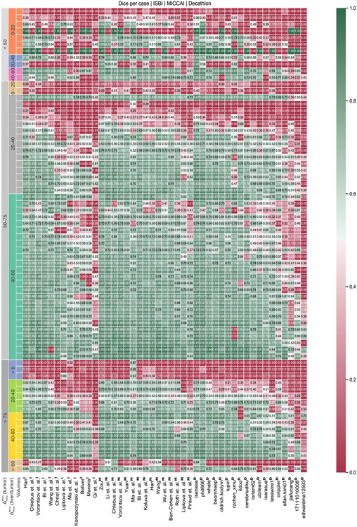
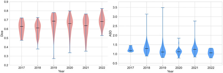
In this work, we report the set-up and results of the Liver Tumor Segmentation Benchmark (LiTS), which was organized in conjunction with the IEEE International Symposium on Biomedical Imaging (ISBI) 2017 and the International Conferences on Medical Image Computing and Computer-Assisted Intervention (MICCAI) 2017 and 2018. The image dataset is diverse and contains primary and secondary tumors with varied sizes and appearances with various lesion-to-background levels (hyper-/hypo-dense), created in collaboration with seven hospitals and research institutions. Seventy-five submitted liver and liver tumor segmentation algorithms were trained on a set of 131 computed tomography (CT) volumes and were tested on 70 unseen test images acquired from different patients. We found that not a single algorithm performed best for both liver and liver tumors in the three events. The best liver segmentation algorithm achieved a Dice score of 0.963, whereas, for tumor segmentation, the best algorithms achieved Dices scores of 0.674 (ISBI 2017), 0.702 (MICCAI 2017), and 0.739 (MICCAI 2018). Retrospectively, we performed additional analysis on liver tumor detection and revealed that not all top-performing segmentation algorithms worked well for tumor detection. The best liver tumor detection method achieved a lesion-wise recall of 0.458 (ISBI 2017), 0.515 (MICCAI 2017), and 0.554 (MICCAI 2018), indicating the need for further research. LiTS remains an active benchmark and resource for research, e.g., contributing the liver-related segmentation tasks in http://medicaldecathlon.com/. In addition, both data and online evaluation are accessible via https://competitions.codalab.org/competitions/17094.
Overview
The claims and statistics of this paper haven’t been checked yet.
Run the check — free, no account needed. It reads the open-access text and checks the sources it cites.
Integrity signals
Registry
Cites
Statistics
Figures
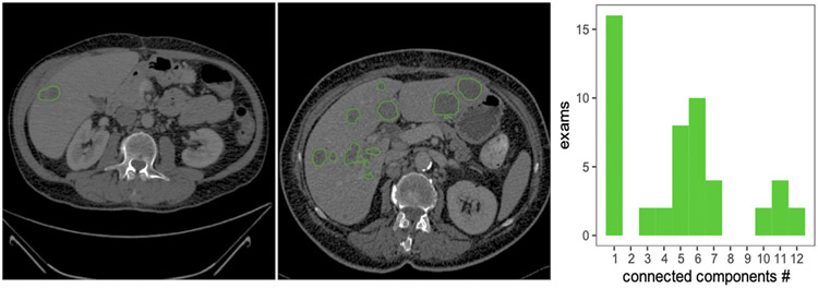
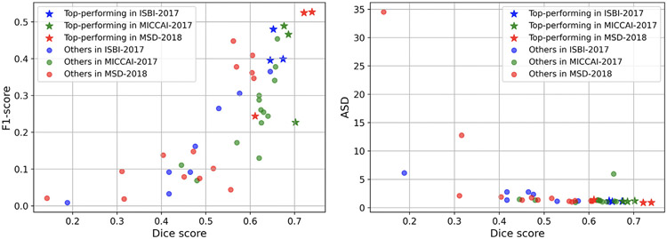
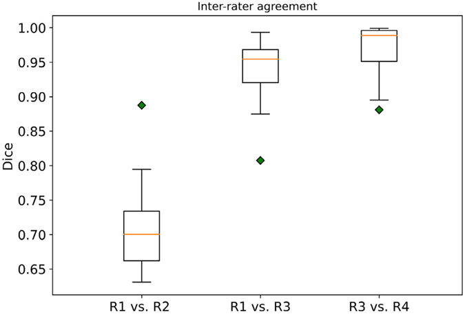
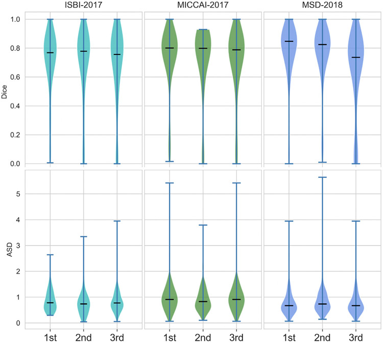
Verification
Claims the paper makes
The claims and statistics of this paper haven’t been checked yet.
Run the check — free, no account needed. It reads the open-access text and checks the sources it cites.
Claims other papers make about this one
Each is checked against this paper’s own text: does it say what the citing paper says it does?
The cited source says what the claim attributes to it.
In the citing paper
“different lesion appearances and annotation targets have been reported across tumor benchmarks [3,8,23]”
From Correction-aware interactive 3D tumor segmentation with sparse and revisable prompts (2026) · cites this paper as [3]
The cited source says what the claim attributes to it.
In the citing paper
“Tumor segmentation is less stable because lesions are not tied to a fixed anatomical shape or location and may be small, heterogeneous, or weakly contrasted [3,8,23].”
From Correction-aware interactive 3D tumor segmentation with sparse and revisable prompts (2026) · cites this paper as [3]
The check wasn’t confident either way, so it gives no verdict — never disputed.
In the citing paper
“The source of the OrganMNIST3D is the same as that of the Organ{A,C,S}MNIST. Instead of 2D images, we directly use the 3D bounding boxes and process the images into 28 × 28 × 28 to perform multi-class classification of 11 body organs.”
From MedMNIST v2 - A large-scale lightweight benchmark for 2D and 3D biomedical image classification (2023) · cites this paper as [30]
Experimental — accuracy not yet measured
Self-check
Percentages, sums and reported differences re-computed from the paper’s own numbers.
The self-check needs the paper’s full text.
Citations
Cites
U-Net: Convolutional Networks for Biomedical Image Segmentation
Ronneberger, O., Fischer, P., & Brox, T. (2015) · Lecture notes in computer science · doi:10.1007/978-3-319-24574-4_28 · Cited by 99,404
New response evaluation criteria in solid tumours: revised RECIST guideline (version 1.1)
Eisenhauer, E. A., Therasse, P., Bogaerts, J., Schwartz, L. H., Sargent, D., Ford, R., Dancey, J., Arbuck, S., Gwyther, S., Mooney, M., et al. (2009) · European Journal of Cancer · doi:10.1016/j.ejca.2008.10.026 · Cited by 29,534
Kass, M., Witkin, A., & Terzopoulos, D. (1988) · International Journal of Computer Vision · doi:10.1007/bf00133570 · Cited by 17,330
A survey on deep learning in medical image analysis
Litjens, G., Kooi, T., Bejnordi, B. E., Setio, A. A. A., Ciompi, F., Ghafoorian, M., van der Laak, J. A. W. M., van Ginneken, B., & Sánchez, C. I. (2017) · Medical Image Analysis · doi:10.1016/j.media.2017.07.005 · Cited by 15,332
Fronts propagating with curvature-dependent speed: Algorithms based on Hamilton-Jacobi formulations
Osher, S., & Sethian, J. A. (1988) · Journal of Computational Physics · doi:10.1016/0021-9991(88)90002-2 · Cited by 14,196
V-Net: Fully Convolutional Neural Networks for Volumetric Medical Image Segmentation
Milletari, F., Navab, N., & Ahmadi, S.-A. (2016) · 2016 Fourth International Conference on 3D Vision (3DV) · doi:10.1109/3dv.2016.79 · Cited by 11,931
Fully convolutional networks for semantic segmentation
Shelhamer, E., Long, J., & Darrell, T. (2017) · IEEE Transactions on Pattern Analysis and Machine Intelligence · doi:10.1109/tpami.2016.2572683 · Cited by 11,898
nnU-Net: a self-configuring method for deep learning-based biomedical image segmentation
Isensee, F., Jaeger, P. F., Kohl, S. A. A., Petersen, J., & Maier-Hein, K. H. (2021) · Nature Methods · doi:10.1038/s41592-020-01008-z · Cited by 10,477
Self-citationYushkevich, P. A., Piven, J., Hazlett, H. C., Smith, R. G., Ho, S., Gee, J. C., & Gerig, G. (2006) · NeuroImage · doi:10.1016/j.neuroimage.2006.01.015 · Cited by 8,868
Active Shape Models-Their Training and Application
Cootes, T. F., Taylor, C. J., Cooper, D. H., & Graham, J. (1995) · Computer Vision and Image Understanding · doi:10.1006/cviu.1995.1004 · Cited by 7,190
Research works printed but not linked: 5
- Ben-Dan, 2008, Liver tumor segmentation in CT images using probabilistic methods, vol. 41, 43
- Deng, X., Du, G., 2008. 3D segmentation in the clinic: A grand challenge II-liver tumor segmentation. In: MICCAI Workshop.
- Erickson, 2016, Radiology data from the cancer genome atlas liver hepatocellular carcinoma [TCGA-LIHC] collection, Cancer Imaging Arch.
- Hann, 2000, Diagnostic imaging approaches and relationship to hepatobiliary cancer staging and therapy, vol. 19, 94
- Heussel, 2007, Follow-up CT measurement of liver malignoma according to RECIST and WHO vs. volumetry, RoFo: Fortschr. Geb. Rontgenstr. Nuklearmed., 179, 958
Cited by
Sorted by citations
Items 401–500 of 1,004
DB-SAM: Delving into High Quality Universal Medical Image Segmentation
Qin, C., Cao, J., Fu, H., Khan, F. S., & Anwer, R. M. (2024) · Lecture notes in computer science · doi:10.1007/978-3-031-72390-2_47 · Cited by 15
Liver segmentation network based on detail enhancement and multi-scale feature fusion
Tinglan, L., Qin, J., Guihe, Q., Shi, W., & Zhang, W. (2025) · Scientific Reports · doi:10.1038/s41598-024-78917-y · Cited by 13
Systematic Review: AI Applications in Liver Imaging with a Focus on Segmentation and Detection
Pomohaci, M. D., Grasu, M. C., Băicoianu-Nițescu, A.-Ş., Enache, R. M., & Lupescu, I. G. (2025) · Life · doi:10.3390/life15020258 · Cited by 13
Variational methods with application to medical image segmentation: A survey
Shu, X., Li, Z., Chang, X., & Yuan, D. (2025) · Neurocomputing · doi:10.1016/j.neucom.2025.130260 · Cited by 15
Boosting segmentation with weak supervision from image-to-image translation.
Vorontsov, E., Molchanov, P. A., Byeon, W., Mello, S. D., Jampani, V., Liu, M.-Y., Kadoury, S., & Kautz, J. (2019) · Cited by 13
Self-citationRaju, A., Ji, Z., Cheng, C., Cai, J., Huang, J., Xiao, J. Z., Lü, L., Liao, C., & Harrison, A. P. (2020) · Lecture notes in computer science · doi:10.1007/978-3-030-59710-8_45 · Cited by 13
Stochastic Planner-Actor-Critic for Unsupervised Deformable Image Registration
Luo, Z., Hu, J., Wang, X., Hu, S., Kong, B., Yin, Y., Song, Q., Wu, X., & Lyu, S. (2022) · Proceedings of the AAAI Conference on Artificial Intelligence · doi:10.1609/aaai.v36i2.20086 · Cited by 14
Vindas, Y., Guépié, B. K., Almar, M., Roux, E., & Delachartre, P. (2022) · Medical Image Analysis · doi:10.1016/j.media.2022.102437 · Cited by 13
Ren, Y., Zou, D., Xu, W., Zhao, X., Lu, W., & He, X. (2023) · Biomedical Signal Processing and Control · doi:10.1016/j.bspc.2023.104591 · Cited by 13
Liu, Y., Yang, F., & Yang, Y. (2023) · Journal of Applied Clinical Medical Physics · doi:10.1002/acm2.13927 · Cited by 14
When liver disease diagnosis encounters deep learning: Analysis, challenges, and prospects
Tian, Y., Liu, M., Sun, Y., & Fu, S. (2023) · iLiver · doi:10.1016/j.iliver.2023.02.002 · Cited by 16
A New Three-stage Curriculum Learning Approach for Deep Network Based Liver Tumor Segmentation
Li, H., Liu, X., Boumaraf, S., Liu, W., Gong, X., & Ma, X. (2020) · International Joint Conference on Neural Networks (IJCNN) · doi:10.1109/ijcnn48605.2020.9206789 · Cited by 13
Self-citationUnsupervised Medical Image Alignment With Curriculum Learning
Burduja, M., & Ionescu, R. T. (2021) · Proceedings - International Conference on Image Processing · doi:10.1109/icip42928.2021.9506067 · Cited by 13
A Review of Generative Adversarial Networks in Cancer Imaging: New Applications, New Solutions.
Osuala, R. O., Kushibar, K., Garrucho, L., Linardos, A., Szafranowska, Z., Klein, S., Glocker, B., Díaz, O., & Lekadir, K. (2021) · Spiral (Imperial College London) · Cited by 13
Efficient Context-Aware Network for Abdominal Multi-organ SegmentationPreprint
Zhang, F., Wang, Y., & Yang, H. (2021) · arXiv (Cornell University) · doi:10.48550/arxiv.2109.10601 · Cited by 13
A Review of Deep Learning‐Based Medical Image Segmentation
Zhang, X., Wang, J., Cheng, X., & Li, J. (2025) · IET Image Processing · doi:10.1049/ipr2.70163 · Cited by 16
Self-citationGe, R., Shi, F., Chen, Y., Tang, S., Zhang, H., Lou, X., Zhao, W., Coatrieux, G., Gao, D., Li, S., et al. (2023) · Biomedical Signal Processing and Control · doi:10.1016/j.bspc.2023.104590 · Cited by 12
Joint Self-Supervised Image-Volume Representation Learning with Intra-inter Contrastive Clustering
Nguyen, D. M. H., Nguyen, H., Mai, T. T. N., Cao, T., Nguyen, B. T., Ho, N., Swoboda, P., Albarqouni, S., Xie, P., & Sonntag, D. (2023) · Proceedings of the AAAI Conference on Artificial Intelligence · doi:10.1609/aaai.v37i12.26687 · Cited by 16
TransLiver: A Hybrid Transformer Model for Multi-phase Liver Lesion Classification
Wang, X., Ying, H., Xu, X., Cai, X., & Zhang, M. (2023) · Lecture notes in computer science · doi:10.1007/978-3-031-43895-0_31 · Cited by 12
Liver Tumor Screening and Diagnosis in CT with Pixel-Lesion-Patient Network
Yan, K., Yin, X., Xia, Y., Wang, F., Wang, S., Gao, Y., Yao, J., Li, C., Bai, X., Zhou, J., et al. (2023) · Lecture notes in computer science · doi:10.1007/978-3-031-43904-9_8 · Cited by 12
Self-citationDifferential CNN and KELM integration for accurate liver cancer detection
Jesi, P. M., & Daniel, V. A. A. (2024) · Biomedical Signal Processing and Control · doi:10.1016/j.bspc.2024.106419 · Cited by 12
HD-Former: A hierarchical dependency Transformer for medical image segmentation
Wu, H., Min, W.-D., Gai, D., Huang, Z., Geng, Y., Wang, Q., & Chen, R. (2024) · Comput. Biol. Medicine · doi:10.1016/j.compbiomed.2024.108671 · Cited by 12
PRISM: A Promptable and Robust Interactive Segmentation Model with Visual Prompts
Li, H., Liu, H., Hu, D., Wang, J., & Oguz, I. (2024) · Lecture Notes in Computer Science · doi:10.1007/978-3-031-72384-1_37 · Cited by 12
Self-citationTowards a general computed tomography image segmentation model for anatomical structures and lesions
Ouyang, X., Gu, D., Li, X., Zhou, W., Chen, Q., Zhan, Y., Zhou, X. S., Shi, F., Xue, Z., & Shen, D. G. (2024) · Communications Engineering · doi:10.1038/s44172-024-00287-0 · Cited by 11
Self-citationSpectral U-Net: Enhancing Medical Image Segmentation via Spectral Decomposition
Peng, Y., Chen, D. Z., & Milan Sonka, A. (2025) · IEEE International Symposium on Biomedical Imaging (ISBI) · doi:10.1109/isbi60581.2025.10981199 · Cited by 12
Cui, L., Xu, M., Liu, C., Liu, T., Yan, X., Zhang, Y., & Yang, X. (2025) · Interdisciplinary Sciences Computational Life Sciences · doi:10.1007/s12539-025-00726-2 · Cited by 12
Self-citationZhang, F., Dvornek, N. C., Yang, J., Chapiro, J., & Duncan, J. S. (2020) · IEEE Transactions on Medical Imaging · doi:10.1109/tmi.2020.2990625 · Cited by 12
Robust Segmentation Models Using an Uncertainty Slice Sampling-Based Annotation Workflow
Chlebus, G., Schenk, A., Hahn, H. K., van Ginneken, B., & Meine, H. (2022) · IEEE Access · doi:10.1109/access.2022.3141021 · Cited by 12
Self-citationSupervised Contrastive Embedding for Medical Image Segmentation
Lee, S.-W., Lee, Y., Lee, G., & Hwang, S. (2021) · IEEE Access · doi:10.1109/access.2021.3118694 · Cited by 12
Review of Generative Adversarial Networks in mono- and cross-modal biomedical image registration
Han, T., Wu, J., Luo, W., Wang, H., Jin, Z., & Qu, L. (2022) · Frontiers in Neuroinformatics · doi:10.3389/fninf.2022.933230 · Cited by 12
Self-citationO.K., S., Riera-Marín, M., Galdran, A., López, J. G., Rodríguez-Comas, J., Piella, G., & Ballester, M. A. G. (2025) · Computerized Medical Imaging and Graphics · doi:10.1016/j.compmedimag.2025.102547 · Cited by 11
UFOS-Net leverages small-scale feature fusion for diabetic foot ulcer segmentation
Jiao, C., Zhao, X., Li, L., Wang, C., & Chen, Y. (2025) · Scientific Reports · doi:10.1038/s41598-025-12442-4 · Cited by 11
Self-citationEskandar, K. (2025) · iLiver · doi:10.1016/j.iliver.2025.100205 · Cited by 14
Karabağ, B., Ayturan, K., & Hardalaç, F. (2026) · Diagnostics · doi:10.3390/diagnostics16050649 · Cited by 14
SDPN: A Slight Dual-Path Network With Local-Global Attention Guided for Medical Image Segmentation
Wang, J., Li, S., Yu, L., Qu, A., Wang, Q., Liu, J., & Wu, Q. (2023) · IEEE Journal of Biomedical and Health Informatics · doi:10.1109/jbhi.2023.3260026 · Cited by 11
Huang, S., Luo, J., Ou, Y., shen, W., Pang, Y., Nie, X., & Zhang, G. (2024) · Journal of Cancer Research and Clinical Oncology · doi:10.1007/s00432-023-05564-7 · Cited by 11
Shape prior-constrained deep learning network for medical image segmentation
Zhang, P., Cheng, Y., & Tamura, S. (2024) · Computers in Biology and Medicine · doi:10.1016/j.compbiomed.2024.108932 · Cited by 11
YOLOv8-Based Frameworks for Liver and Tumor Segmentation Task on LiTS
Randar, S., Shah, V., Kulkarni, H., Suryawanshi, Y., Joshi, A. D., & Sawant, S. T. (2024) · SN Computer Science · doi:10.1007/s42979-024-03097-5 · Cited by 11
Kang, M., Ting, C., Ting, F. F., & Phan, C.-W. R. (2024) · Proceedings - International Conference on Image Processing · doi:10.1109/icip51287.2024.10647768 · Cited by 11
Silva-Rodríguez, J., Dolz, J., & Ayed, I. B. (2025) · Medical Image Analysis · doi:10.1016/j.media.2025.103596 · Cited by 15
Structure-Aware Registration Network for Liver DCE-CT Images
Xue, P., Zhang, J., Ma, L., Liu, M., Gu, Y., Huang, J., Liu, F., Pan, Y., Cao, X., & Shen, D. G. (2024) · IEEE Journal of Biomedical and Health Informatics · doi:10.1109/jbhi.2024.3350166 · Cited by 11
Cascaded Dilated Deep Residual Network for Volumetric Liver Segmentation From CT Image
Mourya, G. K., Gogoi, M., Talbar, S. N., Dutande, P. V., & Baid, U. (2020) · International Journal of E-Health and Medical Communications · doi:10.4018/ijehmc.2021010103 · Cited by 11
Multi-Slice Dense-Sparse Learning for Efficient Liver and Tumor Segmentation
Zhao, Z., Ma, Z., Liu, Y., Zeng, Z., & Chow, P. K. H. (2021) · Annual International Conference of the IEEE Engineering in Medicine and Biology Society (EMBC) · doi:10.1109/embc46164.2021.9629698 · Cited by 11
Learning Consistency- and Discrepancy-Context for 2D Organ Segmentation
Li, L., Lian, S., Luo, Z., Li, S., Wang, B., & Li, S. (2021) · Lecture notes in computer science · doi:10.1007/978-3-030-87193-2_25 · Cited by 11
CyTran: Cycle-Consistent Transformers for Non-Contrast to Contrast CT Translation.Preprint
Ristea, N.-C., Ionescu, A.-I., Savencu, O., Georgescu, M.-I., Verga, N. I., Khan, F. S., & Ionescu, R. T. (2021) · arXiv (Cornell University) · Cited by 11
Deep learning-assisted multiple organ segmentation from whole-body CT imagesPreprint
Salimi, Y., Shiri, I., Mansouri, Z., & Zaidi, H. (2023) · medRxiv · doi:10.1101/2023.10.20.23297331 · Cited by 11
A comprehensive and easy-to-use multi-domain multi-task medical imaging meta-dataset
Woerner, S., Jaques, A., & Baumgartner, C. F. (2025) · Scientific Data · doi:10.1038/s41597-025-04866-4 · Cited by 11
A comprehensive analysis of Mamba for 3D volumetric medical image segmentation
Wang, C., Xie, Y., Chen, Q., Zhou, Y., & Wu, Q. (2025) · Pattern Recognition · doi:10.1016/j.patcog.2025.112701 · Cited by 10
Self-citationLifting wavelet transform-guided network with histogram attention for liver segmentation in CT scans
Liu, H., Sun, W., Fu, Y., Zhang, S., Jin, J., Fang, J., & Wang, B. (2026) · Information Fusion · doi:10.1016/j.inffus.2026.104153 · Cited by 10
Diao, Z., Jiang, H., & Zhou, Y. (2023) · Computerized Medical Imaging and Graphics · doi:10.1016/j.compmedimag.2023.102217 · Cited by 10
Wendler, T., Kreissl, M. C., Schemmer, B., Rogasch, J. M. M., & Benetti, F. D. (2023) · Nuklearmedizin - NuclearMedicine · doi:10.1055/a-2200-2145 · Cited by 10
Customized m-RCNN and hybrid deep classifier for liver cancer segmentation and classification
Khan, R., Su, L., Zaman, A., Hassan, H., Kang, Y., & Huang, B. (2024) · Heliyon · doi:10.1016/j.heliyon.2024.e30528 · Cited by 10
CCSI: Continual Class-Specific Impression for data-free class incremental learning
Ayromlou, S., Tsang, T. S. M., Abolmaesumi, P., & Li, X. (2024) · Medical Image Analysis · doi:10.1016/j.media.2024.103239 · Cited by 10
Self-citationLin, H., Zhao, M., Zhu, L., Pei, X., Wu, H., Zhang, L., & Li, Y. (2024) · Frontiers in Oncology · doi:10.3389/fonc.2024.1423774 · Cited by 10
Self-citationHu, Z., Chen, H., Long, H., Ren, X., & Mei, W. (2024) · Biomedical Signal Processing and Control · doi:10.1016/j.bspc.2024.106861 · Cited by 10
Self-citationKim, S., Park, H., Chikontwe, P., Kang, M., Jin, K. H., Adeli, E., Pohl, K. M., & Park, S. H. (2025) · IEEE Transactions on Medical Imaging · doi:10.1109/tmi.2025.3525581 · Cited by 10
Self-citationJonske, F., Kim, M., Nasca, E., Evers, J., Haubold, J., Hosch, R., Nensa, F., Kamp, M., Seibold, C., Egger, J., et al. (2025) · Computer Methods and Programs in Biomedicine · doi:10.1016/j.cmpb.2025.108634 · Cited by 10
Multi-resolution based dual-channel UNet with cross clique for medical image dense prediction
Zhou, X., Jin, G., Liu, Y., Li, J., Wang, J., Ying, S., Zheng, Y., & Shi, J. (2025) · Expert Systems with Applications · doi:10.1016/j.eswa.2025.127190 · Cited by 10
Self-citationMulti-AD: cross-domain unsupervised anomaly detection for medical and industrial applications
Rahmaniar, W., & Suzuki, K. (2025) · Pattern Recognition · doi:10.1016/j.patcog.2025.112486 · Cited by 10
Rangraz, E. J., Tang, X., Laeken, C. V., Maleux, G. A., Dekervel, J., Cutsem, E. V., Verslype, C., Baete, K., Nuyts, J. L., & Deroose, C. M. (2020) · EJNMMI Research · doi:10.1186/s13550-020-00675-5 · Cited by 10
2D medical image segmentation via learning multi-scale contextual dependencies
Pang, S., Du, A., Yu, Z., & Orgun, M. A. (2021) · Methods · doi:10.1016/j.ymeth.2021.05.015 · Cited by 10
Ananda, S., Iwamoto, Y., Han, X., Lin, L., Hu, H., & Chen, Y. (2022) · Annual International Conference of the IEEE Engineering in Medicine and Biology Society (EMBC) · doi:10.1109/embc48229.2022.9871188 · Cited by 10
Self-citationZhou, X., Li, Z., & Tong, T. (2022) · IEEE International Conference on Bioinformatics and Biomedicine (BIBM) · doi:10.1109/bibm55620.2022.9995371 · Cited by 11
Liu, P., Du, J., & Vong, C. (2023) · Neural Networks · doi:10.1016/j.neunet.2023.04.023 · Cited by 10
Scaling up 3D Kernels with Bayesian Frequency Re-parameterization for Medical Image Segmentation
Lee, H. H., Liu, Q., Bao, S., Yang, Q., Yu, X., Cai, L. Y., Li, T. Z., Huo, Y., Koutsoukos, X., & Landman, B. A. (2023) · Lecture notes in computer science · doi:10.1007/978-3-031-43901-8_60 · Cited by 10
Liu, X., Ono, K., & Bise, R. (2024) · Image and Vision Computing · doi:10.1016/j.imavis.2024.105056 · Cited by 14
Modanwal, G., Al-Kindi, S. G., Walker, J. R., Dhamdhere, R., Yuan, L., Ji, M., Lu, C. Y., Fu, P., Rajagopalan, S., & Madabhushi, A. (2022) · EBioMedicine · doi:10.1016/j.ebiom.2022.104315 · Cited by 10
Liver Lesion Segmentation with slice-wise 2D Tiramisu and Tversky loss functionPreprint
Roth, K., Konopczyński, T., & Hesser, J. (2019) · arXiv (Cornell University) · doi:10.48550/arxiv.1905.03639 · Cited by 10
Self-citationHuo, Y., Cai, J., Cheng, C., Raju, A., Yan, K., Landman, B. A., Xiao, J. Z., Lü, L., Liao, C., & Harrison, A. P. (2020) · arXiv (Cornell University) · doi:10.48550/arxiv.2006.15691 · Cited by 10
Generative Adversarial U-Net for Domain-free Medical Image AugmentationPreprint
Chen, X., Li, Y., Yao, L., Adeli, E., & Zhang, Y. (2021) · arXiv (Cornell University) · doi:10.48550/arxiv.2101.04793 · Cited by 14
Li, D., Yang, J., Kreis, K., Torralba, A. B., & Fidler, S. (2021) · arXiv (Cornell University) · doi:10.48550/arxiv.2104.05833 · Cited by 10
Fragemann, J., Ardizzone, L., Egger, J., Kleesiek, J., & Workshop, M. (2022) · doi:10.36227/techrxiv.19364897 · Cited by 10
mlf-core: a framework for deterministic machine learning
Heumos, L., Ehmele, P., Cuellar, L. K., Menden, K., Miller, E., Lemke, S., Gabernet, G., & Nahnsen, S. (2023) · Bioinformatics · doi:10.1093/bioinformatics/btad164 · Cited by 11
A Coarse-to-Fine Fusion Network for Small Liver Tumor Detection and Segmentation: A Real-World Study
Wu, S., Yu, H., Li, C., Zheng, R., Xia, X., Wang, C., & Wang, H. (2023) · Diagnostics · doi:10.3390/diagnostics13152504 · Cited by 9
Self-citationZero-shot Unsupervised Transfer Instance Segmentation
Shin, G., Albanie, S., & Xie, W. (2023) · IEEE/CVF Conference on Computer Vision and Pattern Recognition Workshops (CVPRW) · doi:10.1109/cvprw59228.2023.00513 · Cited by 9
Huang, Z., Yang, X. N., Huang, S., & Guo, L. (2023) · Bioengineering · doi:10.3390/bioengineering10101119 · Cited by 9
Self-citationDeep learning application for abdominal organs segmentation on 0.35 T MR-Linac images
Zhou, Y., Lalande, A., Chevalier, C., Baude, J., Aubignac, L., Boudet, J., & Bessières, I. (2024) · Frontiers in Oncology · doi:10.3389/fonc.2023.1285924 · Cited by 9
Improving abdominal image segmentation with overcomplete shape priors
Sadikine, A., Badic, B., Tasu, J., Noblet, V., Ballet, P., Visvikis, D., & Conze, P.-H. (2024) · Computerized Medical Imaging and Graphics · doi:10.1016/j.compmedimag.2024.102356 · Cited by 9
Deep Learning Framework for Liver Tumor Segmentation
Gupta, K., Aggarwal, S., Jha, A. K., Habib, A., Jagtap, J., Kolhar, S., Patil, S., Kotecha, K. V., & Choudhury, T. (2024) · EAI Endorsed Transactions on Pervasive Health and Technology · doi:10.4108/eetpht.10.5561 · Cited by 9
Patel, N., Celaya, A., Eltaher, M., Glenn, R., Savannah, K. J. B., Brock, K. K., Sanchez, J. I., Calderone, T. L., Cleere, D. W., Elsaiey, A., et al. (2024) · Scientific Reports · doi:10.1038/s41598-024-71674-y · Cited by 9
Robust and smooth Couinaud segmentation via anatomical structure-guided point-voxel network
Zhang, X., Ali, S., Liu, T., Zhao, X. Y., Cui, Z., Han, M., Ma, S., Zhu, J., Wang, L., Wang, L., et al. (2024) · Computers in Biology and Medicine · doi:10.1016/j.compbiomed.2024.109202 · Cited by 10
Self-citationArtificial intelligence for detection and characterization of focal hepatic lesions: a review
Anta, J. A., Moreno‐Vedia, J., López, J. G., Ríos-Vives, M. Á., Munuera, J., & Rodríguez‐Comas, J. (2024) · Abdominal Radiology · doi:10.1007/s00261-024-04597-x · Cited by 9
Liao, J., Wang, H., Gu, H., & Cai, Y. (2024) · PLoS ONE · doi:10.1371/journal.pone.0312105 · Cited by 12
Boosting Convolution With Efficient MLP-Permutation for Volumetric Medical Image Segmentation
Lin, Y., Fang, X., Zhang, D., Cheng, K.-T. T., & Chen, H. (2025) · IEEE Transactions on Medical Imaging · doi:10.1109/tmi.2025.3530113 · Cited by 10
Self-citationLMTTM-VMI: Linked Memory Token Turing Machine for 3D volumetric medical image classification
Wei, H., Yang, Y., Sun, S., Feng, M., Wang, R., & Han, X. (2025) · Computer Methods and Programs in Biomedicine · doi:10.1016/j.cmpb.2025.108640 · Cited by 9
Self-citationLi, C., Zhou, S., Kong, J., Qi, L., & Xue, H. (2025) · IEEE International Conference on Acoustics Speech and Signal Processing · doi:10.1109/icassp49660.2025.10888834 · Cited by 9
An advanced transformer framework for liver tumor segmentation using MRI images
Sivanagaraju, P., Venkataramana, S., & Reddy, P. V. G. D. P. (2025) · Biomedical Signal Processing and Control · doi:10.1016/j.bspc.2025.107808 · Cited by 9
Song, Z., Wu, W., & Wu, S. (2025) · Sensors · doi:10.3390/s25061814 · Cited by 9
FourierLoss: Shape-aware loss function with Fourier descriptors
Erden, M. B., Cansız, S., Çakı, O., Khattak, H., Etiz, D., Yakar, M. C., Duruer, K., Barut, B., & Gündüz-Demir, Ç. (2025) · Neurocomputing · doi:10.1016/j.neucom.2025.130155 · Cited by 9
Alzubi, T. M., & Mukhtar, H. (2025) · IEEE Access · doi:10.1109/access.2025.3569073 · Cited by 9
FGS-FL: Enhancing federated learning with differential privacy via flat gradient stream
Hu, J., & Zhang, H. (2025) · Expert Systems with Applications · doi:10.1016/j.eswa.2025.128273 · Cited by 9
Bi, Y., Huang, L., Clarenbach, R., Ghotbi, R., Karlas, A., Navab, N., & Jiang, Z. (2025) · Medical Image Analysis · doi:10.1016/j.media.2025.103737 · Cited by 14
Clustering-guided contrastive prototype learning: Towards semi-supervised medical image segmentation
Lv, Z., Wu, Z., & Zhu, J. (2025) · Pattern Recognition · doi:10.1016/j.patcog.2025.112321 · Cited by 9
A Review on the Applications of GANs for 3D Medical Image Analysis
Usama, Z., Alavi, A., & Chan, J. (2025) · Applied Sciences · doi:10.3390/app152011219 · Cited by 11
Large-scale generative tumor synthesis in computed tomography images for improving tumor recognition
Wu, L., Zhuang, J., Zhou, Y., He, S., Ma, J., Luo, L., Wang, X., Ni, X., Zhong, X., Wu, M., et al. (2025) · Nature Communications · doi:10.1038/s41467-025-66071-6 · Cited by 13
Self-citationThe Growing Role for Semantic Segmentation in Urology
Rickman, J., Struyk, G., Simpson, B., Byun, B. C., & Papanikolopoulos, N. P. (2021) · European Urology Focus · doi:10.1016/j.euf.2021.07.017 · Cited by 9
Liver Multi-class Tumour Segmentation and Detection Based on Hyperion Pre-trained Models
(2022) · International journal of intelligent engineering and systems · doi:10.22266/ijies2022.1231.36 · Cited by 9
Attention-based CT scan interpolation for lesion segmentation of colorectal liver metastases
Hamghalam, M., Do, R., & Simpson, A. L. (2023) · SPIE Medical Imaging: Biomedical Applications in Molecular, Structural, and Functional Imaging · doi:10.1117/12.2656072 · Cited by 9
Optimization of U-shaped pure transformer medical image segmentation network
Dan, Y., Jin, W., Wang, Z., & Sun, C. (2023) · PeerJ Computer Science · doi:10.7717/peerj-cs.1515 · Cited by 9
Masked image modeling-based boundary reconstruction for 3D medical image segmentation
Liu, C., Cheng, Y., & Tamura, S. (2023) · Computers in Biology and Medicine · doi:10.1016/j.compbiomed.2023.107526 · Cited by 10
Metadata
- Authors
- Bilic, P., Christ, P., Li, H. B., Vorontsov, E., Ben-Cohen, A., Kaissis, G., Szeskin, A., Jacobs, C., Mamani, G. E. H., Chartrand, G., Lohöfer, F., Holch, J. W., Sommer, W., Hofmann, F., Hostettler, A., Lev-Cohain, N., Drozdzal, M., Amitai, M. M., Vivanti, R., Sosna, J., Ezhov, I., Sekuboyina, A., Navarro, F., Kofler, F., Paetzold, J. C., Shit, S., Hu, X., Lipková, J., Rempfler, M., Piraud, M., Kirschke, J., Wiestler, B., Zhang, Z., Hülsemeyer, C., Beetz, M., Ettlinger, F., Antonelli, M., Bae, W., Bellver, M., Bi, L., Chen, H., Chlebus, G., Dam, E. B., Dou, Q., Fu, C.-W., Georgescu, B., Giró-i-Nieto, X., Gruen, F., Han, X., Heng, P.-A., Hesser, J., Moltz, J. H., Igel, C., Isensee, F., Jäger, P., Jia, F., Kaluva, K. C., Khened, M., Kim, I., Kim, J.-H., Kim, S., Kohl, S., Konopczynski, T., Kori, A., Krishnamurthi, G., Li, F., Li, H., Li, J., Li, X., Lowengrub, J., Ma, J., Maier-Hein, K., Maninis, K.-K., Meine, H., Merhof, D., Pai, A., Perslev, M., Petersen, J., Pont-Tuset, J., Qi, J., Qi, X., Rippel, O., Roth, K., Sarasua, I., Schenk, A., Shen, Z., Torres, J., Wachinger, C., Wang, C., Weninger, L., Wu, J., Xu, D., Yang, X., Yu, S. C.-H., Yuan, Y., Yue, M., Zhang, L., Cardoso, J., Bakas, S., Braren, R., Heinemann, V., Pal, C., Tang, A., Kadoury, S., Soler, L., van Ginneken, B., Greenspan, H., Joskowicz, L., & Menze, B.
- Affiliations
- Technical University of Munich (DE)
- Guangdong University of Foreign Studies (CN)
- Polytechnique Montréal (CA)
- Tel Aviv University (IL)
- TUM Klinikum (DE)
- Hebrew University of Jerusalem (IL)
- Radboud University Medical Center (NL)
- Université de Montréal (CA)
- German Cancer Research Center (DE)
- LMU Klinikum (DE)
- Institut de Recherche contre les Cancers de l’Appareil Digestif (FR)
- Rafael Advanced Defense Systems (Israel) (IL)
- Hadassah Medical Center (IL)
- University of Zurich (CH)
- Helmholtz Munich (DE)
- Chinese University of Hong Kong (HK)
- Brigham and Women's Hospital (US)
- ETH Zurich (CH)
- Nanjing Drum Tower Hospital (CN)
- King's College London (GB)
- Hong Kong University of Science and Technology (HK)
- Barcelona Supercomputing Center (ES)
- The University of Sydney (AU)
- University of Copenhagen (DK)
- Siemens Healthcare (United States) (US)
- Heidelberg University (DE)
- University of North Carolina at Chapel Hill (US)
- Fraunhofer Institute for Digital Medicine (DE)
- Chinese Academy of Sciences (CN)
- Indian Institute of Technology Madras (IN)
- Samsung Medical Center (KR)
- Kakao Brain (South Korea) (KR)
- University of California, Irvine (US)
- University of Bremen (DE)
- RWTH Aachen University (DE)
- University of Hong Kong (HK)
- University of Tübingen (DE)
- Medizinische Hochschule Hannover (DE)
- University of Illinois Urbana-Champaign (US)
- KTH Royal Institute of Technology (SE)
- Nvidia (United States) (US)
- Nanjing University (CN)
- Icahn School of Medicine at Mount Sinai (US)
- Corresponding author
- Li, Hongwei Bran
- Li, Hongchao
- ORCID
- Li, Hongwei Bran
- Vorontsov, Eugene
- Kaissis, Georgios
- Szeskin, Adi
- Jacobs, Colin
- Mamani, Gabriel Efrain Humpire
- Chartrand, Gabriel
- Lohöfer, Fabian
- Holch, Julian Walter
- Hostettler, Alexandre
- Drozdzal, Michal
- Amitai, Michal Marianne
- Sosna, Jacob
- Ezhov, Ivan
- Sekuboyina, Anjany
- Navarro, Fernando
- Kofler, Florian
- Paetzold, Johannes C.
- Shit, Suprosanna
- Hu, Xiaobin
- Lipková, Jana
- Piraud, Marie
- Kirschke, Jan
- Wiestler, Benedikt
- Zhang, Zhiheng
- Beetz, Marcel
- Antonelli, Michela
- Bae, Woong
- Bellver, Míriam
- Chen, Hao
- Chlebus, Grzegorz
- Dam, Erik B.
- Dou, Qi
- Fu, Chi-Wing
- Georgescu, Bogdan
- Giró-i-Nieto, Xavier
- Han, Xu
- Heng, Pheng-Ann
- Hesser, Jürgen
- Moltz, Jan Hendrik
- Igel, Christian
- Isensee, Fabian
- Jäger, Paul
- Jia, Fucang
- Kaluva, Krishna Chaitanya
- Khened, Mahendra
- Kim, Jae-Hun
- Kim, Sungwoong
- Kori, Avinash
- Krishnamurthi, Ganapathy
- Li, Fan
- Li, Hongchao
- Li, Junbo
- Li, Xiaomeng
- Lowengrub, John
- Ma, Jun
- Maier-Hein, Klaus
- Maninis, Kevis-Kokitsi
- Meine, Hans
- Merhof, Dorit
- Pai, Akshay
- Perslev, Mathias
- Petersen, Jens
- Pont-Tuset, Jordi
- Qi, Xiaojuan
- Rippel, Oliver
- Roth, Karsten
- Schenk, Andrea
- Shen, Zengming
- Torres, Jordi
- Wachinger, Christian
- Wang, Chunliang
- Weninger, Leon
- Xu, Daguang
- Yang, Xiaoping
- Yu, Simon Chun-Ho
- Yuan, Yading
- Zhang, Liping
- Bakas, Spyridon
- Braren, Rickmer
- Heinemann, Volker
- Pal, Christopher
- Tang, An
- Kadoury, Samuel
- van Ginneken, Bram
- Greenspan, Hayit
- Menze, Bjoern
- Venue
- Medical Image Analysis
- Publisher
- Elsevier BV
- Published
- 2023-02
- Volume
- 84
- Pages
- 102680
- ISSN
- 1361-8415 (ISSN-L)
- PMC
- PMC10631490
- OpenAlex
- W2910094941
- Cited by
- 1,412
- References
- 146
- Citations by year
- 2026: 259 · 2025: 337 · 2024: 311 · 2023: 195
- FWCI
- 77.21
- Type
- journal article
- Language
- en
- Open access
- hybrid
- doi.org
- Licence
- Locations
- Medical Image Analysis (publishedVersion, cc-by-nc-nd)
- PubMed (publishedVersion)
- Research at the University of Copenhagen (University of Copenhagen) (publishedVersion, cc-by-nc-nd)
- PubMed Central (acceptedVersion, cc-by-nc-nd)
- Zurich Open Repository and Archive (University of Zurich) (acceptedVersion, cc-by-nc-nd)
- arXiv (Cornell University) (submittedVersion, cc-by-nc-nd)
- Rare & Special e-Zone (The Hong Kong University of Science and Technology) (submittedVersion)
- Rare & Special e-Zone (The Hong Kong University of Science and Technology) (submittedVersion, cc-by-nc-sa)
- eScholarship (California Digital Library) (submittedVersion, cc-by-nc-nd)
- Radboud Repository (Radboud University) (submittedVersion, cc-by-nc-sa)
- IRIS eCampus Telematic University (Università degli Studi eCampus) (submittedVersion)
- mediaTUM (Technical University of Munich) (submittedVersion)
- Site cant be reached (submittedVersion, cc-by-nc-nd)
- Fraunhofer-Publica (Fraunhofer-Gesellschaft) (submittedVersion)
- PolyPublie (École Polytechnique de Montréal) (submittedVersion)
- PuSH - Publication Server of Helmholtz Zentrum München (submittedVersion, cc-by-nc-nd)
- UPCommons institutional repository (Universitat Politècnica de Catalunya) (submittedVersion, cc-by-nc-nd)
- Repository for Publications and Research Data (ETH Zurich) (submittedVersion, cc-by-nc-nd)
- Zurich Open Repository and Archive (University of Zurich) (submittedVersion, cc-by-nc-nd)
- Radboud Repository (Radboud University) (submittedVersion, other-oa)
- arXiv (Cornell University)
- ETH Zürich Research Collection
- Zurich Open Repository and Archive (University of Zurich)
- Funders
- Deutsche Forschungsgemeinschaft (SFB824, GSC81, subproject B12)
- Universität Zürich
- Fondation de l'Association des radiologistes du Québec (34939)
- National Institutes of Health (1s10od023495-01)
- Fonds de Recherche du Québec - Santé (34939, Junior 2)
- National Cancer Institute (U01CA242871)
- International Graduate School of Science and Engineering (GSC 81)
- Topics
- Physical Sciences › Computer Science › Computer Vision and Pattern Recognition › Medical Image Segmentation Techniques
- Physical Sciences › Computer Science › Computer Vision and Pattern Recognition › Brain Tumor Detection and Classification
- Health Sciences › Medicine › Radiology, Nuclear Medicine and Imaging › Radiomics and Machine Learning in Medical Imaging
- Keywords
- liver tumor segmentation, liver segmentation, liver cancer, computed tomography, segmentation benchmark, primary liver tumors, secondary liver tumors, lesion detection, medical image segmentation
- MeSH terms
- Algorithms, Humans, Image Processing, Computer-Assisted, Liver, Liver Neoplasms, Retrospective Studies, Benchmarking
About these checks
Registry, retraction, Cites and statistics results are computed, not judged, with one exception: a printed reference that no index lists is read once by a model before it counts as a citation error. Claims are extracted by a model and kept only when their quote appears verbatim in the text; each cited source is then checked, with a model’s help, for whether it says what the claim relies on. Those verdicts can be wrong — read the claim’s quote and the cited source before relying on one.
Statistics and arithmetic are recomputed, not judged.
Confidence bands are shown only for fields calibrated on our annotated test set. None is calibrated yet, so no band is shown.
What counts as a claim ›How we measure confidence ›How we check this ›
Are you an author, or do you see a problem with this page? Request a review
