

The DOI matches its Crossref record; 37 of the 37 references it cites were found in a registry or an index.
Astrocyte IKKβ/NF-κB signaling is required for diet-induced obesity and hypothalamic inflammation
Open accessAbstract
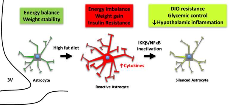
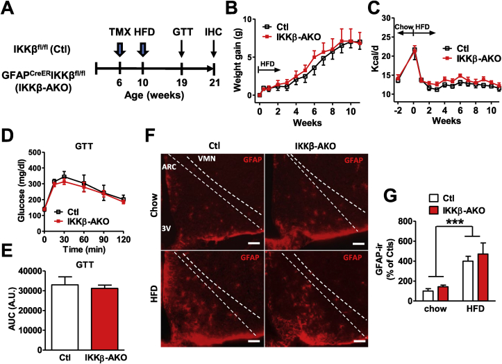
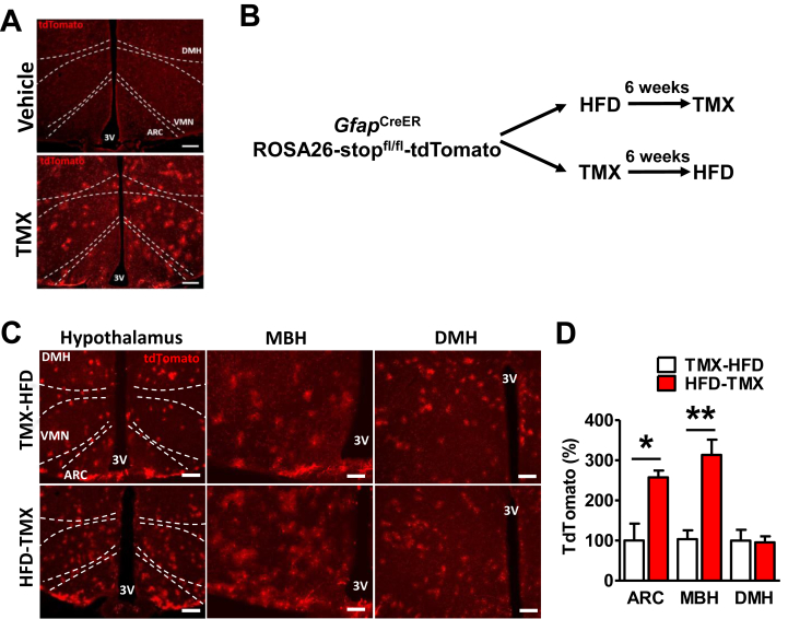
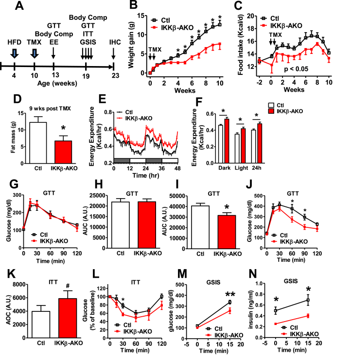
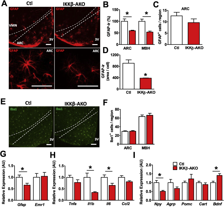
Objective Obesity and high fat diet (HFD) consumption in rodents is associated with hypothalamic inflammation and reactive gliosis. While neuronal inflammation promotes HFD-induced metabolic dysfunction, the role of astrocyte activation in susceptibility to hypothalamic inflammation and diet-induced obesity (DIO) remains uncertain. Methods Metabolic phenotyping, immunohistochemical analyses, and biochemical analyses were performed on HFD-fed mice with a tamoxifen-inducible astrocyte-specific knockout of IKKβ ( Gfap CreER Ikbkb fl/fl , IKKβ-AKO), an essential cofactor of NF-κB-mediated inflammation. Results IKKβ-AKO mice with tamoxifen-induced IKKβ deletion prior to HFD exposure showed equivalent HFD-induced weight gain and glucose intolerance as Ikbkb fl/fl littermate controls. In Gfap CreER TdTomato marker mice treated using the same protocol, minimal Cre-mediated recombination was observed in the mediobasal hypothalamus (MBH). By contrast, mice pretreated with 6 weeks of HFD exposure prior to tamoxifen administration showed substantially increased recombination throughout the MBH. Remarkably, this treatment approach protected IKKβ-AKO mice from further weight gain through an immediate reduction of food intake and increase of energy expenditure. Astrocyte IKKβ deletion after HFD exposure—but not before—also reduced glucose intolerance and insulin resistance, likely as a consequence of lower adiposity. Finally, both hypothalamic inflammation and astrocytosis were reduced in HFD-fed IKKβ-AKO mice. Conclusions These data support a requirement for astrocytic inflammatory signaling in HFD-induced hyperphagia and DIO susceptibility that may provide a novel target for obesity therapeutics.
Overview
The claims and statistics of this paper haven’t been checked yet.
Run the check — free, no account needed. It reads the open-access text and checks the sources it cites.
Integrity signals
Registry
Cites
Statistics
Figures
Verification
Claims the paper makes
The claims and statistics of this paper haven’t been checked yet.
Run the check — free, no account needed. It reads the open-access text and checks the sources it cites.
Claims other papers make about this one
Each is checked against this paper’s own text: does it say what the citing paper says it does?
In the citing paper
“Reversing neuroinflammation via targeted genetic [96,97], pharmacological [98,99], or dietary [49,100] approaches that rescue behavioral anomalies offers convincing support for a causal role of neuroinflammation in obesity-induced neuropathophysiology.”
From The menace of obesity to depression and anxiety prevalence (2022) · cites this paper as [97]
Self-check
Percentages, sums and reported differences re-computed from the paper’s own numbers.
The self-check needs the paper’s full text.
Citations
Cites
Inflammation and metabolic disorders
Hotamisligil, G. S. (2006) · Nature · doi:10.1038/nature05485 · Cited by 9,199
Astrocytes: Biology and pathology
Sofroniew, M. V., & Vinters, H. V. (2010) · Acta Neuropathologica · doi:10.1007/s00401-009-0619-8 · Cited by 5,196
Inflammatory links between obesity and metabolic disease
Lumeng, C. N., & Saltiel, A. R. (2011) · Journal of Clinical Investigation · doi:10.1172/jci57132 · Cited by 2,319
Obesity is associated with hypothalamic injury in rodents and humans.
Thaler, J. P., Yi, C.-X., Schur, E. A., Guyenet, S. J., Hwang, B. H., Dietrich, M. O., Zhao, X., Sarruf, D. A., Izgur, V., Maravilla, K. R., et al. (2012) · Journal of Clinical Investigation · doi:10.1172/jci59660 · Cited by 1,857
Hypothalamic IKKβ/NF-κB and ER Stress Link Overnutrition to Energy Imbalance and Obesity
Zhang, X., Zhang, G., Zhang, H., Karin, M., Bai, H., & Cai, D. (2008) · Cell · doi:10.1016/j.cell.2008.07.043 · Cited by 1,350
De Souza, C. T., Araujo, E. P., Bordin, S., Ashimine, R., Zollner, R. L., Boschero, A. C., Saad, M. J. A., & Velloso, L. A. (2005) · Endocrinology · doi:10.1210/en.2004-1520 · Cited by 1,171
Astrocytes as determinants of disease progression in inherited amyotrophic lateral sclerosis
Yamanaka, K., Chun, S. J., Boillee, S., Fujimori-Tonou, N., Yamashita, H., Gutmann, D. H., Takahashi, R., Misawa, H., & Cleveland, D. W. (2008) · Nature Neuroscience · doi:10.1038/nn2047 · Cited by 1,140
Milanski, M., Degasperi, G., Coope, A., Morari, J., Denis, R., Cintra, D. E., Tsukumo, D. M. L., Anhe, G., Amaral, M. E., Takahashi, H. K., et al. (2009) · The Journal of Neuroscience · doi:10.1523/jneurosci.2760-08.2009 · Cited by 1,125
Henry, C. J., Huang, Y., Wynne, A., Hanke, M., Himler, J., Bailey, M. T., Sheridan, J. F., & Godbout, J. P. (2008) · Journal of Neuroinflammation · doi:10.1186/1742-2094-5-15 · Cited by 678
Valdearcos, M., Robblee, M. M., Benjamin, D. I., Nomura, D. K., Xu, A. W., & Koliwad, S. K. (2014) · Cell Reports · doi:10.1016/j.celrep.2014.11.018 · Cited by 629
Posey, K. A., Clegg, D. J., Printz, R. L., Byun, J., Morton, G. J., Vivekanandan-Giri, A., Pennathur, S., Baskin, D. G., Heinecke, J. W., Woods, S. C., et al. (2009) · American Journal of Physiology-Endocrinology and Metabolism · doi:10.1152/ajpendo.90377.2008 · Cited by 586
Astrocytic Insulin Signaling Couples Brain Glucose Uptake with Nutrient Availability
García‐Cáceres, C., Quarta, C., Varela, L., Gao, Y., Gruber, T., Legutko, B., Jastroch, M., Johansson, P., Ninkovic, J., Yi, C., et al. (2016) · Cell · doi:10.1016/j.cell.2016.07.028 · Cited by 533
Kleinridders, A., Schenten, D., Könner, A. C., Belgardt, B. F., Mauer, J., Okamura, T., Wunderlich, F. T., Medzhitov, R., & Brüning, J. C. (2009) · Cell Metabolism · doi:10.1016/j.cmet.2009.08.013 · Cited by 483
Kaltschmidt, B., & Kaltschmidt, C. (2009) · Cold Spring Harbor Perspectives in Biology · doi:10.1101/cshperspect.a001271 · Cited by 392
Unger, T. J., Calderon, G. A., Bradley, L., Sena‐Esteves, M., & Rios, M. (2007) · Journal of Neuroscience · doi:10.1523/jneurosci.3308-07.2007 · Cited by 385
Leptin signaling in astrocytes regulates hypothalamic neuronal circuits and feeding
Kim, J. G., Suyama, S., Koch, M., Jin, S., Argente-Arizon, P., Argente, J., Liu, Z.-W., Zimmer, M. R., Jeong, J. K., Szigeti-Buck, K., et al. (2014) · Nature Neuroscience · doi:10.1038/nn.3725 · Cited by 336
Beckervordersandforth, R., Tripathi, P., Ninkovic, J., Bayam, E., Lepier, A., Stempfhuber, B., Kirchhoff, F., Hirrlinger, J., Haslinger, A., Lie, D. C., et al. (2010) · Cell Stem Cell · doi:10.1016/j.stem.2010.11.017 · Cited by 335
Astrogliosis: An integral player in the pathogenesis of Alzheimer's disease
Osborn, L. M., Kamphuis, W., Wadman, W. J., & Hol, E. M. (2016) · Progress in Neurobiology · doi:10.1016/j.pneurobio.2016.01.001 · Cited by 312
Early Postnatal Astroglial Cells Produce Multilineage Precursors and Neural Stem Cells In Vivo
Ganat, Y. M., Silbereis, J. C., Cave, C., Ngu, H., Anderson, G. M., Ohkubo, Y., Ment, L. R., & Vaccarino, F. M. (2006) · Journal of Neuroscience · doi:10.1523/jneurosci.2532-06.2006 · Cited by 264
Astrocytes Control Food Intake by Inhibiting AGRP Neuron Activity via Adenosine A1 Receptors
Yang, L., Qi, Y., & Yang, Y. (2015) · Cell Reports · doi:10.1016/j.celrep.2015.04.002 · Cited by 248
Buckman, L. B., Hasty, A. H., Flaherty, D. K., Buckman, C. T., Thompson, M. M., Matlock, B. K., Weller, K., & Ellacott, K. L. J. (2014) · Brain Behavior and Immunity · doi:10.1016/j.bbi.2013.06.007 · Cited by 216
IKKβ Is Required for Peripheral B Cell Survival and Proliferation
Li, Z., Omori, S. A., Labuda, T., Karin, M., & Rickert, R. C. (2003) · The Journal of Immunology · doi:10.4049/jimmunol.170.9.4630 · Cited by 189
Inhibiting microglia expansion prevents diet-induced hypothalamic and peripheral inflammation
André, C., Guzman-Quevedo, O., Rey, C., Rémus-Borel, J., Clark, S., Castellanos-Jankiewicz, A., Ladeveze, E., Leste-Lasserre, T., Nadjar, A., Abrous, D. N., et al. (2017) · Diabetes · doi:10.2337/db16-0586 · Cited by 182
Hypothalamic innate immune reaction in obesity
Kälin, S., Heppner, F. L., Bechmann, I., Prinz, M., Tschöp, M. H., & Yi, C.-X. (2015) · Nature Reviews Endocrinology · doi:10.1038/nrendo.2015.48 · Cited by 167
BDNF AND NGF TREATMENT IN LESIONED RATS
Lapchak, P. A., & Hefti, F. F. (1992) · Neuroreport · doi:10.1097/00001756-199205000-00007 · Cited by 156
Schur, E. A., Melhorn, S. J., Oh, S.-K., Lacy, J. M., Berkseth, K. E., Guyenet, S. J., Sonnen, J. A., Tyagi, V., Leon, M. R. B. D., Leon, B. D., et al. (2015) · Obesity · doi:10.1002/oby.21248 · Cited by 155
Regional astrogliosis in the mouse hypothalamus in response to obesity
Buckman, L. B., Thompson, M. M., Moreno, H. N., & Ellacott, K. L. J. (2012) · The Journal of Comparative Neurology · doi:10.1002/cne.23233 · Cited by 151
Buckman, L. B., Thompson, M. M., Lippert, R. N., Blackwell, T. S., Yull, F. E., & Ellacott, K. L. J. (2014) · Molecular Metabolism · doi:10.1016/j.molmet.2014.10.001 · Cited by 138
Astrocytes Regulate GLP-1 Receptor-Mediated Effects on Energy Balance
Reiner, D. J., Mietlicki-Baase, E. G., McGrath, L. E., Zimmer, D. J., Bence, K. K., Sousa, G. L., Konanur, V. R., Krawczyk, J., Burk, D. H., Kanoski, S. E., et al. (2016) · The Journal of Neuroscience · doi:10.1523/jneurosci.3579-15.2016 · Cited by 133
Benzler, J., Ganjam, G. K., Pretz, D., Oelkrug, R., Koch, C. E., Legler, K., Stöhr, S., Culmsee, C., Williams, L. M., & Tups, A. (2015) · Diabetes · doi:10.2337/db14-0093 · Cited by 129
Berkseth, K. E., Guyenet, S. J., Melhorn, S. J., Lee, D., Thaler, J. P., Schur, E. A., & Schwartz, M. W. (2014) · Endocrinology · doi:10.1210/en.2014-1121 · Cited by 110
Direct modulation of GFAP-expressing glia in the arcuate nucleus bi-directionally regulates feeding
Chen, N., 杉原, 弘., Kim, J., Fu, Z., Barak, B., Sur, M., Feng, G., & Han, W. (2016) · eLife · doi:10.7554/elife.18716 · Cited by 110
Glia: silent partners in energy homeostasis and obesity pathogenesis
Douglass, J. D., Dorfman, M. D., & Thaler, J. P. (2016) · Diabetologia · doi:10.1007/s00125-016-4181-3 · Cited by 99
Ghrelin Regulates Glucose and Glutamate Transporters in Hypothalamic Astrocytes
Fuente-Martín, E., García-Cáceres, C., Argente-Arizón, P., Díaz, F., Granado, M., Freire-Regatillo, A., Castro-González, D., Ceballos, M. L., Frago, L. M., Dickson, S. L., et al. (2016) · Scientific Reports · doi:10.1038/srep23673 · Cited by 91
Longer T2relaxation time is a marker of hypothalamic gliosis in mice with diet-induced obesity
Lee, D., Thaler, J. P., Berkseth, K. E., Melhorn, S. J., Schwartz, M. W., & Schur, E. A. (2013) · American Journal of Physiology-Endocrinology and Metabolism · doi:10.1152/ajpendo.00020.2013 · Cited by 51
High-fat diet feeding causes rapid, non-apoptotic cleavage of caspase-3 in astrocytes
Guyenet, S. J., Hồng, N. T., Hwang, B. H., Schwartz, M. W., Baskin, D. G., & Thaler, J. P. (2013) · Brain Research · doi:10.1016/j.brainres.2013.03.033 · Cited by 44
Moon, Y., Kim, H. J., Kim, J. Y., Kim, H., Kim, W. R., & Sun, W. (2011) · Animal Cells and Systems · doi:10.1080/19768354.2011.611254 · Cited by 9
Cited by
Sorted by citations
Valdearcos, M., Douglass, J. D., Robblee, M. M., Dorfman, M. D., Stifler, D. R., Bennett, M. L., Gerritse, I., Fasnacht, R., Barres, B. A., Thaler, J. P., et al. (2017) · Cell Metabolism · doi:10.1016/j.cmet.2017.05.015 · Cited by 477
Self-citationThe menace of obesity to depression and anxiety prevalence
Fulton, S., Décarie-Spain, L., Fioramonti, X., Guiard, B., & Nakajima, S. (2022) · Trends in Endocrinology & Metabolism · doi:10.1016/j.tem.2021.10.005 · Cited by 441
“Reversing neuroinflammation via targeted genetic [96,97], pharmacological [98,99], or dietary [49,100] approaches that rescue behavioral anomalies offers convincing support for a causal role of neuroinflammation in obesity-induced neuropathophysiology.”
BackgroundRole of astrocytes, microglia, and tanycytes in brain control of systemic metabolism
García‐Cáceres, C., Balland, É., Prévot, V., Luquet, S. H., Woods, S. C., Koch, M., Horváth, T. L., Yi, C., Chowen, J. A., Verkhratsky, A., et al. (2018) · Nature Neuroscience · doi:10.1038/s41593-018-0286-y · Cited by 343
The Versatile Tanycyte: A Hypothalamic Integrator of Reproduction and Energy Metabolism
Prévot, V., Dehouck, B., Sharif, A., Ciofi, P., Giacobini, P., & Clasadonte, J. (2018) · Endocrine Reviews · doi:10.1210/er.2017-00235 · Cited by 299
The melanocortin pathway and control of appetite-progress and therapeutic implications
Baldini, G., & Phelan, K. D. (2019) · Journal of Endocrinology · doi:10.1530/joe-18-0596 · Cited by 300
Metadata
- Authors
- Douglass, J. D., Dorfman, M. D., Fasnacht, R., Shaffer, L. D., & Thaler, J. P.
- Affiliations
- University of Washington (US)
- Corresponding author
- Thaler, J.P.
- Venue
- Molecular Metabolism
- Publisher
- Elsevier BV
- Published
- 2017-04
- Volume
- 6
- Issue
- 4
- Pages
- 366-373
- ISSN
- 2212-8778 (ISSN-L)
- PMC
- PMC5369266
- OpenAlex
- W2581154578
- Cited by
- 248
- References
- 37
- Citations by year
- 2026: 12 · 2025: 23 · 2024: 28 · 2023: 34 · 2022: 32 · 2021: 31 · 2020: 31 · 2019: 26 · 2018: 20 · 2017: 11
- FWCI
- 14.72
- Type
- journal article
- Language
- en
- Open access
- gold
- doi.org
- Licence
- Locations
- Molecular Metabolism (publishedVersion, cc-by-nc-nd)
- PubMed Central (publishedVersion, cc-by-nc-nd)
- PubMed (publishedVersion)
- DOAJ (DOAJ: Directory of Open Access Journals) (submittedVersion, cc-by-sa)
- Funders
- American Diabetes Association (1-14-ACE-51)
- NIDDK (K08 DK088872, T32 DK7247-37, F32 DK108473-01)
- NIH (DK035816)
- Diabetes Research Center (DK017047)
- Topics
- Life Sciences › Neuroscience › Endocrine and Autonomic Systems › Regulation of Appetite and Obesity
- Health Sciences › Medicine › Endocrinology, Diabetes and Metabolism › Adipokines, Inflammation, and Metabolic Diseases
- Life Sciences › Neuroscience › Neurology › Neuroinflammation and Neurodegeneration Mechanisms
- Keywords
- diet-induced obesity, hypothalamic inflammation, astrocytes, IKKβ, NF-κB signaling, astrocyte-specific knockout, high-fat diet, impaired glucose tolerance, insulin resistance, hyperphagia, energy expenditure, reactive gliosis, astrocyte activation, mediobasal hypothalamus, obesity treatment
- MeSH terms
- Animals, Astrocytes, Cells, Cultured, Gliosis, Hypothalamus, Inflammation, Male, Mice, Inbred C57BL, Obesity, Signal Transduction, NF-kappa B, Mice, I-kappa B Kinase, Diet, High-Fat
About these checks
Registry, retraction, Cites and statistics results are computed, not judged, with one exception: a printed reference that no index lists is read once by a model before it counts as a citation error. Claims are extracted by a model and kept only when their quote appears verbatim in the text; each cited source is then checked, with a model’s help, for whether it says what the claim relies on. Those verdicts can be wrong — read the claim’s quote and the cited source before relying on one.
Statistics and arithmetic are recomputed, not judged.
Confidence bands are shown only for fields calibrated on our annotated test set. None is calibrated yet, so no band is shown.
What counts as a claim ›How we measure confidence ›How we check this ›
Are you an author, or do you see a problem with this page? Request a review
