

The DOI matches its Crossref record; 11 of the 11 references it cites were found in a registry or an index.
Cysteine Dioxygenase 1 Mediates Erastin-Induced Ferroptosis in Human Gastric Cancer Cells
Overview
The claims and statistics of this paper haven’t been checked yet.
Run the check — free, no account needed. It reads the open-access text and checks the sources it cites.
Integrity signals
Registry
Cites
Statistics
Figures
Verification
Claims the paper makes
The claims and statistics of this paper haven’t been checked yet.
Run the check — free, no account needed. It reads the open-access text and checks the sources it cites.
Claims other papers make about this one
Each is checked against this paper’s own text: does it say what the citing paper says it does?
In the citing paper
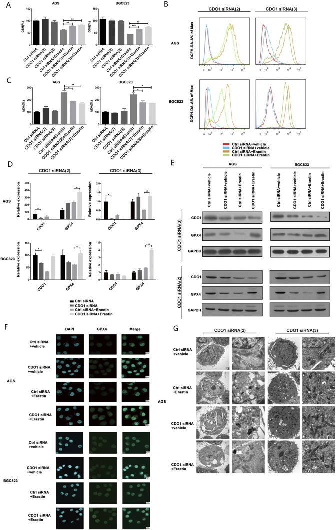
“The level of GSH is apparently decreased in the process of erastin-induced ferroptosis in human gastric cancer cells (Hao et al., 2017).”
From MiR-378a-3p/ SLC7A11 regulate ferroptosis in nerve injury induced by lead exposure (2022) · cites this paper as Hao et al., 2017
Self-check
Percentages, sums and reported differences re-computed from the paper’s own numbers.
The self-check needs the paper’s full text.
Citations
Cites
Regulation of Ferroptotic Cancer Cell Death by GPX4
Yang, W. S., SriRamaratnam, R., Welsch, M. E., Shimada, K., Skouta, R., Viswanathan, V. S., Cheah, J. H., Clemons, P. A., Shamji, A. F., Clish, C. B., et al. (2014) · Cell · doi:10.1016/j.cell.2013.12.010 · Cited by 7,713
Inactivation of the ferroptosis regulator Gpx4 triggers acute renal failure in mice
Friedmann Angeli, J. P., Schneider, M., Proneth, B., Tyurina, Y. Y., Tyurin, V. A., Hammond, V. J., Herbach, N., Aichler, M., Walch, A., Eggenhofer, E., et al. (2014) · Nature Cell Biology · doi:10.1038/ncb3064 · Cited by 3,732
Ferroptosis: process and function
Xie, Y., Hou, W., Song, X., Yu, Y., Huang, J., Sun, X., Kang, R., & Tang, D. (2016) · Cell Death & Differentiation · doi:10.1038/cdd.2015.158 · Cited by 3,727
Sun, X., Ou, Z., Chen, R., Niu, X., Chen, D., Kang, R., & Tang, D. (2016) · Hepatology · doi:10.1002/hep.28251 · Cited by 2,012
Regulated necrosis: the expanding network of non-apoptotic cell death pathways
Berghe, T. V., Linkermann, A., Jouan-Lanhouet, S., Walczak, H., & Vandenabeele, P. (2014) · Nature Reviews Molecular Cell Biology · doi:10.1038/nrm3737 · Cited by 1,735
RAS–RAF–MEK-dependent oxidative cell death involving voltage-dependent anion channels
Yagoda, N., von Rechenberg, M., Zaganjor, E., Bauer, A. J., Yang, W. S., Fridman, D. J., Wolpaw, A. J., Smukste, I., Peltier, J. M., Boniface, J. J., et al. (2007) · Nature · doi:10.1038/nature05859 · Cited by 1,631
Ferrostatins Inhibit Oxidative Lipid Damage and Cell Death in Diverse Disease Models
Skouta, R., Dixon, S. J., Wang, J., Dunn, D. E., Orman, M., Shimada, K., Rosenberg, P. A., Lo, D. C., Weinberg, J. M., Linkermann, A., et al. (2014) · Journal of the American Chemical Society · doi:10.1021/ja411006a · Cited by 1,146 · cited 2×
“The existence of ferroptosis has been confirmed in various tumorous cells such as fibrosarcoma [11], lung cancer [12], osteosarcoma [13], kidney cancer [9], and prostate cancer cells [8,14].”
BackgroundHeme oxygenase-1 accelerates erastin-induced ferroptotic cell death
Kwon, M.-Y., Park, E., Lee, S.-J., & Chung, S. W. (2015) · Oncotarget · doi:10.18632/oncotarget.5162 · Cited by 612
Identification of artesunate as a specific activator of ferroptosis in pancreatic cancer cells
Eling, N., Reuter, L., Hazin, J., Hamacher-Brady, A., & Brady, N. R. (2015) · Oncoscience · doi:10.18632/oncoscience.160 · Cited by 524
Louandre, C., Marcq, I., Bouhlal, H., Lachaier, E., Godin, C., Saidak, Z., François, C., Chatelain, D., Debuysscher, V., Barbare, J.-C., et al. (2015) · Cancer Letters · doi:10.1016/j.canlet.2014.11.014 · Cited by 374
Metabolism and iron signaling in ferroptotic cell death
Gao, M., Monian, P., & Jiang, X. (2015) · Oncotarget · doi:10.18632/oncotarget.5671 · Cited by 71 · cited 1×
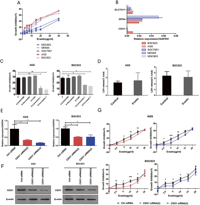
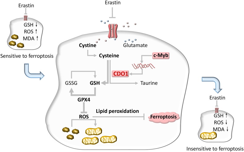
“Regulation of CDO1 on Ferroptosis Biomarkers Previous studies demonstrated that erastin targets system XC − and inhibits cystine uptake, subsequently leading to increased ferroptotic events including GSH depletion, GPX4 inactivation, ROS increases, and changes in mitochondrial morphology [9,10,12,23].”
Background
Research works printed but not linked: 39
- Kaczmarek, 2013, Necroptosis: the release of damage-associated molecular patterns and its physiological relevance, Immunity, 38, 209, 10.1016/j.immuni.2013.02.003 · DOI · Paper page
- Suzanne, 2013, Shaping organisms with apoptosis, Cell Death Differ, 20, 669, 10.1038/cdd.2013.11 · DOI
- Dixon, 2012, Ferroptosis: an iron-dependent form of nonapoptotic cell death, Cell, 149, 1060, 10.1016/j.cell.2012.03.042 · DOI · Paper page
- Yang, 2016, Ferroptosis: death by lipid peroxidation, Trends Cell Biol, 26, 165, 10.1016/j.tcb.2015.10.014 · DOI · Paper page
- Zhao, 2017, ROS signaling under metabolic stress: cross-talk between AMPK and AKT pathway, Mol Cancer, 16, 10.1186/s12943-017-0648-1 · DOI · Paper page
- Conrad, 2015, Glutathione peroxidase 4 (Gpx4) and ferroptosis: what's so special about it?, Mol Cell Oncol, 2, e995047, 10.4161/23723556.2014.995047 · DOI
- Cao, 2016, Mechanisms of ferroptosis, Cell Mol Life Sci, 73, 2195, 10.1007/s00018-016-2194-1 · DOI · Paper page
- Jurkowska, 2015, Propargylglycine inhibits hypotaurine/taurine synthesis and elevates cystathionine and homocysteine concentrations in primary mouse hepatocytes, Amino Acids, 47, 1215, 10.1007/s00726-015-1948-7 · DOI
- Gao, 2015, Glutaminolysis and transferrin regulate ferroptosis, Mol Cell, 59, 298, 10.1016/j.molcel.2015.06.011 · DOI · Paper page
- Stipanuk, 2009, Cysteine dioxygenase: a robust system for regulation of cellular cysteine levels, Amino Acids, 37, 55, 10.1007/s00726-008-0202-y · DOI
- Lu, 2009, Regulation of glutathione synthesis, Mol Aspects Med, 30, 42, 10.1016/j.mam.2008.05.005 · DOI
- Hayano, 2016, Loss of cysteinyl-tRNA synthetase (CARS) induces the transsulfuration pathway and inhibits ferroptosis induced by cystine deprivation, Cell Death Differ, 23, 270, 10.1038/cdd.2015.93 · DOI
- Yu, 2016, Crosstalk between cystine and glutathione is critical for the regulation of amino acid signaling pathways and ferroptosis, Sci Rep, 6, 30033, 10.1038/srep30033 · DOI
- Sun, 2015, HSPB1 as a novel regulator of ferroptotic cancer cell death, Oncogene, 34, 5617, 10.1038/onc.2015.32 · DOI · Paper page
- Yuan, 2016, CISD1 inhibits ferroptosis by protection against mitochondrial lipid peroxidation, Biochem Biophys Res Commun, 478, 838, 10.1016/j.bbrc.2016.08.034 · DOI
- Tsikas, 2017, Assessment of lipid peroxidation by measuring malondialdehyde (MDA) and relatives in biological samples: analytical and biological challenges, Anal Biochem, 524, 13, 10.1016/j.ab.2016.10.021 · DOI · Paper page
- Yahya, 1996, Antibodies against malondialdehyde (MDA) in MRL/lpr/lpr mice: evidence for an autoimmune mechanism involving lipid peroxidation, J Autoimmun, 9, 3, 10.1006/jaut.1996.0002 · DOI
- Dolma, 2003, Identification of genotype-selective antitumor agents using synthetic lethal chemical screening in engineered human tumor cells, Cancer Cell, 3, 285, 10.1016/S1535-6108(03)00050-3 · DOI · Paper page
- Vargova, 2011, MYB transcriptionally regulates the miR-155 host gene in chronic lymphocytic leukemia, Blood, 117, 3816, 10.1182/blood-2010-05-285064 · DOI
- Baer, 1992, Defective c-myc and c-myb RNA turnover in acute myeloid leukemia cells, Blood, 79, 1319, 10.1182/blood.V79.5.1319.1319 · DOI
- Wan, 2007, Benzene's metabolites alter c-MYB activity via reactive oxygen species in HD3 cells, Toxicol Appl Pharmacol, 222, 180, 10.1016/j.taap.2007.04.016 · DOI
- Booken, 2008, Sezary syndrome is a unique cutaneous T-cell lymphoma as identified by an expanded gene signature including diagnostic marker molecules CDO1 and DNM3, Leukemia, 22, 393, 10.1038/sj.leu.2405044 · DOI
- Ramsden, 1997, Human cysteine dioxygenase type I (CDO-I; EC 1.13.11.20): 5′ flanking region and intron-exon structure of the gene, Mol Pathol, 50, 269, 10.1136/mp.50.5.269 · DOI
- Rhodes, 2014, Pooled analysis of iron-related genes in Parkinson's disease: association with transferrin, Neurobiol Dis, 62, 172, 10.1016/j.nbd.2013.09.019 · DOI
- Greenshields, 2017, Contribution of reactive oxygen species to ovarian cancer cell growth arrest and killing by the anti-malarial drug artesunate, Mol Carcinog, 56, 75, 10.1002/mc.22474 · DOI
- McBean, 2002, Cerebral cystine uptake: a tale of two transporters, Trends Pharmacol Sci, 23, 299, 10.1016/S0165-6147(02)02060-6 · DOI
- Polewski, 2016, Increased expression of system xc- in glioblastoma confers an altered metabolic state and temozolomide resistance, Mol Cancer Res, 14, 1229, 10.1158/1541-7786.MCR-16-0028 · DOI
- Lo, 2008, The xc- cystine/glutamate antiporter: a mediator of pancreatic cancer growth with a role in drug resistance, Br J Cancer, 99, 464, 10.1038/sj.bjc.6604485 · DOI
- Gout, 2003, In vitro studies on the lymphoma growth-inhibitory activity of sulfasalazine, Anticancer Drugs, 14, 21, 10.1097/00001813-200301000-00004 · DOI · Paper page
- Han, 2006, The taurine transporter: mechanisms of regulation, Acta Physiol (Oxf), 187, 61, 10.1111/j.1748-1716.2006.01573.x · DOI
- Jeschke, 2013, Frequent inactivation of cysteine dioxygenase type 1 contributes to survival of breast cancer cells and resistance to anthracyclines, Clin Cancer Res, 19, 3201, 10.1158/1078-0432.CCR-12-3751 · DOI
- Brait, 2012, Cysteine dioxygenase 1 is a tumor suppressor gene silenced by promoter methylation in multiple human cancers, PLoS One, 7, e44951, 10.1371/journal.pone.0044951 · DOI
- Kwon, 2015, Epigenetic regulation of the novel tumor suppressor cysteine dioxygenase 1 in esophageal squamous cell carcinoma, Tumour Biol, 36, 7449, 10.1007/s13277-015-3443-x · DOI
- Ushiku, 2017, Promoter DNA methylation of CDO1 gene and its clinical significance in esophageal squamous cell carcinoma, Dis Esophagus, 30, 1
- Nakano, 2016, Dysregulation of c-Myb pathway by aberrant expression of proto-oncogene MYB provides the basis for malignancy in adult T-cell leukemia/lymphoma cells, Clin Cancer Res, 22, 5915, 10.1158/1078-0432.CCR-15-1739 · DOI
- Minekura, 2001, Genomic organization and transcription units of the human acyl-CoA synthetase 3 gene, Gene, 278, 185, 10.1016/S0378-1119(01)00714-4 · DOI
- Lee, 2016, C-myb regulates autophagy for pulp vitality in glucose oxidative stress, J Dent Res, 95, 430, 10.1177/0022034515622139 · DOI
- Guehmann, 1992, Reduction of a conserved Cys is essential for Myb DNA-binding, Nucleic Acids Res, 20, 2279, 10.1093/nar/20.9.2279 · DOI
- Chen, 2015, Myeloid zinc-finger 1 (MZF-1) suppresses prostate tumor growth through enforcing ferroportin-conducted iron egress, Oncogene, 34, 3839, 10.1038/onc.2014.310 · DOI
Cited by
Sorted by citations
Ferroptosis: past, present and future
Li, J., Cao, F., Yin, H.-L., Huang, Z.-J., Lin, Z.-T., Mao, N., Sun, B., & Wang, G. (2020) · Cell Death & Disease · doi:10.1038/s41419-020-2298-2 · Cited by 4,171
The application of Ferroptosis in diseases
Qiu, Y., Cao, Y., Cao, W., Jia, Y., & Lu, N. (2020) · Pharmacological Research · doi:10.1016/j.phrs.2020.104919 · Cited by 415
MiR-378a-3p/ SLC7A11 regulate ferroptosis in nerve injury induced by lead exposure
Wang, W., Shi, F., Cui, J., Pang, S., Zheng, G., & Zhang, Y. (2022) · Ecotoxicology and Environmental Safety · doi:10.1016/j.ecoenv.2022.113639 · Cited by 47
Metadata
- Authors
- Hao, S., Yu, J., He, W., Huang, Q., Zhao, Y., Liang, B., Zhang, S., Wen, Z., Dong, S., Rao, J., Liao, W., & Shi, M.
- Venue
- Neoplasia
- Publisher
- Elsevier BV
- Published
- 2017-12
- Volume
- 19
- Issue
- 12
- Pages
- 1022-1032
- ISSN
- 1476-5586
- PMC
- PMC5686465
- Cited by
- 276
- References
- 11
- Language
- en
- Funders
- National Natural Science Foundation of China (81472317)
- Science and Technology Planning Project of Guangdong Province (2016A020215232)
- CSCO-Merck Serono Oncology Research Fund (Y-MX2014-048))
- Special Foundation of National Clinical Specialties of China
About these checks
Registry, retraction, Cites and statistics results are computed, not judged, with one exception: a printed reference that no index lists is read once by a model before it counts as a citation error. Claims are extracted by a model and kept only when their quote appears verbatim in the text; each cited source is then checked, with a model’s help, for whether it says what the claim relies on. Those verdicts can be wrong — read the claim’s quote and the cited source before relying on one.
Statistics and arithmetic are recomputed, not judged.
Confidence bands are shown only for fields calibrated on our annotated test set. None is calibrated yet, so no band is shown.
What counts as a claim ›How we measure confidence ›How we check this ›
Are you an author, or do you see a problem with this page? Request a review
