

The DOI matches its Crossref record; 31 of the 31 references it cites were found in a registry or an index.
Increased in vivo glial activation in patients with amyotrophic lateral sclerosis: Assessed with [11C]-PBR28
Open accessAbstract
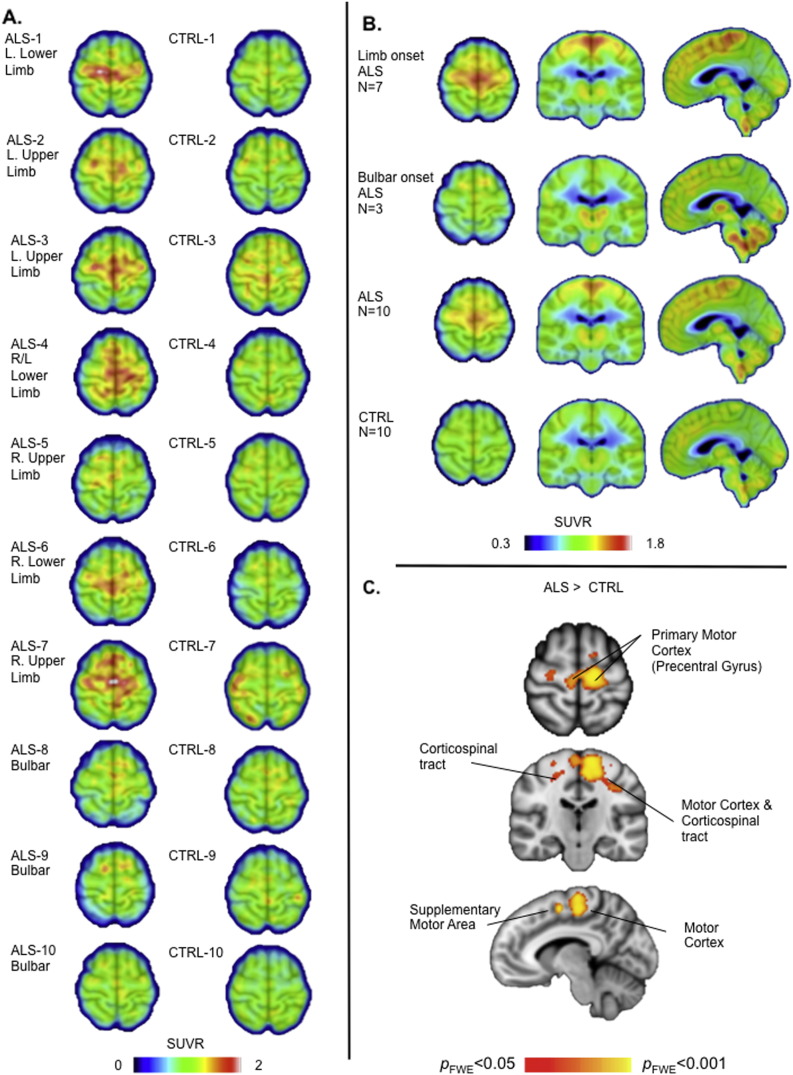
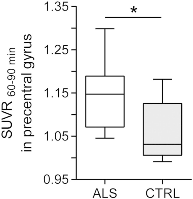
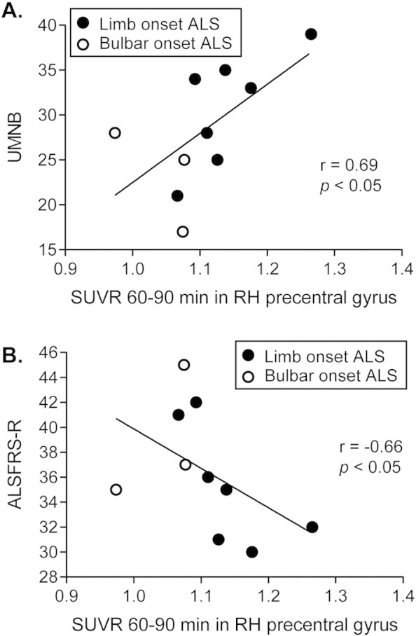
Evidence from human post mortem, in vivo and animal model studies implicates the neuroimmune system and activated microglia in the pathology of amyotrophic lateral sclerosis. The study aim was to further evaluate in vivo neuroinflammation in individuals with amyotrophic lateral sclerosis using [(11)C]-PBR28 positron emission tomography. Ten patients with amyotrophic lateral sclerosis (seven males, three females, 38-68 years) and ten age- and [(11)C]-PBR28 binding affinity-matched healthy volunteers (six males, four females, 33-65 years) completed a positron emission tomography scan. Standardized uptake values were calculated from 60 to 90 min post-injection and normalized to whole brain mean. Voxel-wise analysis showed increased binding in the motor cortices and corticospinal tracts in patients with amyotrophic lateral sclerosis compared to healthy controls (p FWE < 0.05). Region of interest analysis revealed increased [(11)C]-PBR28 binding in the precentral gyrus in patients (normalized standardized uptake value = 1.15) compared to controls (1.03, p < 0.05). In patients those values were positively correlated with upper motor neuron burden scores (r = 0.69, p < 0.05), and negatively correlated with the amyotrophic lateral sclerosis functional rating scale (r = -0.66, p < 0.05). Increased in vivo glial activation in motor cortices, that correlates with phenotype, complements previous histopathological reports. Further studies will determine the role of [(11)C]-PBR28 as a marker of treatments that target neuroinflammation.
Overview
The claims and statistics of this paper haven’t been checked yet.
Run the check — free, no account needed. It reads the open-access text and checks the sources it cites.
Integrity signals
Registry
Cites
Statistics
Figures
Verification
Claims the paper makes
The claims and statistics of this paper haven’t been checked yet.
Run the check — free, no account needed. It reads the open-access text and checks the sources it cites.
Claims other papers make about this one
Each is checked against this paper’s own text: does it say what the citing paper says it does?
The source backs part of the claim, not all of it — or backs it for a different population, condition or comparison.
In the citing paper
“Microglial activation in middle frontal and superior or middle temporal gyrus regions was also highly found in vivo PET imaging studies in humans”
From Microglia in neurodegenerative diseases: mechanism and potential therapeutic targets (2023) · cites this paper as [312]
Experimental — accuracy not yet measured
Self-check
Percentages, sums and reported differences re-computed from the paper’s own numbers.
The self-check needs the paper’s full text.
Citations
Cites
Polazzi, E., Mengoni, I., Caprini, M., Peña‐Altamira, E., Kurtys, E., & Monti, B. (2012) · Neurosignals · doi:10.1159/000337115 · Cited by 9,023
Nonparametric permutation tests for functional neuroimaging: A primer with examples
Nichols, T. E., & Holmes, A. P. (2002) · Human Brain Mapping · doi:10.1002/hbm.1058 · Cited by 6,256
El Escorial revisited: revised criteria for the diagnosis of amyotrophic lateral sclerosis
Brooks, B. R., Miller, R. G., Swash, M., & Munsat, T. L. (2000) · Amyotrophic Lateral Sclerosis and Other Motor Neuron Disorders · doi:10.1080/146608200300079536 · Cited by 5,433 · cited 2×
“The protocol was approved by the Institutional Review Board and the Radioactive Drug Research Committee.”
MethodCedarbaum, J. M., Stambler, N., Malta, E., Fuller, C., Hilt, D., Thurmond, B., & Nakanishi, A. (1999) · Journal of the Neurological Sciences · doi:10.1016/s0022-510x(99)00210-5 · Cited by 3,307 · cited 6×
“The clinical outcomes obtained from the participants with ALS included the revised ALS functional rating scale (ALSFRS-R) (Cedarbaum et al., 1999), upper motor neuron burden scale (UMNB) (Ellis et al.”
MethodInfluentialDose-ranging study of riluzole in amyotrophic lateral sclerosis
Amyotrophic Lateral Sclerosis/Riluzole Study Group II, Lacomblez, L., Bensimon, G., Meininger, V., Leigh, P. N., & Guillet, P. (1996) · The Lancet · doi:10.1016/s0140-6736(96)91680-3 · Cited by 1,303 · cited 1×
“Riluzole, the only FDA-approved treatment provides a modest survival benefit (Lacomblez et al., 1996), S-R, amyotrophic lateralsclerorate; MR,magnetic resonance; ron emission tomography; SUV, in;UMNB, uppermotorneuron search Institute, Massachusetts on, 02114 MA, USA.”
BackgroundOwen, D. R., Yeo, A. J., Gunn, R. N., Song, K., Wadsworth, G., Lewis, A., Rhodes, C., Pulford, D. J., Bennacef, I., Parker, C. A., et al. (2012) · Journal of Cerebral Blood Flow & Metabolism · doi:10.1038/jcbfm.2011.147 · Cited by 779 · cited 2×
“[11C]-PBR28, along with all second-generation TSPO radiotracers to date, has differential binding affinity to TSPO depending on an Ala147Thr polymorphism in the TSPO gene with Ala/Ala leading to high-, Ala/Thr to mixed-, and Thr/Thr to low-affinity binding (Kreisl et al., 2013; Owen et al., 2012).”
BackgroundTurner, M. R., Cagnin, A., Turkheimer, F. E., Miller, C. C. J., Shaw, C. E., Brooks, D. J., Leigh, P. N., & Banati, R. B. (2004) · Neurobiology of Disease · doi:10.1016/j.nbd.2003.12.012 · Cited by 737 · cited 6×
“This finding is consistent with the first microglial PET study in ALS patients (Turner et al., 2004), as well as histopathological studies reporting increased activated microglia near degenerating motor neurons (Brettschneider et al., 2012; Henkel et al., 2004; Kawamata et al., 1992), suggesting…”
BackgroundResultInfluentialMolecular pathways of motor neuron injury in amyotrophic lateral sclerosis
Ferraiuolo, L., Kirby, J., Grierson, A. J., Sendtner, M., & Shaw, P. J. (2011) · Nature Reviews Neurology · doi:10.1038/nrneurol.2011.152 · Cited by 615 · cited 4×
“This is an open access article under but there are no available treatments that prevent or stop disease progression and current animal models have not yet successfully predicted treatment response in people (Atassi et al., 2012; Ferraiuolo et al., 2011).”
BackgroundMethodInfluentialHenkel-Tigges, J., Engelhardt, J., Siklós, L., Simpson, E. P., Kim, S. H., Pan, T., Goodman, J. C., Siddique, T., Beers, D. R., & Appel, S. H. (2003) · Annals of Neurology · doi:10.1002/ana.10805 · Cited by 576
Immunologic reactions in amyotrophic lateral sclerosis brain and spinal cord tissue.
Kawamata, T., Akiyama, H., Yamada, T., & McGeer, P. L. (1992) · PubMed Central · Cited by 546
Cited by
Sorted by citations
Items 101–200 of 231
Viviano, M., Barresi, E., Siméon, F. G., Costa, B., Taliani, S., Settimo, F. D., Pike, V. W., & Castellano, S. (2022) · Current Medicinal Chemistry · doi:10.2174/0929867329666220329204054 · Cited by 35
Brody, A. L., Gehlbach, D. L., Garcia, L. Y., Enoki, R., Hoh, C. K., Vera, D. R., Kotta, K. K., London, E. D., Okita, K., Nurmi, E. L., et al. (2018) · Psychopharmacology · doi:10.1007/s00213-018-5077-3 · Cited by 32
“…regions for PET data analysis (Colasanti et al. 2016; Coughlin et al. 2014; Hamelin et al. 2016; Kreisl et al. 2016; Lyoo et al. 2015; Zurcher et al. 2015) to minimize potential confounds, but this method would not have been appropriate here due to the hypothesized and confirmed…”
Giridharan, V. V., Collodel, A., Generoso, J. S., Scaini, G., Wassather, R., Selvaraj, S., Hasbun, R., Dal‐Pizzol, F., Petronilho, F. C., & Barichello, T. (2020) · Journal of Neuroinflammation · doi:10.1186/s12974-019-1692-0 · Cited by 32
Liu, E., Karpf, L., & Bohl, D. (2021) · Frontiers in Molecular Neuroscience · doi:10.3389/fnmol.2021.767041 · Cited by 32
“…Kreisl, 2017) only to the motor cortex regions with positive correlations with the Upper Motor Neuron Burden Scale (UMNB) and negative ones with the Amyotrophic Lateral Sclerosis Functional Rating Scale – Revised (ALSFRS-R) (Zürcher et al., 2015; Alshikho et al., 2016, 2018; Ratai et al., 2018).”
BackgroundMethodInfluentialPET Imaging of Human Brown Adipose Tissue with the TSPO Tracer [11C]PBR28
Ran, C., Albrecht, D., Bredella, M. A., Yang, J., Yang, J., Liang, S. H., Cypess, A. M., Loggia, M. L., Atassi, N., & Moore, A. V. (2017) · Molecular Imaging and Biology · doi:10.1007/s11307-017-1129-z · Cited by 31
“In this retrospective analysis, we present imaging data from three healthy volunteers, who were imaged under thermoneutral conditions with [(11)C]PBR28, a TSPO PET tracer that has been widely used for both preclinical and clinical investigations [44, 50].”
Self-citationChoi, S. Y., Lee, J., Chung, A.-Y., Jo, Y., Shin, J., Park, H., Kim, H., Lopez-Gonzalez, R., Ryu, J. R., & Sun, W. (2020) · Cell Death and Disease · doi:10.1038/s41419-020-03102-8 · Cited by 30
Glial Cell Dysfunction in C9orf72-Related Amyotrophic Lateral Sclerosis and Frontotemporal Dementia
Ghasemi, M., Keyhanian, K., & Douthwright, C. L. (2021) · Cells · doi:10.3390/cells10020249 · Cited by 35
Nair, A., Veronese, M., Xu, X., Curtis, C. J. C., Turkheimer, F., Howard, R. J., & Reeves, S. (2016) · EJNMMI Research · doi:10.1186/s13550-016-0226-3 · Cited by 32
Edlow, B. L., Bodien, Y. G., Baxter, T., Belanger, H. G., Cali, R. J., Deary, K. B., Fischl, B. R., Foulkes, A. S., Gilmore, N., Greve, D. N., et al. (2022) · Journal of Neurotrauma · doi:10.1089/neu.2022.0030 · Cited by 30
“50 Additionally, TSPO PET identifies microglial activation, 52 a pathophysiological process that may contribute to the astroglial scarring observed postmortem in brain tissue of military personnel with a history of blast exposure.”
Self-citationThe translocator protein (18 kDa) and its role in neuropsychiatric disorders
Barichello, T., Simões, L. R., Collodel, A., Giridharan, V. V., Dal‐Pizzol, F., Macêdo, D. S., & Quevedo, J. (2017) · Neuroscience & Biobehavioral Reviews · doi:10.1016/j.neubiorev.2017.10.010 · Cited by 30
Current and emerging ALS biomarkers: utility and potential in clinical trials
Taga, A., & Maragakis, N. J. (2018) · Expert Review of Neurotherapeutics · doi:10.1080/14737175.2018.1530987 · Cited by 29
Microglial crosstalk with astrocytes and immune cells in amyotrophic lateral sclerosis
Calafatti, M., Cocozza, G., Limatola, C., & Garofalo, S. (2023) · Frontiers in Immunology · doi:10.3389/fimmu.2023.1223096 · Cited by 29
“Microglia increase the expression of CD14, CD18, SR-A, and CD68 in ALS spinal cord, and CD68 microglial cells are detected in close proximity toMNs (66), and in the brain of ALS patients using Positron emission tomography (PET) imaging (67, 68).”
MethodNeuroinflammatory Pathways in the ALS-FTD Continuum: A Focus on Genetic Variants
Marchi, F. D., Tondo, G., Corrado, L., Menegon, F., Aprile, D., Anselmi, M., D’Alfonso, S., Comi, C., & Mazzini, L. (2023) · Genes · doi:10.3390/genes14081658 · Cited by 31
Russmann, V., Brendel, M., Mille, E., Helm-Vicidomini, A., Beck, R., Gunther, L. M., Lindner, S., Rominger, A., Keck, M. V., Salvamoser, J. D., et al. (2017) · NeuroImage Clinical · doi:10.1016/j.nicl.2017.04.003 · Cited by 27
Neuroinflammation and Neurodegeneration
Morales, I., Farias, G. A., Cortés, N., & Maccioni, R. B. (2016) · InTech eBooks · doi:10.5772/64545 · Cited by 34
PET Neurochemical Imaging Modes
Placzek, M. S., Zhao, W., Wey, H., Morin, T., & Hooker, J. M. (2015) · Seminars in Nuclear Medicine · doi:10.1053/j.semnuclmed.2015.09.001 · Cited by 26
“A primary example is amyloid aggregate imaging, which has been reviewed extensively.26-28 Another example is the measurement of translocator protein (TSPO) expression with PET, which has been used as a marker of neuroinflammation for several diseases including Alzheimer disease,29 Parkinson disease,30 Huntington disea…”
Self-citationIs Targeting the Inflammasome a Way Forward for Neuroscience Drug Discovery?
Swanton, T., Cook, J., Beswick, J. A., Freeman, S., Lawrence, C. B., & Brough, D. (2018) · SLAS DISCOVERY · doi:10.1177/2472555218786210 · Cited by 26
Perani, D., Iaccarino, L., Lammertsma, A. A., Windhorst, A. D., Edison, P., Boellaard, R., Hansson, O., Nordberg, A. K., Jacobs, A. H., & IMBI Project (2019) · Alzheimer s & Dementia · doi:10.1016/j.jalz.2019.02.004 · Cited by 25
Ferraiuolo, L., & Maragakis, N. J. (2021) · Neuroscience Letters · doi:10.1016/j.neulet.2021.135911 · Cited by 28
Hedde, J. R., Hanks, A. N., Schmidt, C. J., & Hughes, Z. A. (2017) · Brain Behavior and Immunity · doi:10.1016/j.bbi.2017.04.015 · Cited by 24
Caldwell, S., & Rothman, D. L. (2021) · Frontiers in Neurology · doi:10.3389/fneur.2021.701170 · Cited by 23
Forsberg, A., Lampa, J., Estelius, J., Červenka, S., Farde, L., Halldin, C., Lekander, M., Höglund, C. O., & Kosek, E. (2019) · Journal of Neuroimmunology · doi:10.1016/j.jneuroim.2019.577000 · Cited by 25
Characterization of cortico-meningeal translocator protein expression in multiple sclerosis
Herranz, E., Treaba, C. A., Barletta, V. T., Mehndiratta, A., Ouellette, R., Sloane, J. A., Ionete, C., Babu, S., Mastantuono, M., Magon, S., et al. (2024) · Brain · doi:10.1093/brain/awae030 · Cited by 22
Self-citationCalcium in the pathomechanism of amyotrophic lateral sclerosis – Taking center stage?
Patai, R., Nógrádi, B., Engelhardt, J. I., & Siklós, L. (2016) · Biochemical and Biophysical Research Communications · doi:10.1016/j.bbrc.2016.08.089 · Cited by 24
Cipollina, G., Serej, A. D., Nolfi, G. D., Gazzano, A., Marsala, A., Spatafora, M. G., & Peviani, M. (2020) · International Journal of Molecular Sciences · doi:10.3390/ijms21217923 · Cited by 20
“However, it must be underlined that the affinity of [11C]-PBR28 for TSPO is significantly affected by a polymorphism in TSPO gene (rs6971) leading to an Ala147-Thr amino-acid substitution [100,101].”
BackgroundDharmadasa, T., Pavey, N., Tu, S., Menon, P., Huynh, W., Mahoney, C., Timmins, H. C., Higashihara, M., van den Bos, M. A. J., Shibuya, K., et al. (2024) · Clinical Neurophysiology · doi:10.1016/j.clinph.2024.04.010 · Cited by 20
Non-invasive estimation of [11C]PBR28 binding potential
Schain, M., Zanderigo, F., Ogden, R. T., & Kreisl, W. C. (2017) · NeuroImage · doi:10.1016/j.neuroimage.2017.12.002 · Cited by 22
“(Dimber et al., 2016; Lyoo et al., 2015; Zurcher et al., 2015).”
Astrocytes in Motor Neuron Diseases
Valori, C. F., Guidotti, G., Brambilla, L., & Rossi, D. (2019) · Advances in experimental medicine and biology · doi:10.1007/978-981-13-9913-8_10 · Cited by 19
Trojsi, F., Nardo, F. D., Siciliano, M., Caiazzo, G., Passaniti, C., D’Alvano, G., Ricciardi, D., Russo, A., Bisecco, A., Lavorgna, L., et al. (2020) · Amyotrophic Lateral Sclerosis and Frontotemporal Degeneration · doi:10.1080/21678421.2020.1813306 · Cited by 20
Bertin, É., Martínez, A., Fayoux, A., Carvalho, K., Carracedo, S., Fernagut, P., Koch‐Nolte, F., Blum, D. L., Bertrand, S. S., & Boué‐Grabot, É. (2022) · Cellular and Molecular Life Sciences · doi:10.1007/s00018-022-04461-5 · Cited by 19
(2023) · Advances in magnetic resonance technology and applications · doi:10.1016/b978-0-323-91771-1.00016-2 · Cited by 19
Ravnik‐Glavač, M., Goričar, K., Vogrinc, D., Koritnik, B., Lavrenčič, J. G., Glavač, D., & Dolžan, V. (2022) · Genes · doi:10.3390/genes13050757 · Cited by 19
“Genes 2022, 13, 757 4 of 16 a ALS-FRS-R, b Slope of the linear regression line for ALS-FRS-R points per month.”
BackgroundInfluentialPET and SPECT Imaging of ALS: An Educational Review
Jamali, A. M., Kethamreddy, M., Burkett, B. J., Port, J. D., & Pandey, M. K. (2023) · Molecular Imaging · doi:10.1155/2023/5864391 · Cited by 18
“found correlations between the uptake from 60 to 90 minutes (“SUV60-90 min”) and many measures, such as a positive correlation with UMNB score and a negative correlation with ALSFRS-R score [50].”
ResultKrull, A. A., Setter, D. O., Gendron, T. F., Hrstka, S., Polzin, M. J., Hart, J., Dudakovic, A., Madigan, N. N., Dietz, A. B., Windebank, A. J., et al. (2021) · Stem Cell Research & Therapy · doi:10.1186/s13287-021-02241-9 · Cited by 16
“Immune system abnormalities [45–47] and neuroinflammation [48, 49] are increasingly recognized as contributors to disease pathology in ALS and are being explored as therapeutic targets in clinical trials.”
BackgroundPredicting disease specific spinal motor neurons and glia in sporadic ALS
Dachet, F., Liu, J., Ravits, J., & Song, F. (2019) · Neurobiology of Disease · doi:10.1016/j.nbd.2019.104523 · Cited by 16
Myeloid and lymphoid expression of C9orf72 regulates IL-17A signaling in mice
Limone, F., Couto, A., Wang, J.-Y., Zhang, Y., McCourt, B. T., Huang, C., Minkin, A., Jani, M., McNeer, S. K., Keaney, J., et al. (2024) · Science Translational Medicine · doi:10.1126/scitranslmed.adg7895 · Cited by 20
Ferrante, M., Inglese, M., Brusaferri, L., Whitehead, A. C., Maccioni, L., Turkheimer, F., Nettis, M. A., Mondelli, V., Howes, O. D., Loggia, M. L., et al. (2024) · Computer Methods and Programs in Biomedicine · doi:10.1016/j.cmpb.2024.108375 · Cited by 16
Self-citationImplications of structural and functional brain changes in amyotrophic lateral sclerosis
Dharmadasa, T., Huynh, W., Tsugawa, J., Shimatani, Y., Ma, Y., & Kiernan, M. C. (2018) · Expert Review of Neurotherapeutics · doi:10.1080/14737175.2018.1464912 · Cited by 16
“Radioligand PET has additionally provided evidence of neuroinflammation in vivo, with increased levels of microglial activation in both the motor cortex and frontal lobe regions of ALS patients [80] strongly correlating with UMN dysfunction.”
BackgroundDowner, O. M., Marcus, R. E. G., Zürcher, N. R., & Hooker, J. M. (2020) · ACS Chemical Neuroscience · doi:10.1021/acschemneuro.0c00362 · Cited by 15
Self-citationArbo, B. D., Marques, C. V., Ruiz-Palmero, I., Ortiz-Rodriguez, A., Ghorbanpoor, S., Arevalo, M., Garcia-Segura, L., & Ribeiro, M. (2016) · Brain Research · doi:10.1016/j.brainres.2015.12.018 · Cited by 2
Barć, K., & Kuźma‐Kozakiewicz, M. (2019) · Neurologia i Neurochirurgia Polska · doi:10.5603/pjnns.a2019.0013 · Cited by 15
“A significantly increased (11)C-PBR28 binding was also observed in the precentral gyrus of ALS patients compared to HCs [143].”
BackgroundMethodInfluentialSari, H., Galbusera, R., Bonnier, G., Lin, Y., Alshelh, Z., Torrado-Carvajal, Á., Mukerji, S. S., Ratai, E., Gandhi, R. T., Chu, J. T., et al. (2022) · Neurology Neuroimmunology & Neuroinflammation · doi:10.1212/nxi.0000000000001144 · Cited by 15
Self-citationIn vivo translocator protein in females with autism spectrum disorder: a pilot study
Tseng, C.-E. J., Canales, C., Marcus, R. E. G., Parmar, A. J., Hightower, B. G., Mullett, J. E., Makary, M. M., Tassone, A. U., Saro, H. K., Hickey, P. C., et al. (2024) · Neuropsychopharmacology · doi:10.1038/s41386-024-01859-6 · Cited by 13
Self-citationCheng, C.-H., Alshelh, Z., Guan, Y., Sullivan, K. A., Loggia, M. L., & Koo, B. (2021) · Brain Behavior & Immunity - Health · doi:10.1016/j.bbih.2021.100364 · Cited by 13
“…using SUVR images from data collected over the 60-90 min post-injection [11C]PBR28 PET interval, was determined from studies on chronic pain and amyotrophic lateral sclerosis, therefore, would require further validation in larger GW veteran cohorts (Loggia et al., 2015; Zurcher et al., 2015).”
MethodSelf-citationYusuying, S., Yusuyin, S., & Cheng, X. (2021) · Neuroscience · doi:10.1016/j.neuroscience.2021.09.024 · Cited by 14
Evidence for brain glial activity in chronic migraine patients: a [11C] PBR28 PET/MR study
Chang, Y. Z., Zhang, X., Xiao, S., Liu, J., Wang, Y., Song, J., Fu, H., Li, Y., Su, H., Yi, H., et al. (2025) · European Journal of Nuclear Medicine and Molecular Imaging · doi:10.1007/s00259-025-07282-3 · Cited by 13
“Voxel-wise standardized uptake value ratio (SUVR) images were generated using the whole brain as the reference region [15, 20, 21 ].”
Balbi, M., Bonanno, G., Bonifacino, T., & Milanese, M. (2023) · International Journal of Molecular Sciences · doi:10.3390/ijms24065240 · Cited by 14
Sun, W., Liu, S., Wei, X., Sun, H., Ma, Z.-W., & Yu, X. (2024) · Journal of Neurology · doi:10.1007/s00415-024-12201-x · Cited by 12
Die hard: necroptosis and its impact on age-dependent neuroinflammatory diseases
Smith, K., Colie, M., Moore, T., & Schisler, J. C. (2024) · Frontiers in Cell Death · doi:10.3389/fceld.2024.1348153 · Cited by 14
Fairley, L. H., Lai, K. O., Grimm, A., Eckert, A., & Barron, A. M. (2024) · Biochimie · doi:10.1016/j.biochi.2024.07.003 · Cited by 14
Dios, A. M., Babu, S., Granucci, E. J., Mueller, K. A., Mills, A. N., Alshikho, M. J., Zürcher, N. R., Cernasov, P., Gilbert, T. M., Glass, J. D., et al. (2019) · Muscle & Nerve · doi:10.1002/mus.26620 · Cited by 12
Self-citationHshieh, T. T., Vasunilashorn, S. M., D’Aquila, M., Arnold, S. E., Dickerson, B. C., Fong, T. G., Jones, R. N., Marcantonio, E. R., Schmitt, E. M., Xu, G., et al. (2019) · Alzheimer s & Dementia Diagnosis Assessment & Disease Monitoring · doi:10.1016/j.dadm.2019.09.004 · Cited by 12
“For analysis of [(11)C]-PBR28 PET data, we will follow established approaches [19,45,48].”
BackgroundMethodSelf-citationCortical glia in SOD1(G93A) mice are subtly affected by ALS-like pathology
Filipi, T., Matusova, Z., Abaffy, P., Vaňátko, O., Turečková, J., Benesova, S., Kubiskova, M., Kirdajová, D., Zahumenský, J., Valihrach, L., et al. (2023) · Scientific Reports · doi:10.1038/s41598-023-33608-y · Cited by 11
Makary, M. M., Weerasekara, A., Rodham, H., Hightower, B. G., Tseng, C.-E. J., Chan, J. G. M., Chew, S., Paganoni, S., Ratai, E., Zürcher, N. R., et al. (2021) · ACS Chemical Neuroscience · doi:10.1021/acschemneuro.0c00772 · Cited by 11
Self-citationLongitudinal evaluation of upper motor neuron burden scales in primary lateral sclerosis
Floeter, M. K., & Wu, T. (2020) · Amyotrophic Lateral Sclerosis and Frontotemporal Degeneration · doi:10.1080/21678421.2020.1790609 · Cited by 11
Role of Autophagy in HIV-1 and Drug Abuse-Mediated Neuroinflammaging
Sil, S., Thangaraj, A., Oladapo, A., Hu, G., Kutchy, N. A., Liao, K., Buch, S. J., & Periyasamy, P. (2022) · Viruses · doi:10.3390/v15010044 · Cited by 10
Quigley, S., Quigg, K. H., & Goutman, S. A. (2025) · CNS Drugs · doi:10.1007/s40263-025-01217-0 · Cited by 10
Jackson, I. M., Carlson, M. L., Beinat, C., Malik, N., Kalita, M., Reyes, S. T., Azevedo, E. C., Nagy, S. C., Alam, I. S., Sharma, R. B., et al. (2023) · ACS Chemical Neuroscience · doi:10.1021/acschemneuro.3c00028 · Cited by 9
Ai, Y., Li, F. Y., Hou, Y., Li, X., Li, W., Qin, K., Suo, X., Lei, D., Shang, H., & Gong, Q. (2023) · Cerebral Cortex · doi:10.1093/cercor/bhad426 · Cited by 9
Hypoxic stress visualized in the cervical spinal cord of ALS patients
Yamashita, T., Hatakeyama, T., Sato, K., Fukui, Y., Hishikawa, N., Takemoto, M., Ohta, Y., Nishiyama, Y., Kawai, N., Tamiya, T., et al. (2020) · Neurological Research · doi:10.1080/01616412.2020.1866383 · Cited by 9
“By using two different radiotracers that bind to activated microglia, (11)C-PK11195 and (11)C-PBR28, neuroinflammation was detected in the motor cortex of ALS patients [7,8].”
BackgroundImaging immunological processes from blood to brain in amyotrophic lateral sclerosis
Amor, S., Nutma, E., Marzin, M. C., & Puentes, F. (2021) · Clinical & Experimental Immunology · doi:10.1111/cei.13660 · Cited by 9
Willekens, S. M. A., Weehaeghe, D. V., Damme, P. V., & Laere, K. V. (2016) · European Journal of Nuclear Medicine and Molecular Imaging · doi:10.1007/s00259-016-3587-y · Cited by 9
Larson, K. C., Martens, L. H., Marconi, M., Dejesus, C., Bruhn, S., Miller, T. A., Tate, B. A., & Levenson, J. M. (2024) · Journal of Neuroinflammation · doi:10.1186/s12974-024-03029-3 · Cited by 9
Flow Cytometry Characterization and Analysis of Glial and Immune Cells from the Spinal Cord
Coser, L. D. O., Comelis, M. T., Matoso, D. E. D. C., Cartarozzi, L. P., & de Oliveira, A. L. R. (2024) · Neuroglia · doi:10.3390/neuroglia5020010 · Cited by 8
Tondo, G., Mazzini, L., Caminiti, S. P., Gallo, C., Matheoud, R., Comi, C., Sacchetti, G. M., Perani, D., & Marchi, F. D. (2024) · Neurobiology of Disease · doi:10.1016/j.nbd.2024.106579 · Cited by 8
“In particular, neuroinflammatory responses have been associated with structural abnormalities, such as cortical thinning and white matter changes in the motor cortices (Alshikho et al., 2018), and with higher UMN burden (Zürcher et al., 2015).”
BackgroundMethodMeasuring disease progression in primary lateral sclerosis
Gilmore, M., Elman, L. B., Babu, S., Andres, P. L., & Floeter, M. K. (2020) · Amyotrophic Lateral Sclerosis and Frontotemporal Degeneration · doi:10.1080/21678421.2020.1837179 · Cited by 7
Does neuroinflammation sustain neurodegeneration in ALS?
Filippi, M., & Agosta, F. (2016) · Neurology · doi:10.1212/wnl.0000000000003441 · Cited by 7
“Increased TSPO expression was subsequently reported in the primary motor cortex, supplementary motor area, as well as temporal cortex of patients with ALS, giving support to a role for inflammatory processes in ALS.(6,7) This piece of evidence is strengthened by the findings that myo-inositol, a spectroscopic marker o…”
ResultInfluentialCurrent neuroprotective therapies and future prospects for motor neuron disease
Evans, L. J., O’Brien, D., & Shaw, P. J. (2024) · International review of neurobiology · doi:10.1016/bs.irn.2024.04.003 · Cited by 5
Peripheral immune markers and amyotrophic lateral sclerosis: a Mendelian randomization study
Hu, Z., Zuo, C., Mao, C., Changhe, S., & Xu, Y. (2023) · Frontiers in Neuroscience · doi:10.3389/fnins.2023.1269354 · Cited by 6
“PET imaging with [11C]-PBR28 has also confirmed neuroinflammation and glial activation in the precentral and paracentral gyrus, the brain regions affected by ALS (Zurcher et al., 2015).”
MethodRadiopharmaceuticals for PET imaging of neuroinflammation
Dupont, A.-C., Guilloteau, D., Kassiou, M., Ribeiro, M. J., Vercouillie, J., Katsifis, A. G., & Arlicot, N. (2016) · Médecine Nucléaire · doi:10.1016/j.mednuc.2016.01.001 · Cited by 6
Arbo, B. D., Vieira-Marques, C., Ruiz-Palmero, I., Ortiz-Rodriguez, A., Arevalo, M., Garcia-Segura, L., & Ribeiro, M. (2017) · Neuroscience Letters · doi:10.1016/j.neulet.2016.12.067 · Cited by 6
Giordani, A., Menziani, M. C., Moresco, R. M., Matarrese, M., Paolino, M., Saletti, M., Giuliani, G., Anzini, M., & Cappelli, A. (2021) · Journal of Medicinal Chemistry · doi:10.1021/acs.jmedchem.1c00379 · Cited by 6
Reversing binding sensitivity to A147T translocator protein
Vo, S. V., Banister, S. D., Freelander, I., Werry, E. L., Reekie, T. A., Ittner, L. M., & Kassiou, M. (2020) · RSC Medicinal Chemistry · doi:10.1039/c9md00580c · Cited by 6
Brody, A. L., Mischel, A. K., Sanavi, A. Y., Wong, A., Bahn, J. H., Minassian, A., Morgan, E. E., Rana, B. K., Hoh, C. K., Vera, D. R., et al. (2024) · Neuropsychopharmacology · doi:10.1038/s41386-024-02035-6 · Cited by 5
A comprehensive update on neuroimaging endpoints in amyotrophic lateral sclerosis
Filippi, M., Ghirelli, A., Spinelli, E. G., & Agosta, F. (2025) · Expert Review of Neurotherapeutics · doi:10.1080/14737175.2025.2470324 · Cited by 5
“…TSPO tracers has consistently been observed in the motor cortex, prefrontal cortex, pons, and thalamus of sporadic ALS patients, corresponding with structural abnormalities in both gray and white matter (Figure 4), and showing significant correlations with clinical measures of UMN burden [41–43].”
The protective effect of PK11195 on D-galactose-induced amnestic mild cognitive impairment in rats
Ye, C., Lin, L., Zhang, P., Chen, Y., Huang, J., & Lin, X. (2020) · Annals of Translational Medicine · doi:10.21037/atm-20-6157 · Cited by 5
“Aβ protein is known to play a major pathological role in the brains of AD patients; astrocytes can induce endogenous TSPO ligands after induction by Aβ protein (21), which means endogenous TSPO ligands can be used as biomarkers.”
BackgroundTSPO PET Imaging as a Biomarker of Neuroinflammation in Neurodegenerative Disorders
Werry, E. L., Bright, F., & Kassiou, M. (2021) · Neuromethods · doi:10.1007/978-1-0716-1712-0_17 · Cited by 5
Toffola, J. D., Ricci, E., Quagliotto, M., Manganotti, P., & Benussi, A. (2025) · Medicina · doi:10.3390/medicina61091685 · Cited by 5
“Furthermore, the use of GABA A receptor ligands has enabled the investigation of inhibitory neurotransmission in ALS, potentially bridging neuroimaging findings with TMS-derived measures of cortical excitability [146,147].”
Cabras, S., Manera, U., Pede, F. D., Zocco, G., Vasta, R., Novara, A., Minerva, E., Matteoni, E., Mattei, F. D., Pellegrino, G., et al. (2025) · Journal of Neurology · doi:10.1007/s00415-025-13501-6 · Cited by 4
“We did not use other UMN scales available in literature (Penn UMN score [37] and MGH score [38]), since we were not able to obtain these total scores from retrospective data due to the high number of missing values.”
Impact of prior exposures on biomarkers of blast during military tactical training
Wang, Z., Sun, S., Liu, Q., Kranfli, A. A., Nemes, J., Sullan, M. J., Hoisington, A. J., Brenner, L. A., Skotak, M., LaValle, C. R., et al. (2025) · Frontiers in Neurology · doi:10.3389/fneur.2025.1589742 · Cited by 5
Masegosa, V. M., Fritz, E., Corvalán, D., Rojas, F., Garcés, P., Navarro, X., Bloms‐Funke, P., van Zundert, B., & Herrando‐Grabulosa, M. (2025) · ACS Chemical Neuroscience · doi:10.1021/acschemneuro.5c00197 · Cited by 4
[ 11 C]PBR28 radiotracer kinetics are not driven by alterations in cerebral blood flow
Sander, C. Y. M., Bovo, S., Torrado-Carvajal, Á., Albrecht, D., Deng, H., Napadow, V., Price, J. C., Hooker, J. M., & Loggia, M. L. (2021) · Journal of Cerebral Blood Flow & Metabolism · doi:10.1177/0271678x211023387 · Cited by 6
Self-citationUpper motor neuron burden measurement in motor neuron diseases: Does one scale fit all?
Babu, S. (2020) · Muscle & Nerve · doi:10.1002/mus.26836 · Cited by 4
Neuroglia in neurodegeneration: Amyotrophic lateral sclerosis and frontotemporal dementia
Marchi, F. D., Spinelli, E. G., & Bendotti, C. (2025) · Handbook of clinical neurology · doi:10.1016/b978-0-443-19102-2.00004-1 · Cited by 4
Mearelli, M., Hirschberg, I., Weissleder, C., Giachino, C., Maria Jose Perez, J., Dubroux, M., Provenzano, F., Rizzuti, M., Ottoboni, L., Sheth, U., et al. (2025) · Glia · doi:10.1002/glia.70080 · Cited by 3
“Notably, autoimmune diseases often precede the onset of ALS/frontotemporal dementia (FTD) (Turner et al. 2013; Miller et al. 2016), and microglial activation at disease onset is correlated with the rate of motor decline (Turner et al. 2004; Zürcher et al. 2015).”
Choroid plexus alterations in autism spectrum disorder: A PET-MRI study
Lila, Y., Tseng, C.-E. J., Johnston, E. G., Canales, C., McDougle, C. J., Hooker, J. M., & Zürcher, N. R. (2025) · Brain Behavior and Immunity · doi:10.1016/j.bbi.2025.106110 · Cited by 3
Self-citationkumari, N., Singh, P., Singh, D., Mishra, A., Kaul, A., Ojha, H., & Tiwari, A. K. (2023) · New Journal of Chemistry · doi:10.1039/d2nj06175a · Cited by 3
Recent advances in combined Positron Emission Tomography and Magnetic Resonance Imaging
Galve, P., Rodríguez-Vila, B., Herraiz, J. L., García‐Vázquez, V., Malpica, N., Udías, J. M., & Torrado-Carvajal, Á. (2024) · Journal of Instrumentation · doi:10.1088/1748-0221/19/01/c01001 · Cited by 3
Zaninotto, A. L., Makary, M. M., Rowe, H. P., Eshghi, M., Tseng, C.-E. J., Chan, J. G. M., Zürcher, N. R., Hooker, J. M., Lewis, A., Keegan, M., et al. (2024) · Frontiers in Neurology · doi:10.3389/fneur.2024.1451177 · Cited by 3
Self-citationToko, M., Ohshita, T., Nakamori, M., Ueno, H., Akiyama, Y., & Maruyama, H. (2024) · Journal of the Neurological Sciences · doi:10.1016/j.jns.2024.123337 · Cited by 3
“We also determined the UMN burden: the United Kingdom upper motor neuron (UK UMN) scale [9,10] and the upper motor neuron burden scale used at the Massachusetts General Hospital (MGH UMNB scale) [9,11].”
Swinnen, B., Laere, K. V., & Damme, P. V. (2016) · InTech eBooks · doi:10.5772/63545 · Cited by 2
“Based on TSPO PET imaging, one study found evidence for increased neuroinflammation in the brainstem of bulbar onset ALS patients, whereas neuro‐ inflammation in the motor cortex seemed to be less pronounced [70].”
Background[11C]PBR‐28 positron emission tomography in multiple sclerosis: Neuroinflammation or otherwise?
Raffel, J., Sridharan, S., & Nicholas, R. S. (2016) · Annals of Neurology · doi:10.1002/ana.24853 · Cited by 3
Jacobsen, A. B., Fanella, G., de Carvalho, M. A., Koltzenburg, M., Santos, M. O., Çengiz, B., Blicher, J. U., Obál, I., Heintzelmann, M. B., Nix, W. A., et al. (2025) · Journal of Neurology · doi:10.1007/s00415-025-12895-7 · Cited by 2
C9orf72 in myeloid cells prevents an inflammatory response to microbial glycogen
McCourt, B. T., Lemr, K., Chakrabarti, S., Woidke, E., Ramaiah, S., Singh, V., Sangwan, N., Brown, J. M., Cominelli, F., Rodriguez‐Palacios, A., et al. (2026) · Cell Reports · doi:10.1016/j.celrep.2025.116906 · Cited by 3
Guo, S., Jin, H., Sun, H., Huang, S., Chen, Y., Chang, Y., Zhang, Y., Ding, L., Chen, S., Fu, C., et al. (2026) · EMBO Molecular Medicine · doi:10.1038/s44321-026-00394-9 · Cited by 3
“Indeed, widespread microglial activation has been well documented in patients with TDP-43 proteinopathies such as amyotrophic lateral sclerosis (ALS) and frontotemporal dementia (FTD) (Cagnin et al, 2004; Togawa et al, 2024; Turner et al, 2004; Zürcher et al, 2015).”
Tang, C., Vanderlinden, G., Schroyen, G., Deprez, S., Laere, K. V., & Koole, M. J. B. (2024) · Journal of Cerebral Blood Flow & Metabolism · doi:10.1177/0271678x241290912 · Cited by 2
Ilieva, H. S., & Maragakis, N. J. (2017) · Advances in neurobiology · doi:10.1007/978-3-319-57193-5_7 · Cited by 2
Turner, M. R., & Pagani, M. (2020) · Springer eBooks · doi:10.1007/978-3-030-53168-3_27 · Cited by 2
Evidence for a Growing Involvement of Glia in Amyotrophic Lateral Sclerosis
Radford, R. A. W., Vidal-Itriago, A., Scherer, N. M., Lee, A., Graeber, M. B., Chung, R. S., & Morsch, M. (2021) · Wiley eBooks · doi:10.1002/9781119745532.ch7 · Cited by 3
The Mitochondrial 18 kDa Translocator Protein as a Biomarker of Radiation-Induced Neuroinflammatory
Родина, А. В. (2024) · Medical Radiology and radiation safety · doi:10.33266/1024-6177-2024-69-3-35-45 · Cited by 1
Metadata
- Authors
- Zürcher, N. R., Loggia, M. L., Lawson, R., Chonde, D. B., Izquierdo-Garcia, D., Yasek, J. E., Akeju, O., Catana, C., Rosen, B. R., Cudkowicz, M. E., Hooker, J. M., & Atassi, N.
- Affiliations
- Harvard University (US)
- Corresponding author
- Hooker, Jacob M.
- Atassi, Nazem
- ORCID
- Venue
- NeuroImage: Clinical
- Publisher
- Elsevier BV
- Published
- 2015
- Volume
- 7
- Pages
- 409-414
- ISSN
- 2213-1582 (ISSN-L)
- PMC
- PMC4310932
- OpenAlex
- W1969387798
- Cited by
- 226
- References
- 31
- Citations by year
- 2026: 6 · 2025: 15 · 2024: 27 · 2023: 15 · 2022: 11 · 2021: 26 · 2020: 25 · 2019: 21 · 2018: 19 · 2017: 27 · 2016: 26 · 2015: 8
- FWCI
- 18.79
- Type
- journal article
- Language
- en
- Open access
- gold
- doi.org
- Licence
- Locations
- NeuroImage Clinical (publishedVersion, cc-by-nc-nd)
- Europe PMC (PubMed Central) (publishedVersion, cc-by-nc-nd)
- PubMed Central (publishedVersion, cc-by-nc-nd)
- PubMed (publishedVersion)
- Digital Access to Scholarship at Harvard (DASH) (Harvard University) (submittedVersion, cc-by)
- DOAJ (DOAJ: Directory of Open Access Journals) (submittedVersion)
- Funders
- Harvard NeuroDiscovery Center
- Muscular Dystrophy Association
- American Academy of Neurology
- Anne Young Fellowship
- NINDS (1K23NS083715-01A1)
- Topics
- Health Sciences › Medicine › Clinical Neurology › Amyotrophic Lateral Sclerosis Research
- Life Sciences › Neuroscience › Neurology › Neuroinflammation and Neurodegeneration Mechanisms
- Life Sciences › Neuroscience › Cellular and Molecular Neuroscience › Alzheimer's disease research and treatments
- Keywords
- amyotrophic lateral sclerosis, glial activation, neuroinflammation, microglial activation, [11C]PBR28, positron emission tomography, motor cortex, corticospinal tract, ALSFRS-R, neuroimmune system
- MeSH terms
- Acetamides, Adult, Aged, Amyotrophic Lateral Sclerosis, Brain, Carbon Radioisotopes, Female, Humans, Male, Middle Aged, Neuroglia, Pyridines, Radiographic Image Interpretation, Computer-Assisted, Radiopharmaceuticals, Positron-Emission Tomography
About these checks
Registry, retraction, Cites and statistics results are computed, not judged, with one exception: a printed reference that no index lists is read once by a model before it counts as a citation error. Claims are extracted by a model and kept only when their quote appears verbatim in the text; each cited source is then checked, with a model’s help, for whether it says what the claim relies on. Those verdicts can be wrong — read the claim’s quote and the cited source before relying on one.
Statistics and arithmetic are recomputed, not judged.
Confidence bands are shown only for fields calibrated on our annotated test set. None is calibrated yet, so no band is shown.
What counts as a claim ›How we measure confidence ›How we check this ›
Are you an author, or do you see a problem with this page? Request a review
