

The DOI matches its Crossref record; 31 of the 31 references it cites were found in a registry or an index.
Increased in vivo glial activation in patients with amyotrophic lateral sclerosis: Assessed with [11C]-PBR28
Open accessAbstract
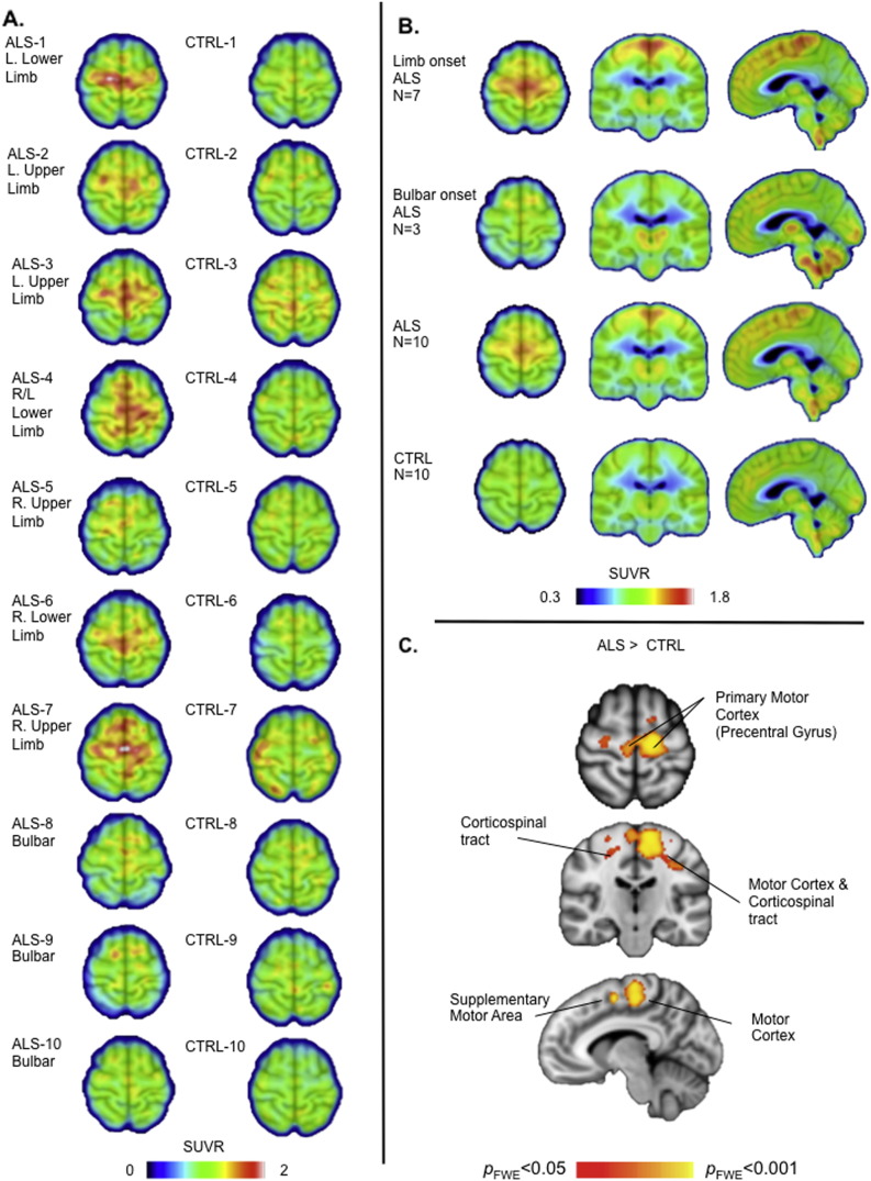
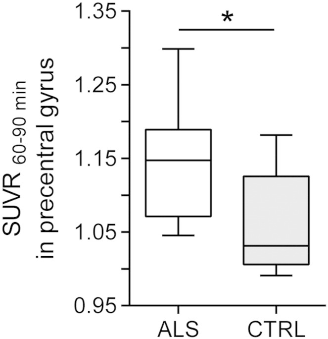
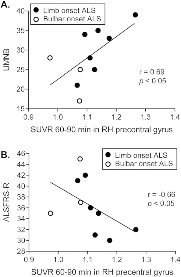
Evidence from human post mortem, in vivo and animal model studies implicates the neuroimmune system and activated microglia in the pathology of amyotrophic lateral sclerosis. The study aim was to further evaluate in vivo neuroinflammation in individuals with amyotrophic lateral sclerosis using [(11)C]-PBR28 positron emission tomography. Ten patients with amyotrophic lateral sclerosis (seven males, three females, 38-68 years) and ten age- and [(11)C]-PBR28 binding affinity-matched healthy volunteers (six males, four females, 33-65 years) completed a positron emission tomography scan. Standardized uptake values were calculated from 60 to 90 min post-injection and normalized to whole brain mean. Voxel-wise analysis showed increased binding in the motor cortices and corticospinal tracts in patients with amyotrophic lateral sclerosis compared to healthy controls (p FWE < 0.05). Region of interest analysis revealed increased [(11)C]-PBR28 binding in the precentral gyrus in patients (normalized standardized uptake value = 1.15) compared to controls (1.03, p < 0.05). In patients those values were positively correlated with upper motor neuron burden scores (r = 0.69, p < 0.05), and negatively correlated with the amyotrophic lateral sclerosis functional rating scale (r = -0.66, p < 0.05). Increased in vivo glial activation in motor cortices, that correlates with phenotype, complements previous histopathological reports. Further studies will determine the role of [(11)C]-PBR28 as a marker of treatments that target neuroinflammation.
Overview
The claims and statistics of this paper haven’t been checked yet.
Run the check — free, no account needed. It reads the open-access text and checks the sources it cites.
Integrity signals
Registry
Cites
Statistics
Figures
Verification
Claims the paper makes
The claims and statistics of this paper haven’t been checked yet.
Run the check — free, no account needed. It reads the open-access text and checks the sources it cites.
Claims other papers make about this one
Each is checked against this paper’s own text: does it say what the citing paper says it does?
The source backs part of the claim, not all of it — or backs it for a different population, condition or comparison.
In the citing paper
“Microglial activation in middle frontal and superior or middle temporal gyrus regions was also highly found in vivo PET imaging studies in humans”
From Microglia in neurodegenerative diseases: mechanism and potential therapeutic targets (2023) · cites this paper as [312]
Experimental — accuracy not yet measured
Self-check
Percentages, sums and reported differences re-computed from the paper’s own numbers.
The self-check needs the paper’s full text.
Citations
Cites
Polazzi, E., Mengoni, I., Caprini, M., Peña‐Altamira, E., Kurtys, E., & Monti, B. (2012) · Neurosignals · doi:10.1159/000337115 · Cited by 9,023
Nonparametric permutation tests for functional neuroimaging: A primer with examples
Nichols, T. E., & Holmes, A. P. (2002) · Human Brain Mapping · doi:10.1002/hbm.1058 · Cited by 6,256
El Escorial revisited: revised criteria for the diagnosis of amyotrophic lateral sclerosis
Brooks, B. R., Miller, R. G., Swash, M., & Munsat, T. L. (2000) · Amyotrophic Lateral Sclerosis and Other Motor Neuron Disorders · doi:10.1080/146608200300079536 · Cited by 5,433 · cited 2×
“The protocol was approved by the Institutional Review Board and the Radioactive Drug Research Committee.”
MethodCedarbaum, J. M., Stambler, N., Malta, E., Fuller, C., Hilt, D., Thurmond, B., & Nakanishi, A. (1999) · Journal of the Neurological Sciences · doi:10.1016/s0022-510x(99)00210-5 · Cited by 3,307 · cited 6×
“The clinical outcomes obtained from the participants with ALS included the revised ALS functional rating scale (ALSFRS-R) (Cedarbaum et al., 1999), upper motor neuron burden scale (UMNB) (Ellis et al.”
MethodInfluentialDose-ranging study of riluzole in amyotrophic lateral sclerosis
Amyotrophic Lateral Sclerosis/Riluzole Study Group II, Lacomblez, L., Bensimon, G., Meininger, V., Leigh, P. N., & Guillet, P. (1996) · The Lancet · doi:10.1016/s0140-6736(96)91680-3 · Cited by 1,303 · cited 1×
“Riluzole, the only FDA-approved treatment provides a modest survival benefit (Lacomblez et al., 1996), S-R, amyotrophic lateralsclerorate; MR,magnetic resonance; ron emission tomography; SUV, in;UMNB, uppermotorneuron search Institute, Massachusetts on, 02114 MA, USA.”
BackgroundOwen, D. R., Yeo, A. J., Gunn, R. N., Song, K., Wadsworth, G., Lewis, A., Rhodes, C., Pulford, D. J., Bennacef, I., Parker, C. A., et al. (2012) · Journal of Cerebral Blood Flow & Metabolism · doi:10.1038/jcbfm.2011.147 · Cited by 779 · cited 2×
“[11C]-PBR28, along with all second-generation TSPO radiotracers to date, has differential binding affinity to TSPO depending on an Ala147Thr polymorphism in the TSPO gene with Ala/Ala leading to high-, Ala/Thr to mixed-, and Thr/Thr to low-affinity binding (Kreisl et al., 2013; Owen et al., 2012).”
BackgroundTurner, M. R., Cagnin, A., Turkheimer, F. E., Miller, C. C. J., Shaw, C. E., Brooks, D. J., Leigh, P. N., & Banati, R. B. (2004) · Neurobiology of Disease · doi:10.1016/j.nbd.2003.12.012 · Cited by 737 · cited 6×
“This finding is consistent with the first microglial PET study in ALS patients (Turner et al., 2004), as well as histopathological studies reporting increased activated microglia near degenerating motor neurons (Brettschneider et al., 2012; Henkel et al., 2004; Kawamata et al., 1992), suggesting…”
BackgroundResultInfluentialMolecular pathways of motor neuron injury in amyotrophic lateral sclerosis
Ferraiuolo, L., Kirby, J., Grierson, A. J., Sendtner, M., & Shaw, P. J. (2011) · Nature Reviews Neurology · doi:10.1038/nrneurol.2011.152 · Cited by 615 · cited 4×
“This is an open access article under but there are no available treatments that prevent or stop disease progression and current animal models have not yet successfully predicted treatment response in people (Atassi et al., 2012; Ferraiuolo et al., 2011).”
BackgroundMethodInfluentialHenkel-Tigges, J., Engelhardt, J., Siklós, L., Simpson, E. P., Kim, S. H., Pan, T., Goodman, J. C., Siddique, T., Beers, D. R., & Appel, S. H. (2003) · Annals of Neurology · doi:10.1002/ana.10805 · Cited by 576
Immunologic reactions in amyotrophic lateral sclerosis brain and spinal cord tissue.
Kawamata, T., Akiyama, H., Yamada, T., & McGeer, P. L. (1992) · PubMed Central · Cited by 546
Evidence for brain glial activation in chronic pain patients
Loggia, M. L., Chonde, D. B., Akeju, O., Arabasz, G., Catana, C., Edwards, R. R., Hill, E., Hsu, S., Izquierdo-Garcia, D., Ji, R.-R., et al. (2015) · Brain · doi:10.1093/brain/awu377 · Cited by 503 · cited 1×
“…registered to MNI (Montreal Neurological Institute) space, spatially smoothed (6 mm full width at half maximum), and intensity-normalized to a mean of 1 (SUVR60–90 min) in order to account for differences in global signal across subjects, as previously done for [11C]-PBR28 (Loggia et al., 2015).”
MethodSelf-citationDiffusion tensor MRI assesses corticospinal tract damage in ALS
Ellis, C. M., Simmons, A., Jones, D. K., Bland, J. D. P., Dawson, J. M., Horsfield, M. A., Williams, S., & Leigh, P. N. (1999) · Neurology · doi:10.1212/wnl.53.5.1051 · Cited by 394 · cited 9×
“Riluzole, the only FDA-approved treatment provides a modest survival benefit (Lacomblez et al., 1996), S-R, amyotrophic lateralsclerorate; MR,magnetic resonance; ron emission tomography; SUV, in;UMNB, uppermotorneuron search Institute, Massachusetts on, 02114 MA, USA.”
BackgroundMethodInfluentialReactive Astrocytes Overexpress TSPO and Are Detected by TSPO Positron Emission Tomography Imaging
Lavisse, S., Guillermier, M., Hérard, A., Petit, F., Delahaye, M., Camp, N. V., Haim, L. B., Lebon, V., Rémy, P., Dollé, F., et al. (2012) · Journal of Neuroscience · doi:10.1523/jneurosci.1487-12.2012 · Cited by 381 · cited 1×
“In patients those values were positively correlated with upper motor neuron burden scores (r = 0.69, p b 0.05), and negatively correlated with the amyotrophic lateral sclerosis functional rating scale (r = –0.66, p b 0.05).”
ResultInfluentialCatana, C., van der Kouwe, A. J. W., Benner, T., Michel, C. J., Hamm, M., Fenchel, M., Fischl, B. R., Rosen, B. R., Schmand, M. J., & Sorensen, A. G. (2010) · Journal of Nuclear Medicine · doi:10.2967/jnumed.109.069112 · Cited by 355 · cited 1×
“This combined MR/PET scanner allowed the simultaneous acquisition of MR and PET data (Catana et al., 2010).”
MethodSelf-citationKreisl, W. C., Fujita, M., Fujimura, Y., Kimura, N., Jenko, K. J., Kannan, P., Hong, J. S., Morse, C. L., Zoghbi, S. S., Gladding, R. L., et al. (2009) · NeuroImage · doi:10.1016/j.neuroimage.2009.11.056 · Cited by 275 · cited 4×
“Published by Elsevier Inc a b s t r a c t a r t i c l e i n f o Article history: Received 26 September 2014 Received in revised form 22 December 2014 Accepted 5 January 2015 Available online 19 January 2015 Keywords: Amyotrophic lateral sclerosis Positron emission tomography [11C]PBR-28 Neuroinflammation Microglia Mot…”
BackgroundResultInfluentialBrettschneider, J., Toledo, J. B., Van Deerlin, V. M., Elman, L., McCluskey, L., Lee, V. M.-Y., & Trojanowski, J. Q. (2012) · PLoS ONE · doi:10.1371/journal.pone.0039216 · Cited by 268 · cited 4×
“In post mortem studies increased activated microglia are correlated with increased upper motor neuron symptoms and faster disease progression (Brettschneider et al., 2012).”
BackgroundInfluentialKreisl, W. C., Jenko, K. J., Hines, C. S., Lyoo, C. H., Corona, W., Morse, C. L., Zoghbi, S. S., Hyde, T. M., Kleinman, J. E., Pike, V. W., et al. (2012) · Journal of Cerebral Blood Flow & Metabolism · doi:10.1038/jcbfm.2012.131 · Cited by 251 · cited 2×
“[11C]-PBR28, along with all second-generation TSPO radiotracers to date, has differential binding affinity to TSPO depending on an Ala147Thr polymorphism in the TSPO gene with Ala/Ala leading to high-, Ala/Thr to mixed-, and Thr/Thr to low-affinity binding (Kreisl et al., 2013; Owen et al., 2012).”
BackgroundMolecular Imaging of Microglial Activation in Amyotrophic Lateral Sclerosis
Corcia, P., Tauber, C., Vercoullie, J., Arlicot, N., Prunier, C., Praline, J., Nicolas, G., Venel, Y., Hommet, C., Baulieu, J.-L., et al. (2012) · PLoS ONE · doi:10.1371/journal.pone.0052941 · Cited by 249
Izquierdo‐Garcia, D., Hansen, A. E., Förster, S., Benoit, D., Schachoff, S., Fürst, S., Chen, K. T., Chonde, D. B., & Catana, C. (2014) · Journal of Nuclear Medicine · doi:10.2967/jnumed.113.136341 · Cited by 218 · cited 1×
“Attenuation correction maps were created using MRbasedmethods (Izquierdo-Garcia et al., 2014).”
MethodSelf-citationRadiotracer synthesis from [11C]-iodomethane: a remarkably simple captive solvent method
Wilson, A. A., García, A., Jin, L., & Houle, S. (2000) · Nuclear Medicine and Biology · doi:10.1016/s0969-8051(00)00132-3 · Cited by 203 · cited 1×
“Briefly, the desmethyl precursor (1.0 mg in 100 µL) was loaded into a 5 mL stainless steel loop for reaction with CH3I using the Wilson captive solvent method (Wilson et al., 2000).”
MethodInfluentialThe microglial-motoneuron dialogue in ALS.
Appel, S. H., Zhao, W., Beers, D. R., & Henkel-Tigges, J. (2011) · Europe PMC (PubMed Central) · Cited by 179
Imaizumi, M., Kim, H. J., Zoghbi, S. S., Briard, E., Hong, J. S., Musachio, J. L., Ruetzler, C. A., Chuang, D., Pike, V. W., Innis, R. B., et al. (2006) · Neuroscience Letters · doi:10.1016/j.neulet.2006.09.093 · Cited by 177 · cited 2×
“[11C]-PBR28 was produced in-house using a procedure modified from the literature (Imaizumi et al., 2007).”
BackgroundMethodDistinct cerebral lesions in sporadic and ‘D90A’ SOD1 ALS: studies with [11C]flumazenil PET
Turner, M. R., Hammers, A., Al‐Chalabi, A., Shaw, C. E., Andersen, P. M., Brooks, D. J., & Leigh, P. N. (2005) · Brain · doi:10.1093/brain/awh509 · Cited by 160 · cited 1×
“…who are pre-symptomatic, but at high risk of developing the disease, such as superoxide dismutase-1 (SOD1) or C9orf72 gene carriers, may also provide important insights in this context, as suggested by a previous PET imaging study investigating pre-symptomatic ALS patients (Turner et al., 2005).”
ResultBrown, A. K., Fujita, M., Fujimura, Y., Liow, J., Stabin, M. G., Ryu, Y. H., Imaizumi, M., Hong, J., Pike, V. W., & Innis, R. B. (2007) · Journal of Nuclear Medicine · doi:10.2967/jnumed.107.044842 · Cited by 152 · cited 2×
“…by binding the 18 kDa translocator protein (TSPO), formerly known as the peripheral benzodiazepine receptor (PBR), which is highly the CC BY-NC-ND license (http://creativecommons.org/licenses/by-nc-nd/4.0/). expressed in activated microglia and astrocytes (Brown et al., 2007; Lavisse et al., 2012).”
BackgroundInfluentialLessons of ALS imaging: Pitfalls and future directions — A critical review
Bede, P., & Hardiman, O. M. (2014) · NeuroImage Clinical · doi:10.1016/j.nicl.2014.02.011 · Cited by 126
Translocator Protein PET Imaging for Glial Activation in Multiple Sclerosis
Oh, U., Fujita, M., Ikonomidou, V. N., Evangelou, I. E., Matsuura, E., Harberts, E. M., Ohayon, J. M., Pike, V. W., Zhang, Y., Zoghbi, S. S., et al. (2010) · Journal of Neuroimmune Pharmacology · doi:10.1007/s11481-010-9243-6 · Cited by 118
Influence of TSPO Genotype on 11C-PBR28 Standardized Uptake Values
Yoder, K. K., Nho, K. T., Risacher, S. L., Kim, S., Shen, L., & Saykin, A. J. (2013) · Journal of Nuclear Medicine · doi:10.2967/jnumed.112.118885 · Cited by 67 · cited 2×
“This binding affinity difference can be detected by standardized uptake value (SUV) measurements (Yoder et al., 2013), and needs to be controlled for in crosssectional study designs.”
BackgroundPET Reveals Inflammation around Calcified Taenia solium Granulomas with Perilesional Edema
Fujita, M., Mahanty, S., Zoghbi, S. S., Araneta, M. D. F., Hong, J. S., Pike, V. W., Innis, R. B., & Nash, T. E. (2013) · PLoS ONE · doi:10.1371/journal.pone.0074052 · Cited by 46 · cited 2×
“…both the two previously published PET studies in ALS using TSPO radiotracers (Corcia et al., 2012; Turner et al., 2004) as well as [11C]PBR28 PET studies in other diseases (Fujita et al., 2013; Oh et al., 2011) have shown that disease-related changes can be observed with comparable sample size.”
BackgroundResultGuiloff, R. J., & Goonetilleke, A. (2001) · Springer eBooks · doi:10.1007/978-1-4471-3787-0 · Cited by 34
Clinical trials in amyotrophic lateral sclerosis
McDermott, C. J. (2019) · Current Opinion in Neurology · doi:10.1097/wco.0000000000000731 · Cited by 22
(2012) · Cambridge University Press eBooks · doi:10.1017/cbo9781139032445 · Cited by 14
Cited by
Sorted by citations
Microglia in neurodegenerative diseases: mechanism and potential therapeutic targets
Gao, C., Jiang, J., Tan, Y., & Chen, S. (2023) · Signal Transduction and Targeted Therapy · doi:10.1038/s41392-023-01588-0 · Cited by 1,616
Mead, R. J., Shan, N., Reiser, H. J., Marshall, F., & Shaw, P. J. (2023) · Nature Reviews Drug Discovery · doi:10.1038/s41573-022-00612-2 · Cited by 505
Radiopharmaceuticals for PET and SPECT Imaging: A Literature Review over the Last Decade
Crișan, G., Moldovean‐Cioroianu, N. S., Timaru, D.-G., Andrieș, G., Cainap, C. I., & Chiş, V. (2022) · International Journal of Molecular Sciences · doi:10.3390/ijms23095023 · Cited by 386
Targeting Oxidative Stress in Central Nervous System Disorders
Patel, M. (2016) · Trends in Pharmacological Sciences · doi:10.1016/j.tips.2016.06.007 · Cited by 312
Brain glial activation in fibromyalgia – A multi-site positron emission tomography investigation
Albrecht, D. S., Forsberg, A., Sandström, A., Bergan, C., Kadetoff, D., Protsenko, E., Lampa, J., Lee, Y. C., Höglund, C. O., Catana, C., et al. (2019) · Brain, Behavior, and Immunity · doi:10.1016/j.bbi.2018.09.018 · Cited by 310
Self-citation
Metadata
- Authors
- Zürcher, N. R., Loggia, M. L., Lawson, R., Chonde, D. B., Izquierdo-Garcia, D., Yasek, J. E., Akeju, O., Catana, C., Rosen, B. R., Cudkowicz, M. E., Hooker, J. M., & Atassi, N.
- Affiliations
- Harvard University (US)
- Corresponding author
- Hooker, Jacob M.
- Atassi, Nazem
- ORCID
- Venue
- NeuroImage: Clinical
- Publisher
- Elsevier BV
- Published
- 2015
- Volume
- 7
- Pages
- 409-414
- ISSN
- 2213-1582 (ISSN-L)
- PMC
- PMC4310932
- OpenAlex
- W1969387798
- Cited by
- 226
- References
- 31
- Citations by year
- 2026: 6 · 2025: 15 · 2024: 27 · 2023: 15 · 2022: 11 · 2021: 26 · 2020: 25 · 2019: 21 · 2018: 19 · 2017: 27 · 2016: 26 · 2015: 8
- FWCI
- 18.79
- Type
- journal article
- Language
- en
- Open access
- gold
- doi.org
- Licence
- Locations
- NeuroImage Clinical (publishedVersion, cc-by-nc-nd)
- Europe PMC (PubMed Central) (publishedVersion, cc-by-nc-nd)
- PubMed Central (publishedVersion, cc-by-nc-nd)
- PubMed (publishedVersion)
- Digital Access to Scholarship at Harvard (DASH) (Harvard University) (submittedVersion, cc-by)
- DOAJ (DOAJ: Directory of Open Access Journals) (submittedVersion)
- Funders
- Harvard NeuroDiscovery Center
- Muscular Dystrophy Association
- American Academy of Neurology
- Anne Young Fellowship
- NINDS (1K23NS083715-01A1)
- Topics
- Health Sciences › Medicine › Clinical Neurology › Amyotrophic Lateral Sclerosis Research
- Life Sciences › Neuroscience › Neurology › Neuroinflammation and Neurodegeneration Mechanisms
- Life Sciences › Neuroscience › Cellular and Molecular Neuroscience › Alzheimer's disease research and treatments
- Keywords
- amyotrophic lateral sclerosis, glial activation, neuroinflammation, microglial activation, [11C]PBR28, positron emission tomography, motor cortex, corticospinal tract, ALSFRS-R, neuroimmune system
- MeSH terms
- Acetamides, Adult, Aged, Amyotrophic Lateral Sclerosis, Brain, Carbon Radioisotopes, Female, Humans, Male, Middle Aged, Neuroglia, Pyridines, Radiographic Image Interpretation, Computer-Assisted, Radiopharmaceuticals, Positron-Emission Tomography
About these checks
Registry, retraction, Cites and statistics results are computed, not judged, with one exception: a printed reference that no index lists is read once by a model before it counts as a citation error. Claims are extracted by a model and kept only when their quote appears verbatim in the text; each cited source is then checked, with a model’s help, for whether it says what the claim relies on. Those verdicts can be wrong — read the claim’s quote and the cited source before relying on one.
Statistics and arithmetic are recomputed, not judged.
Confidence bands are shown only for fields calibrated on our annotated test set. None is calibrated yet, so no band is shown.
What counts as a claim ›How we measure confidence ›How we check this ›
Are you an author, or do you see a problem with this page? Request a review
