

The DOI matches its Crossref record; 31 of the 31 references it cites were found in a registry or an index.
Increased in vivo glial activation in patients with amyotrophic lateral sclerosis: Assessed with [11C]-PBR28
Open accessAbstract
Evidence from human post mortem, in vivo and animal model studies implicates the neuroimmune system and activated microglia in the pathology of amyotrophic lateral sclerosis. The study aim was to further evaluate in vivo neuroinflammation in individuals with amyotrophic lateral sclerosis using [(11)C]-PBR28 positron emission tomography. Ten patients with amyotrophic lateral sclerosis (seven males, three females, 38-68 years) and ten age- and [(11)C]-PBR28 binding affinity-matched healthy volunteers (six males, four females, 33-65 years) completed a positron emission tomography scan. Standardized uptake values were calculated from 60 to 90 min post-injection and normalized to whole brain mean. Voxel-wise analysis showed increased binding in the motor cortices and corticospinal tracts in patients with amyotrophic lateral sclerosis compared to healthy controls (p FWE < 0.05). Region of interest analysis revealed increased [(11)C]-PBR28 binding in the precentral gyrus in patients (normalized standardized uptake value = 1.15) compared to controls (1.03, p < 0.05). In patients those values were positively correlated with upper motor neuron burden scores (r = 0.69, p < 0.05), and negatively correlated with the amyotrophic lateral sclerosis functional rating scale (r = -0.66, p < 0.05). Increased in vivo glial activation in motor cortices, that correlates with phenotype, complements previous histopathological reports. Further studies will determine the role of [(11)C]-PBR28 as a marker of treatments that target neuroinflammation.
Overview
The claims and statistics of this paper haven’t been checked yet.
Run the check — free, no account needed. It reads the open-access text and checks the sources it cites.
Integrity signals
Registry
Cites
Statistics
Figures
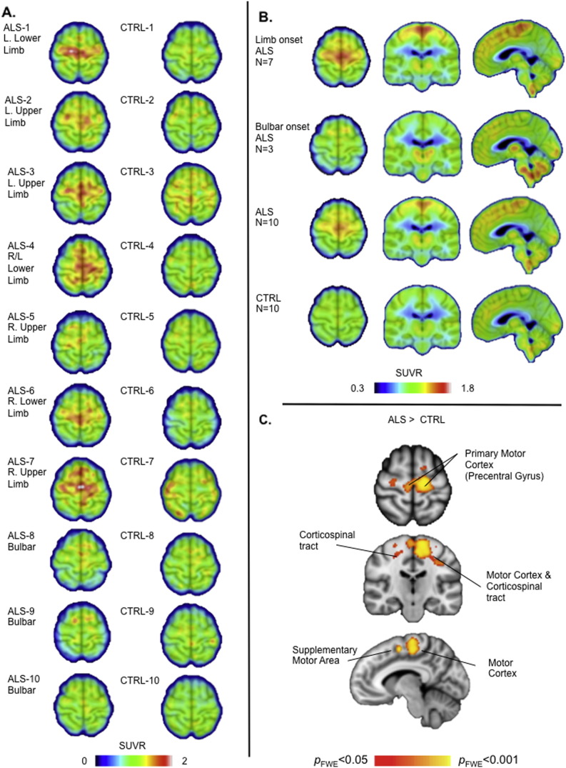
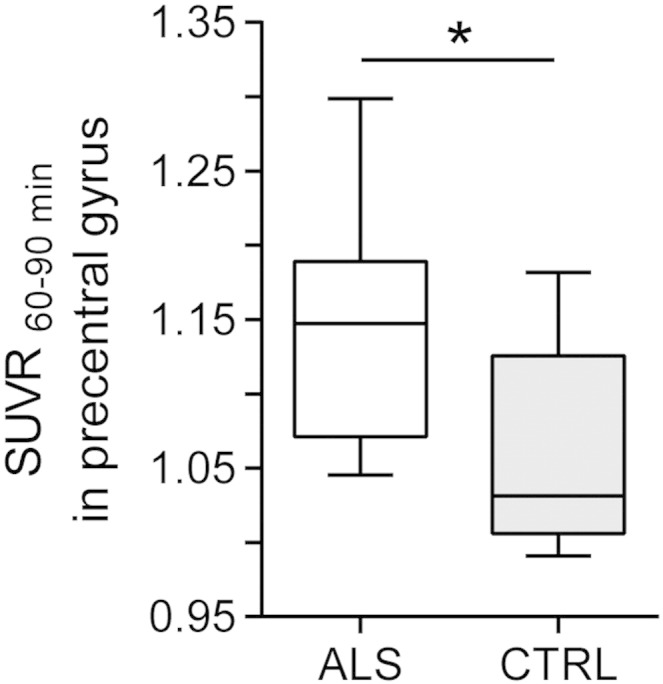
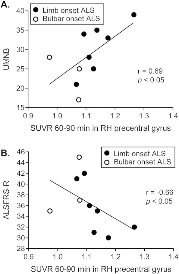
Verification
Claims the paper makes
The claims and statistics of this paper haven’t been checked yet.
Run the check — free, no account needed. It reads the open-access text and checks the sources it cites.
Claims other papers make about this one
Each is checked against this paper’s own text: does it say what the citing paper says it does?
The source backs part of the claim, not all of it — or backs it for a different population, condition or comparison.
In the citing paper
“Microglial activation in middle frontal and superior or middle temporal gyrus regions was also highly found in vivo PET imaging studies in humans”
From Microglia in neurodegenerative diseases: mechanism and potential therapeutic targets (2023) · cites this paper as [312]
Experimental — accuracy not yet measured
Self-check
Percentages, sums and reported differences re-computed from the paper’s own numbers.
The self-check needs the paper’s full text.
Citations
Cites
Polazzi, E., Mengoni, I., Caprini, M., Peña‐Altamira, E., Kurtys, E., & Monti, B. (2012) · Neurosignals · doi:10.1159/000337115 · Cited by 9,023
Nonparametric permutation tests for functional neuroimaging: A primer with examples
Nichols, T. E., & Holmes, A. P. (2002) · Human Brain Mapping · doi:10.1002/hbm.1058 · Cited by 6,256
El Escorial revisited: revised criteria for the diagnosis of amyotrophic lateral sclerosis
Brooks, B. R., Miller, R. G., Swash, M., & Munsat, T. L. (2000) · Amyotrophic Lateral Sclerosis and Other Motor Neuron Disorders · doi:10.1080/146608200300079536 · Cited by 5,433 · cited 2×
“The protocol was approved by the Institutional Review Board and the Radioactive Drug Research Committee.”
MethodCedarbaum, J. M., Stambler, N., Malta, E., Fuller, C., Hilt, D., Thurmond, B., & Nakanishi, A. (1999) · Journal of the Neurological Sciences · doi:10.1016/s0022-510x(99)00210-5 · Cited by 3,307 · cited 6×
“The clinical outcomes obtained from the participants with ALS included the revised ALS functional rating scale (ALSFRS-R) (Cedarbaum et al., 1999), upper motor neuron burden scale (UMNB) (Ellis et al.”
MethodInfluentialDose-ranging study of riluzole in amyotrophic lateral sclerosis
Amyotrophic Lateral Sclerosis/Riluzole Study Group II, Lacomblez, L., Bensimon, G., Meininger, V., Leigh, P. N., & Guillet, P. (1996) · The Lancet · doi:10.1016/s0140-6736(96)91680-3 · Cited by 1,303 · cited 1×
“Riluzole, the only FDA-approved treatment provides a modest survival benefit (Lacomblez et al., 1996), S-R, amyotrophic lateralsclerorate; MR,magnetic resonance; ron emission tomography; SUV, in;UMNB, uppermotorneuron search Institute, Massachusetts on, 02114 MA, USA.”
BackgroundOwen, D. R., Yeo, A. J., Gunn, R. N., Song, K., Wadsworth, G., Lewis, A., Rhodes, C., Pulford, D. J., Bennacef, I., Parker, C. A., et al. (2012) · Journal of Cerebral Blood Flow & Metabolism · doi:10.1038/jcbfm.2011.147 · Cited by 779 · cited 2×
“[11C]-PBR28, along with all second-generation TSPO radiotracers to date, has differential binding affinity to TSPO depending on an Ala147Thr polymorphism in the TSPO gene with Ala/Ala leading to high-, Ala/Thr to mixed-, and Thr/Thr to low-affinity binding (Kreisl et al., 2013; Owen et al., 2012).”
BackgroundTurner, M. R., Cagnin, A., Turkheimer, F. E., Miller, C. C. J., Shaw, C. E., Brooks, D. J., Leigh, P. N., & Banati, R. B. (2004) · Neurobiology of Disease · doi:10.1016/j.nbd.2003.12.012 · Cited by 737 · cited 6×
“This finding is consistent with the first microglial PET study in ALS patients (Turner et al., 2004), as well as histopathological studies reporting increased activated microglia near degenerating motor neurons (Brettschneider et al., 2012; Henkel et al., 2004; Kawamata et al., 1992), suggesting…”
BackgroundResultInfluentialMolecular pathways of motor neuron injury in amyotrophic lateral sclerosis
Ferraiuolo, L., Kirby, J., Grierson, A. J., Sendtner, M., & Shaw, P. J. (2011) · Nature Reviews Neurology · doi:10.1038/nrneurol.2011.152 · Cited by 615 · cited 4×
“This is an open access article under but there are no available treatments that prevent or stop disease progression and current animal models have not yet successfully predicted treatment response in people (Atassi et al., 2012; Ferraiuolo et al., 2011).”
BackgroundMethodInfluentialHenkel-Tigges, J., Engelhardt, J., Siklós, L., Simpson, E. P., Kim, S. H., Pan, T., Goodman, J. C., Siddique, T., Beers, D. R., & Appel, S. H. (2003) · Annals of Neurology · doi:10.1002/ana.10805 · Cited by 576
Immunologic reactions in amyotrophic lateral sclerosis brain and spinal cord tissue.
Kawamata, T., Akiyama, H., Yamada, T., & McGeer, P. L. (1992) · PubMed Central · Cited by 546
Cited by
Sorted by citations
Microglia in neurodegenerative diseases: mechanism and potential therapeutic targets
Gao, C., Jiang, J., Tan, Y., & Chen, S. (2023) · Signal Transduction and Targeted Therapy · doi:10.1038/s41392-023-01588-0 · Cited by 1,616
Mead, R. J., Shan, N., Reiser, H. J., Marshall, F., & Shaw, P. J. (2023) · Nature Reviews Drug Discovery · doi:10.1038/s41573-022-00612-2 · Cited by 505
Radiopharmaceuticals for PET and SPECT Imaging: A Literature Review over the Last Decade
Crișan, G., Moldovean‐Cioroianu, N. S., Timaru, D.-G., Andrieș, G., Cainap, C. I., & Chiş, V. (2022) · International Journal of Molecular Sciences · doi:10.3390/ijms23095023 · Cited by 386
Targeting Oxidative Stress in Central Nervous System Disorders
Patel, M. (2016) · Trends in Pharmacological Sciences · doi:10.1016/j.tips.2016.06.007 · Cited by 312
Brain glial activation in fibromyalgia – A multi-site positron emission tomography investigation
Albrecht, D. S., Forsberg, A., Sandström, A., Bergan, C., Kadetoff, D., Protsenko, E., Lampa, J., Lee, Y. C., Höglund, C. O., Catana, C., et al. (2019) · Brain, Behavior, and Immunity · doi:10.1016/j.bbi.2018.09.018 · Cited by 310
Self-citationNeuroinflammation in Neurodegenerative Disorders-a Review
Schain, M., & Kreisl, W. C. (2017) · Current Neurology and Neuroscience Reports · doi:10.1007/s11910-017-0733-2 · Cited by 301
Spiller, K. J., Restrepo, C. R., Khan, T., Dominique, M. A., Fang, T. C., Canter, R. G., Roberts, C. J., Miller, K. R., Ransohoff, R. M., Trojanowski, J. Q., et al. (2018) · Nature Neuroscience · doi:10.1038/s41593-018-0083-7 · Cited by 301
Radiopharmaceuticals and their applications in medicine
Zhang, S., Wang, X., Gao, X., Chen, X., Li, L., Li, G., Liu, C., Miao, Y., Wang, R., & Hu, K. (2025) · Signal Transduction and Targeted Therapy · doi:10.1038/s41392-024-02041-6 · Cited by 346
Werry, E. L., Bright, F., Piguet, O., Ittner, L. M., Halliday, G. M., Hodges, J. R., Kiernan, M. C., Loy, C. T., Kril, J. J., & Kassiou, M. (2019) · International Journal of Molecular Sciences · doi:10.3390/ijms20133161 · Cited by 298
PET imaging of neuroinflammation in neurological disorders
Kreisl, W. C., Kim, M.-J., Coughlin, J. M., Henter, I. D., Owen, D. R., & Innis, R. B. (2020) · The Lancet Neurology · doi:10.1016/s1474-4422(20)30346-x · Cited by 254
In Vivo Imaging of Human Neuroinflammation
Albrecht, D., Granziera, C., Hooker, J. M., & Loggia, M. L. (2016) · ACS Chemical Neuroscience · doi:10.1021/acschemneuro.6b00056 · Cited by 212
“Brain 138, 604−615, 44 with permission from Oxford University Press; and Zurcher et al. (2015) Increased in vivo glial activation in patients with amyotrophic lateral sclerosis: Assessed with [11C]-PBR28.”
Self-citationNeuroinflammatory component of gray matter pathology in multiple sclerosis
Herranz, E., Giannì, C., Louapre, C., Treaba, C. A., Govindarajan, S. T., Ouellette, R., Loggia, M. L., Sloane, J. A., Madigan, N. K., Izquierdo‐Garcia, D., et al. (2016) · Annals of Neurology · doi:10.1002/ana.24791 · Cited by 209
“In humans, (11)CPBR28 SUVR has also been used to reliably distinguish healthy individuals from cases with neurological disorders.(27,49,50) This method is expected to improve subject tolerability by allowing shorter scan time and not requiring arterial catheterization, making it less invasive and more feasible for cli…”
MethodSelf-citationThe impact of high and low dose ionising radiation on the central nervous system
Betlazar, C., Middleton, R. J., Banati, R. B., & Liu, G.-J. (2016) · Redox Biology · doi:10.1016/j.redox.2016.08.002 · Cited by 196
Recent developments on PET radiotracers for TSPO and their applications in neuroimaging
Zhang, L., Hu, K., Shao, T., Hou, L., Zhang, S., Ye, W., Josephson, L., Meyer, J. H., Zhang, M., Vasdev, N., et al. (2020) · Acta Pharmaceutica Sinica B · doi:10.1016/j.apsb.2020.08.006 · Cited by 195
Dupont, A.-C., Largeau, B., Santiago Ribeiro, M., Guilloteau, D., Tronel, C., & Arlicot, N. (2017) · International Journal of Molecular Sciences · doi:10.3390/ijms18040785 · Cited by 182
Glia Crosstalk in Neuroinflammatory Diseases
Bernaus, A., Blanco, S., & Sevilla, A. (2020) · Frontiers in Cellular Neuroscience · doi:10.3389/fncel.2020.00209 · Cited by 157
Neuroinflammation of the spinal cord and nerve roots in chronic radicular pain patients
Albrecht, D., Ahmed, S. U., Kettner, N. W., Borra, R., Cohen‐Adad, J., Deng, H., Houle, T. T., Opalacz, A., Roth, S., Melo, M. F. V., et al. (2018) · Pain · doi:10.1097/j.pain.0000000000001171 · Cited by 155
Self-citationNon-neuronal cells in amyotrophic lateral sclerosis — from pathogenesis to biomarkers
Vahsen, B. F., Gray, E., Thompson, A. G., Ansorge, O., Anthony, D. C., Cowley, S. A., Talbot, K., & Turner, M. R. (2021) · Nature Reviews Neurology · doi:10.1038/s41582-021-00487-8 · Cited by 147
Glial Contribution to Excitatory and Inhibitory Synapse Loss in Neurodegeneration
Henstridge, C. M., Tzioras, M., & Paolicelli, R. C. (2019) · Frontiers in Cellular Neuroscience · doi:10.3389/fncel.2019.00063 · Cited by 155
Imaging of neuroinflammation in migraine with aura
Albrecht, D., Mainero, C., Ichijo, E., Ward, N., Granziera, C., Zürcher, N. R., Akeju, O., Bonnier, G., Price, J. C., Hooker, J. M., et al. (2019) · Neurology · doi:10.1212/wnl.0000000000007371 · Cited by 134
“In order to account for this variability, several groups have reduced variability by normalizing PET signal to uptake in whole brain or whole gray matter.(5,12,16,17) While this approach may improve the detection of focal effects by robustly reducing between-participant variability, it may be not be appropriate when t…”
BackgroundMethodSelf-citationBiomarkers in Motor Neuron Disease: A State of the Art Review
Verber, N. S., Shepheard, S. R., Sassani, M., McDonough, H. E., Moore, S. A., Alix, J. J. P., Wilkinson, I. D., Jenkins, T. M., & Shaw, P. J. (2019) · Frontiers in Neurology · doi:10.3389/fneur.2019.00291 · Cited by 130
The human gut microbiota in people with amyotrophic lateral sclerosis
Nicholson, K., Bjornevik, K., Abu-Ali, G., Chan, J., Cortese, M., Dedi, B., Jeon, M., Xavier, R., Huttenhower, C., Ascherio, A., et al. (2021) · Amyotrophic Lateral Sclerosis and Frontotemporal Degeneration · doi:10.1080/21678421.2020.1828475 · Cited by 123
In-vivo imaging of neuroinflammation in veterans with Gulf War illness
Alshelh, Z., Albrecht, D., Bergan, C., Akeju, O., Clauw, D. J., Conboy, L. A., Edwards, R. R., Kim, M., Lee, Y., Protsenko, E., et al. (2020) · Brain Behavior and Immunity · doi:10.1016/j.bbi.2020.01.020 · Cited by 122
“…patients demonstrates increased TSPO signal in the primary motor cortex (Alshikho et al., 2016; Alshikho et al., 2018; Paganoni et al., 2018; Zurcher et al., 2015), a region where glial activation can be documented histologically (Hudson et al., 1993; Kawamata et al., 1992; Rothstein et…”
BackgroundMethodSelf-citationALS biomarkers for therapy development: State of the field and future directions
Benatar, M. G., Boylan, K., Jeromin, A., Rutkove, S. B., Berry, J. D., Atassi, N., & Bruijn, L. I. (2015) · Muscle & Nerve · doi:10.1002/mus.24979 · Cited by 116
“In addition, the degree of binding in the PCG correlated negatively with the functional status and positively with upper motor neuron signs.(115) Based on these findings, TSPO PET imaging is a promising molecular imaging modality that could serve as a pharmacodynamic biomarker for ALS therapies that target neuroinflam…”
BackgroundSelf-citationAdvances in molecular pathology, diagnosis, and treatment of amyotrophic lateral sclerosis
Ilieva, H., Vullaganti, M., & Kwan, J. (2023) · BMJ · doi:10.1136/bmj-2023-075037 · Cited by 111
The immune response of the human brain to abdominal surgery
Forsberg, A., Červenka, S., Fagerlund, M. J., Rasmussen, L. S., Zetterberg, H., Harris, H. E., Stridh, P., Christensson, E., Granström, A., Schening, A., et al. (2017) · Annals of Neurology · doi:10.1002/ana.24909 · Cited by 103
PDE4B as a microglia target to reduce neuroinflammation
Pearse, D. D., & Hughes, Z. A. (2016) · Glia · doi:10.1002/glia.22986 · Cited by 98
Alshikho, M. J., Zürcher, N. R., Loggia, M. L., Cernasov, P., Reynolds, B., Pijanowski, O., Chonde, D. B., Izquierdo‐Garcia, D., Mainero, C., Catana, C., et al. (2018) · Annals of Neurology · doi:10.1002/ana.25251 · Cited by 99
Self-citationNeuroimaging Endpoints in Amyotrophic Lateral Sclerosis
Menke, R., Agosta, F., Großkreutz, J., Filippi, M., & Turner, M. R. (2016) · Neurotherapeutics · doi:10.1007/s13311-016-0484-9 · Cited by 99
“Additionally, ligand PET has provided in vivo evidence for activated microglia in ALS in motor and frontal lobe regions [89, 90], widespread reductions in flumazenil binding [91] in keeping with loss of inhibitory neuronal influences [92], and profoundly reduced binding of a ligand with serotonin receptor affinity in…”
Biomarkers in Neurodegenerative Diseases
Jeromin, A., & Bowser, R. (2017) · Advances in Neurobiology · doi:10.1007/978-3-319-57193-5_20 · Cited by 92
Glial activation colocalizes with structural abnormalities in amyotrophic lateral sclerosis
Alshikho, M. J., Zürcher, N. R., Loggia, M. L., Cernasov, P., Chonde, D. B., Izquierdo‐Garcia, D., Yasek, J. E., Akeju, O., Catana, C., Rosen, B. R., et al. (2016) · Neurology · doi:10.1212/wnl.0000000000003427 · Cited by 90
Self-citationClinical and Radiological Markers of Extra-Motor Deficits in Amyotrophic Lateral Sclerosis
Christidi, F., Karavasilis, E., Rentzos, M., Kelekis, N., Evdokimidis, I., & Bede, P. (2018) · Frontiers in Neurology · doi:10.3389/fneur.2018.01005 · Cited by 94
Augmented expression of TSPO after intracerebral hemorrhage: a role in inflammation?
Bonsack, F., Alleyne, C. H., & Sukumari‐Ramesh, S. (2016) · Journal of Neuroinflammation · doi:10.1186/s12974-016-0619-2 · Cited by 98
Best, L. A., Ghadery, C., Pavese, N., Tai, Y., & Strafella, A. P. (2019) · Current Neurology and Neuroscience Reports · doi:10.1007/s11910-019-0934-y · Cited by 93
Inflammation, Immunity, and amyotrophic lateral sclerosis: I. Etiology and pathology
Lyon, M. S., Wosiski‐Kuhn, M., Gillespie, R., Caress, J. B., & Milligan, C. (2018) · Muscle & Nerve · doi:10.1002/mus.26289 · Cited by 94
Neuroinflammation in Huntington’s Disease: New Insights with 11 C-PBR28 PET/MRI
Lois, C., González, I., Izquierdo-García, D., Zürcher, N. R., Wilkens, P., Loggia, M. L., Hooker, J. M., & Rosas, H. D. (2018) · ACS Chemical Neuroscience · doi:10.1021/acschemneuro.8b00072 · Cited by 84
Self-citationNeuroimmune signatures in chronic low back pain subtypes
Alshelh, Z., Brusaferri, L., Saha, A., Morrissey, E. J., Knight, P. C., Kim, M., Zhang, Y., Hooker, J. M., Albrecht, D., Torrado-Carvajal, Á., et al. (2021) · Brain · doi:10.1093/brain/awab336 · Cited by 86
Self-citationSilencing strategies for therapy of SOD1-mediated ALS
van Zundert, B., & Brown, R. H. (2016) · Neuroscience Letters · doi:10.1016/j.neulet.2016.07.059 · Cited by 81
Vu, L. T., An, J., Kovalik, T., Gendron, T. F., Petrucelli, L., & Bowser, R. P. (2020) · Journal of Neurology Neurosurgery & Psychiatry · doi:10.1136/jnnp-2019-321916 · Cited by 85
PET neuroimaging reveals histone deacetylase dysregulation in schizophrenia
Gilbert, T. M., Zürcher, N. R., Wu, C. J., Bhanot, A., Hightower, B. G., Kim, M., Albrecht, D., Wey, H., Schroeder, F. A., Rodriguez-Thompson, A., et al. (2018) · Journal of Clinical Investigation · doi:10.1172/jci123743 · Cited by 78
Self-citationExploring the genetics and non-cell autonomous mechanisms underlying ALS/FTLD
Chen, H., Kankel, M. W., Su, S. C., Han, S., & Ofengeim, D. (2018) · Cell Death and Differentiation · doi:10.1038/s41418-018-0060-4 · Cited by 74
MRI/MRS in neuroinflammation: methodology and applications
Quarantelli, M. (2015) · Clinical and Translational Imaging · doi:10.1007/s40336-015-0142-y · Cited by 76
“demonstrate subtle but significant glial activation, correlating with pain severity, in specific brain regions in patients with chronic back pain [118] and in the motor cortices and corticospinal tracts of patients with amyotrophic lateral sclerosis [119].”
BackgroundThe neuroinflammatory component of negative affect in patients with chronic pain
Albrecht, D., Kim, M., Akeju, O., Torrado-Carvajal, Á., Edwards, R. R., Zhang, Y., Bergan, C., Protsenko, E., Kucyi, A., Wasan, A. D., et al. (2019) · Molecular Psychiatry · doi:10.1038/s41380-019-0433-1 · Cited by 80
Self-citationNeuroimaging in amyotrophic lateral sclerosis: current and emerging uses
Agosta, F., Spinelli, E. G., & Filippi, M. (2018) · Expert Review of Neurotherapeutics · doi:10.1080/14737175.2018.1463160 · Cited by 73
“tex, extra-motor frontotemporal areas, thalamus, midbrain, pons, and subcortical WM of patients with ALS [73,74].”
BackgroundImplications of Microglia in Amyotrophic Lateral Sclerosis and Frontotemporal Dementia
Haukedal, H., & Freude, K. (2019) · Journal of Molecular Biology · doi:10.1016/j.jmb.2019.02.004 · Cited by 68
Masrori, P., Beckers, J., Gossye, H., & Damme, P. V. (2022) · Molecular Neurodegeneration · doi:10.1186/s13024-022-00525-z · Cited by 80
What Does Imaging Reveal About the Pathology of Amyotrophic Lateral Sclerosis?
Turner, M. R., & Verstraete, E. (2015) · Current Neurology and Neuroscience Reports · doi:10.1007/s11910-015-0569-6 · Cited by 71
In vivo PET imaging of neuroinflammation in Alzheimer’s disease
Lagarde, J., Sarazin, M., & Bottlaender, M. (2017) · Journal of Neural Transmission · doi:10.1007/s00702-017-1731-x · Cited by 79
Protein aggregation and therapeutic strategies in SOD1- and TDP-43- linked ALS
Tsekrekou, M., Giannakou, M., Papanikolopoulou, K., & Σκρέτας, Γ. (2024) · Frontiers in Molecular Biosciences · doi:10.3389/fmolb.2024.1383453 · Cited by 71
Update on recent advances in amyotrophic lateral sclerosis
Riva, N., Domi, T., Pozzi, L., Lunetta, C., Schito, P., Spinelli, E. G., Cabras, S., Matteoni, E., Consonni, M., Bella, E. D., et al. (2024) · Journal of Neurology · doi:10.1007/s00415-024-12435-9 · Cited by 74
Zürcher, N. R., Loggia, M. L., Mullett, J. E., Tseng, C.-E. J., Bhanot, A., Richey, L., Hightower, B. G., Wu, C. J., Parmar, A. J., Butterfield, R. I., et al. (2020) · Molecular Psychiatry · doi:10.1038/s41380-020-0682-z · Cited by 72
Self-citationHunter, M., Spiller, K. J., Dominique, M. A., Xu, H. W., Hunter, F. W., Fang, T., Canter, R. G., Roberts, C. J., Ransohoff, R. M., Trojanowski, J. Q., et al. (2021) · Acta Neuropathologica Communications · doi:10.1186/s40478-021-01239-x · Cited by 68
Pathology of callosal damage in ALS: An ex-vivo, 7 T diffusion tensor MRI study
Cárdenas, A. M., Sarlls, J. E., Kwan, J. Y., Bageac, D. V., Gala, Z. S., Danielian, L. E., Ray‐Chaudhury, A., Wang, H., Miller, K. L., Foxley, S., et al. (2017) · NeuroImage Clinical · doi:10.1016/j.nicl.2017.04.024 · Cited by 64
Ma, L., Zhang, H., Liu, N., Wang, P., Guo, W.-Z., Fu, Q., Jiao, L.-B., Ma, Y., & Mi, W. (2016) · Brain Research Bulletin · doi:10.1016/j.brainresbull.2016.02.001 · Cited by 66
Brody, A. L., Hubert, R., Enoki, R., Garcia, L. Y., Mamoun, M. S., Okita, K., London, E. D., Nurmi, E. L., Seaman, L. C., & Mandelkern, M. A. (2017) · Neuropsychopharmacology · doi:10.1038/npp.2017.48 · Cited by 67
“…Suridjan et al, 2015; Varrone et al, 2015; Yasuno et al, 2008, 2012), Lewy body dementia (Surendranathan et al, 2015), amyotrophic lateral sclerosis (Zurcher et al, 2015), stroke (Lartey et al, 2014), and non-smokers with major depression (Setiawan et al, 2015), but not Parkinson’s disease…”
BackgroundMethodImmune mechanisms and shared immune targets in neurodegenerative diseases
Weiner, H. L. (2024) · Nature Reviews Neurology · doi:10.1038/s41582-024-01046-7 · Cited by 72
“TSPO PET has revealed microglial activation 50,51 in cortical and subcortical structures in the brain of people with ALS, and this activation was also apparent in people who were carriers of SOD1 mutations but were presymptomatic 52 .”
Ratai, E., Alshikho, M. J., Zürcher, N. R., Loggia, M. L., Cebulla, C. L., Cernasov, P., Reynolds, B., Fish, J., Seth, R., Babu, S., et al. (2018) · NeuroImage Clinical · doi:10.1016/j.nicl.2018.08.007 · Cited by 62
Self-citationUltra high-field (7tesla) magnetic resonance spectroscopy in Amyotrophic Lateral Sclerosis
Atassi, N., Xu, M., Triantafyllou, C., Keil, B. R., Lawson, R. J., Cernasov, P., Ratti, E., Long, C. J., Paganoni, S., Murphy, A., et al. (2017) · PLoS ONE · doi:10.1371/journal.pone.0177680 · Cited by 57
Self-citationTSPO Versus P2X7 as a Target for Neuroinflammation: An In Vitro and In Vivo Study
Weehaeghe, D. V., Schoor, E. V., Vocht, J. D., Koole, M. J. B., Attili, B. N. N. R., Celen, S., Declercq, L., Thal, D. R., Damme, P. V., Bormans, G. M., et al. (2019) · Journal of Nuclear Medicine · doi:10.2967/jnumed.119.231985 · Cited by 58
The involvement of NLRP3 inflammasome in the treatment of neurodegenerative diseases
Feng, Y.-S., Tan, Z., Wu, L., Dong, F., & Zhang, F. (2021) · Biomedicine & Pharmacotherapy · doi:10.1016/j.biopha.2021.111428 · Cited by 59
11 C‐PK11195 PET–based molecular study of microglia activation in SOD1 amyotrophic lateral sclerosis
Tondo, G., Iaccarino, L., Cerami, C., Vanoli, G. E., Presotto, L., Masiello, V., Coliva, A., Salvi, F., Bartolomei, I., Mosca, L., et al. (2020) · Annals of Clinical and Translational Neurology · doi:10.1002/acn3.51112 · Cited by 56
“More recent studies involving second-generation TSPO tracers, that is, (18)F-DPA-714 and (11)C-PBR28, confirmed increased microglial activation in primary motor and supplementary motor cortices, which correlated with markers of degeneration in the same regions, including atrophy and white matter changes,(65) and with…”
BackgroundSelvaraj, S., Bloomfield, P. S., Cao, B., Veronese, M., Turkheimer, F., & Howes, O. D. (2017) · Schizophrenia Research · doi:10.1016/j.schres.2017.08.063 · Cited by 55
“…inter- subject blood binding, typical of TSPO targeting tracers (Albrecht et al., 2016; Lockhart et al., 2003; Turkheimer et al., 2015b), regional distribution volume estimates were normalized for whole brain activity in agreement with previous studies (Loggia et al., 2015; Zürcher et al., 2015).”
ResultMarshall, J., Nietupski, J. B., Park, H. J., Cao, J. H., Bangari, D. S., Silvescu, C. I., Wilper, T., Randall, K. L., Tietz, D., Wang, B., et al. (2019) · Molecular Therapy · doi:10.1016/j.ymthe.2019.05.018 · Cited by 55
Neuroepigenetic signatures of age and sex in the living human brain
Gilbert, T. M., Zürcher, N. R., Catanese, M. C., Tseng, C.-E. J., Biase, M. A. D., Lyall, A. E., Hightower, B. G., Parmar, A. J., Bhanot, A., Wu, C. J., et al. (2019) · Nature Communications · doi:10.1038/s41467-019-11031-0 · Cited by 55
Self-citationInterplay between immunity and amyotrophic lateral sclerosis: Clinical impact
Marchi, F. D., Munitić, I., Amedei, A., Berry, J. D., Feldman, E. L., Aronica, E., Nardo, G., Weehaeghe, D. V., Niccolai, E., Prtenjača, N., et al. (2021) · Neuroscience & Biobehavioral Reviews · doi:10.1016/j.neubiorev.2021.06.027 · Cited by 52
Berry, J. D., Paganoni, S., Atassi, N., Macklin, E. A., Goyal, N., Rivner, M. H., Simpson, E. P., Appel, S. H., Grasso, D., Mejia, N. I., et al. (2017) · Muscle & Nerve · doi:10.1002/mus.25733 · Cited by 53
Self-citationEvidence for an early innate immune response in the motor cortex of ALS
Jara, J. H., Genç, B., Stanford, M. J., Pytel, P., Roos, R. P., Weıntraub, S., Mesulam, M. M., Bigio, E. H., Miller, R. J., & Özdi̇nler, P. H. (2017) · Journal of Neuroinflammation · doi:10.1186/s12974-017-0896-4 · Cited by 58
Principles of Simultaneous PET/MR Imaging
Catana, C. (2017) · Magnetic Resonance Imaging Clinics of North America · doi:10.1016/j.mric.2017.01.002 · Cited by 49
Self-citationGargiulo, S., Anzilotti, S., Coda, A. R. D., Gramanzini, M., Greco, A., Panico, M., Vinciguerra, A., Zannetti, A., Vicidomini, C., Dollé, F., et al. (2016) · European Journal of Nuclear Medicine and Molecular Imaging · doi:10.1007/s00259-016-3311-y · Cited by 48
“Moreover, our findings are in agreement with those of previous studies in humans showing an increased [(11)C](R)PK11195 PET signal in the pons of ALS patients [22], and with those of a more recent study showing increased PBR28 uptake in the brainstem of individuals with bulbar-onset ALS [24].”
BackgroundResultKinetic modeling and parameter estimation of TSPO PET imaging in the human brain
Wimberley, C., Lavisse, S., Hillmer, A. T., Hinz, R., Turkheimer, F., & Zanotti‐Fregonara, P. (2021) · European Journal of Nuclear Medicine and Molecular Imaging · doi:10.1007/s00259-021-05248-9 · Cited by 52
Chauveau, F., Becker, G., & Boutin, H. (2021) · European Journal of Nuclear Medicine and Molecular Imaging · doi:10.1007/s00259-021-05425-w · Cited by 54
Inflammation and neuronal death in the motor cortex of the wobbler mouse, an ALS animal model
Dahlke, C., Saberi, D., Ott, B., Brand‐Saberi, B., Schmitt‐John, T., & Theiß, C. (2015) · Journal of Neuroinflammation · doi:10.1186/s12974-015-0435-0 · Cited by 47
“Prolonged glial cell accumulation surrounding motor neurons in the spinal cord of wobbler mice, rats, and mice expressing the ALS-linked SOD1 mutation and even in post mortem studies is also well established to proceed the degeneration and subsequent paralysis of affected animals and ALS patients [34, 39, 40].”
BackgroundA PET/CT approach to spinal cord metabolism in amyotrophic lateral sclerosis
Marini, C., Cistaro, A., Campi, C., Calvo, A., Caponnetto, C., Nobili, F. M., Fania, P., Beltrametti, M. C., Moglia, C., Novi, G., et al. (2016) · European Journal of Nuclear Medicine and Molecular Imaging · doi:10.1007/s00259-016-3440-3 · Cited by 47
Pseudoreference Regions for Glial Imaging with 11C-PBR28: Investigation in 2 Clinical Cohorts
Albrecht, D., Normandin, M. D., Shcherbinin, S., Wooten, D., Schwarz, A. J., Zürcher, N. R., Barth, V., Guehl, N. J., Akeju, O., Atassi, N., et al. (2017) · Journal of Nuclear Medicine · doi:10.2967/jnumed.116.178335 · Cited by 50
“WB voxelwise SUVR analyses were also performed for comparison with the SUVRWB data previously reported (14,17).”
BackgroundMethodInfluentialSelf-citationAmyotrophic lateral sclerosis: The complement and inflammatory hypothesis
Kjældgaard, A.-L., Pilely, K., Olsen, K. S., Pedersen, S. W., Lauritsen, A. Ø., Møller, K., & Garred, P. (2018) · Molecular Immunology · doi:10.1016/j.molimm.2018.06.007 · Cited by 45
Thalamic neuroinflammation as a reproducible and discriminating signature for chronic low back pain
Torrado-Carvajal, Á., Toschi, N., Albrecht, D., Chang, K., Akeju, O., Kim, M., Edwards, R. R., Zhang, Y., Hooker, J. M., Duggento, A., et al. (2020) · Pain · doi:10.1097/j.pain.0000000000002108 · Cited by 46
Self-citationNeuroinflammation in amyotrophic lateral sclerosis: pathogenic insights and therapeutic implications
Calma, A. D., Pavey, N., Menon, P., & Vucic, S. (2024) · Current Opinion in Neurology · doi:10.1097/wco.0000000000001279 · Cited by 48
Ultra-High Field Proton MR Spectroscopy in Early-Stage Amyotrophic Lateral Sclerosis
Cheong, I., Marjańska, M., Deelchand, D. K., Eberly, L. E., Walk, D., & Öz, G. (2017) · Neurochemical Research · doi:10.1007/s11064-017-2248-2 · Cited by 48
Considerations for Amyotrophic Lateral Sclerosis (ALS) Clinical Trial Design
Fournier, C. N. (2022) · Neurotherapeutics · doi:10.1007/s13311-022-01271-2 · Cited by 45
The Skeletal Muscle Emerges as a New Disease Target in Amyotrophic Lateral Sclerosis
Pikatza‐Menoio, O., Elicegui, A., Bengoetxea, X., Naldaiz‐Gastesi, N., de Munaín, A. L., Gereñu, G., Gil-Bea, F. J., & Alonso‐Martín, S. (2021) · Journal of Personalized Medicine · doi:10.3390/jpm11070671 · Cited by 50
“As an example, ALS patients carrying SOD1 mutations displayed microglial activation [35,36].”
BackgroundVarghese, A. M., Ghosh, M., Bhagat, S. K., Vijayalakshmi, K., Preethish‐Kumar, V., Vengalil, S., Pradeep-Chandra-Reddy, C., Nashi, S., Polavarapu, K., Sharma, M., et al. (2020) · Journal of Neuroinflammation · doi:10.1186/s12974-020-01909-y · Cited by 47
Kosek, E., Martinsen, S., Gerdle, B. U. C., Mannerkorpi, K., Löfgren, M., Bilevičiūtė-Ljungar, I., Fransson, P., Schalling, M., Ingvar, M., Ernberg, M. M., et al. (2016) · Brain Behavior and Immunity · doi:10.1016/j.bbi.2016.07.150 · Cited by 45
Positron Emission Tomography Molecular Imaging Biomarkers for Amyotrophic Lateral Sclerosis
Chew, S., & Atassi, N. (2019) · Frontiers in Neurology · doi:10.3389/fneur.2019.00135 · Cited by 43
Self-citationSupervised clustering for TSPO PET imaging
Schubert, J., Tonietto, M., Turkheimer, F., Zanotti-Fregonara, P., & Veronese, M. (2021) · European Journal of Nuclear Medicine and Molecular Imaging · doi:10.1007/s00259-021-05309-z · Cited by 40
Crisafulli, S. G., Brajkovic, S., Mis, M. S. C., Parente, V., & Corti, S. P. (2017) · Molecular Neurobiology · doi:10.1007/s12035-017-0532-4 · Cited by 41
Wang, W., Zhang, L., Zhang, X., Xue, R., Li, L., Zhao, W., Fu, Q., Mi, W., & Li, Y. (2016) · Frontiers in Pharmacology · doi:10.3389/fphar.2016.00384 · Cited by 40
“…been applied in the assessment of neuroinflammation in patients with several neuropathological conditions that are accompanied by cognitive impairment (Batarseh and Papadopoulos, 2010; Colasanti et al., 2014; Rissanen et al., 2014; Suridjan et al., 2015; Zurcher et al., 2015; Wang et al., 2016).”
BackgroundImaging in amyotrophic lateral sclerosis: MRI and PET
Kassubek, J., & Pagani, M. (2019) · Current Opinion in Neurology · doi:10.1097/wco.0000000000000728 · Cited by 40
Gliosis and Neurodegenerative Diseases: The Role of PET and MR Imaging
Cavaliere, C., Tramontano, L., Fiorenza, D., Alfano, V., Aiello, M., & Salvatore, M. (2020) · Frontiers in Cellular Neuroscience · doi:10.3389/fncel.2020.00075 · Cited by 41
Frontotemporal Pathology in Motor Neuron Disease Phenotypes: Insights From Neuroimaging
McKenna, M. C., Corcia, P., Couratier, P., Siah, W. F., Pradat, P., & Bede, P. (2021) · Frontiers in Neurology · doi:10.3389/fneur.2021.723450 · Cited by 40
Ibudilast (MN-166) in amyotrophic lateral sclerosis- an open label, safety and pharmacodynamic trial
Babu, S., Hightower, B. G., Chan, J. G. M., Zürcher, N. R., Kivisäkk, P., Tseng, C.-E. J., Sanders, D. L., Robichaud, A., Banno, H., Evora, A., et al. (2021) · NeuroImage Clinical · doi:10.1016/j.nicl.2021.102672 · Cited by 39
Self-citationClinical Applications of Small-molecule PET Radiotracers: Current Progress and Future Outlook
Vāvere, A. L., & Scott, P. J. H. (2017) · Seminars in Nuclear Medicine · doi:10.1053/j.semnuclmed.2017.05.001 · Cited by 39
Imaging Translocator Protein as a Biomarker of Neuroinflammation in Dementia
Kreisl, W. C., Henter, I. D., & Innis, R. B. (2017) · Advances in pharmacology · doi:10.1016/bs.apha.2017.08.004 · Cited by 41
“A uthor M anuscript A uthor M anuscript A uthor M anuscript A uthor M anuscript approaches with a different radioligand without first validating it against kinetic modeling (Bloomfield et al., 2016; Zurcher et al., 2015).”
Brownell, A., Kuruppu, D., Kil, K.-E., Jokivarsi, K. T., Poutiainen, P., Zhu, A., & Maxwell, M. (2015) · Journal of Neuroinflammation · doi:10.1186/s12974-015-0439-9 · Cited by 37
A pilot trial of RNS60 in amyotrophic lateral sclerosis
Paganoni, S., Alshikho, M. J., Luppino, S. D., Chan, J. G. M., Pothier, L. M., Schoenfeld, D., Andres, P. L., Babu, S., Zürcher, N. R., Loggia, M. L., et al. (2018) · Muscle & Nerve · doi:10.1002/mus.26385 · Cited by 39
“Neuroinflammation is increasingly implicated in ALS pathogenesis, including activation of circulating monocytes, lymphocytic infiltration,(14) and microglial activation in the central nervous system (CNS).(7,8) Mouse models of ALS show that activation of microglia and influx of T-lymphocytes into the CNS occur before…”
InfluentialSelf-citationA Novel Interaction of Translocator Protein 18 kDa (TSPO) with NADPH Oxidase in Microglia
Loth, M. K., Guariglia, S. R., Ré, D. B., Perez, J., de Paiva, V. N., McGlothan, J. L., Chambers, J. W., Azzam, D. J., & Guilarte, T. R. (2020) · Molecular Neurobiology · doi:10.1007/s12035-020-02042-w · Cited by 35
Scott, G., Mahmud, M., Owen, D. R., & Johnson, M. R. (2016) · Seizure · doi:10.1016/j.seizure.2016.10.023 · Cited by 37
Imaging of glia activation in people with primary lateral sclerosis
Paganoni, S., Alshikho, M. J., Zürcher, N. R., Cernasov, P., Babu, S., Loggia, M. L., Chan, J. G. M., Chonde, D. B., Izquierdo‐Garcia, D., Catana, C., et al. (2017) · NeuroImage Clinical · doi:10.1016/j.nicl.2017.10.024 · Cited by 36
“Abbreviations: Ala = alanine; ALSFRS-R = amyotrophic lateral sclerosis functional rating scale revised; HC = healthy control; PLS = primary lateral sclerosis; SD = standard deviation; Thr = threonine; TSPO = translocator protein; UMNB = upper motor neuron burden scale.”
InfluentialSelf-citationArbo, B. D., Benetti, F., García‐Segura, L. M., & Ribeiro, M. F. M. (2015) · The Journal of Steroid Biochemistry and Molecular Biology · doi:10.1016/j.jsbmb.2015.07.007 · Cited by 40
“injury and inflammation, as well as in several neur opathological conditions including stroke and neurodegenerative disorders such as Alzh eimer’s disease (AD), Parkinson’s disease, Huntington’s disease, multiple sclerosis a nd amyotrophic lateral sclerosis [2, 41-43].”
Njiwa, J. Y., Costes, N., Bouillot, C., Bouvard, S., Fieux, S., Becker, G., Levigoureux, E., Kocevar, G., Stamile, C., Langlois, J., et al. (2016) · Journal of Cerebral Blood Flow & Metabolism · doi:10.1177/0271678x16653615 · Cited by 35
Weehaeghe, D. V., Babu, S., Vocht, J. D., Zürcher, N. R., Chew, S., Tseng, C.-E. J., Loggia, M. L., Koole, M. J. B., Rezaei, A., Schramm, G., et al. (2020) · Journal of Nuclear Medicine · doi:10.2967/jnumed.119.241059 · Cited by 34
“Details on the PET acquisition were previously published (7).”
BackgroundSelf-citation
Metadata
- Authors
- Zürcher, N. R., Loggia, M. L., Lawson, R., Chonde, D. B., Izquierdo-Garcia, D., Yasek, J. E., Akeju, O., Catana, C., Rosen, B. R., Cudkowicz, M. E., Hooker, J. M., & Atassi, N.
- Affiliations
- Harvard University (US)
- Corresponding author
- Hooker, Jacob M.
- Atassi, Nazem
- ORCID
- Venue
- NeuroImage: Clinical
- Publisher
- Elsevier BV
- Published
- 2015
- Volume
- 7
- Pages
- 409-414
- ISSN
- 2213-1582 (ISSN-L)
- PMC
- PMC4310932
- OpenAlex
- W1969387798
- Cited by
- 226
- References
- 31
- Citations by year
- 2026: 6 · 2025: 15 · 2024: 27 · 2023: 15 · 2022: 11 · 2021: 26 · 2020: 25 · 2019: 21 · 2018: 19 · 2017: 27 · 2016: 26 · 2015: 8
- FWCI
- 18.79
- Type
- journal article
- Language
- en
- Open access
- gold
- doi.org
- Licence
- Locations
- NeuroImage Clinical (publishedVersion, cc-by-nc-nd)
- Europe PMC (PubMed Central) (publishedVersion, cc-by-nc-nd)
- PubMed Central (publishedVersion, cc-by-nc-nd)
- PubMed (publishedVersion)
- Digital Access to Scholarship at Harvard (DASH) (Harvard University) (submittedVersion, cc-by)
- DOAJ (DOAJ: Directory of Open Access Journals) (submittedVersion)
- Funders
- Harvard NeuroDiscovery Center
- Muscular Dystrophy Association
- American Academy of Neurology
- Anne Young Fellowship
- NINDS (1K23NS083715-01A1)
- Topics
- Health Sciences › Medicine › Clinical Neurology › Amyotrophic Lateral Sclerosis Research
- Life Sciences › Neuroscience › Neurology › Neuroinflammation and Neurodegeneration Mechanisms
- Life Sciences › Neuroscience › Cellular and Molecular Neuroscience › Alzheimer's disease research and treatments
- Keywords
- amyotrophic lateral sclerosis, glial activation, neuroinflammation, microglial activation, [11C]PBR28, positron emission tomography, motor cortex, corticospinal tract, ALSFRS-R, neuroimmune system
- MeSH terms
- Acetamides, Adult, Aged, Amyotrophic Lateral Sclerosis, Brain, Carbon Radioisotopes, Female, Humans, Male, Middle Aged, Neuroglia, Pyridines, Radiographic Image Interpretation, Computer-Assisted, Radiopharmaceuticals, Positron-Emission Tomography
About these checks
Registry, retraction, Cites and statistics results are computed, not judged, with one exception: a printed reference that no index lists is read once by a model before it counts as a citation error. Claims are extracted by a model and kept only when their quote appears verbatim in the text; each cited source is then checked, with a model’s help, for whether it says what the claim relies on. Those verdicts can be wrong — read the claim’s quote and the cited source before relying on one.
Statistics and arithmetic are recomputed, not judged.
Confidence bands are shown only for fields calibrated on our annotated test set. None is calibrated yet, so no band is shown.
What counts as a claim ›How we measure confidence ›How we check this ›
Are you an author, or do you see a problem with this page? Request a review
