

The DOI matches its Crossref record; 39 of the 40 references it cites were found in a registry or an index.
Glutathione peroxidase 4 and vitamin E cooperatively prevent hepatocellular degeneration
Open accessAbstract
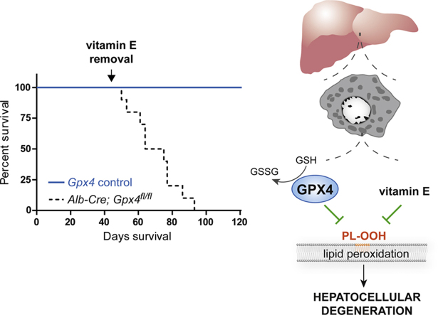
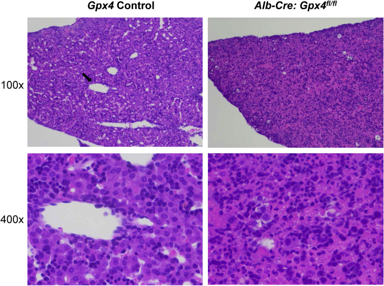
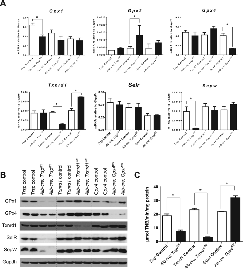
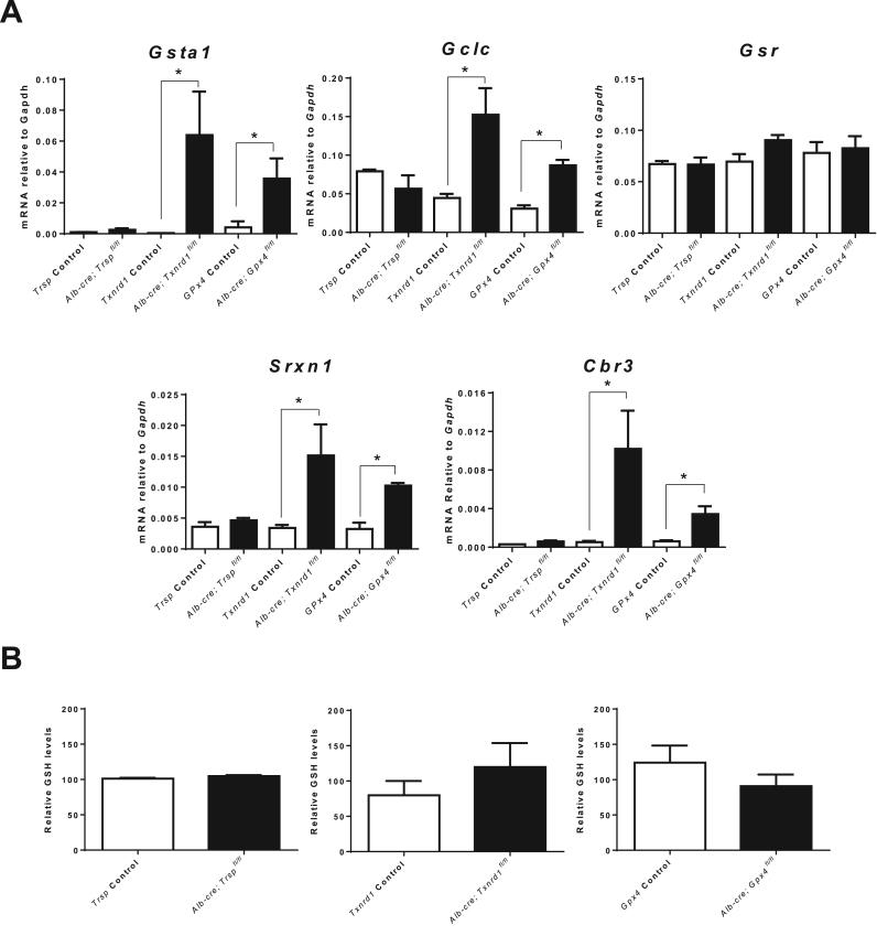
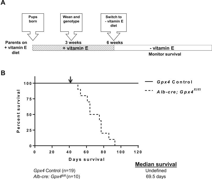
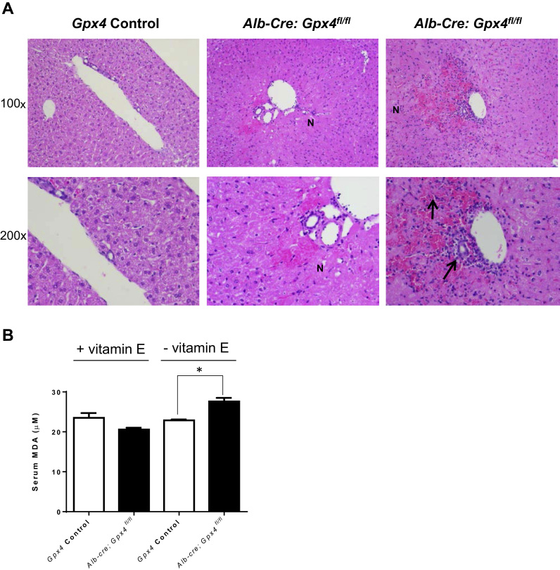
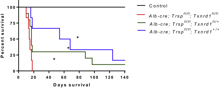
The selenoenzyme glutathione peroxidase 4 (Gpx4) is an essential mammalian glutathione peroxidase, which protects cells against detrimental lipid peroxidation and governs a novel form of regulated necrotic cell death, called ferroptosis. To study the relevance of Gpx4 and of another vitally important selenoprotein, cytosolic thioredoxin reductase (Txnrd1), for liver function, mice with conditional deletion of Gpx4 in hepatocytes were studied, along with those lacking Txnrd1 and selenocysteine (Sec) tRNA ( Trsp ) in hepatocytes. Unlike Txnrd1 - and Trsp -deficient mice, Gpx4 −/− mice died shortly after birth and presented extensive hepatocyte degeneration. Similar to Txnrd1 -deficient livers, Gpx4 −/− livers manifested upregulation of nuclear factor (erythroid-derived)-like 2 (Nrf2) response genes. Remarkably, Gpx4 −/− pups born from mothers fed a vitamin E-enriched diet survived, yet this protection was reversible as subsequent vitamin E deprivation caused death of Gpx4 -deficient mice ~4 weeks thereafter. Abrogation of selenoprotein expression in Gpx4 −/− mice did not result in viable mice, indicating that the combined deficiency aggravated the loss of Gpx4 in liver. By contrast, combined Trsp / Txnrd1 -deficient mice were born, but had significantly shorter lifespans than either single knockout, suggesting that Txnrd1 plays an important role in supporting liver function of mice lacking Trsp . In sum our study demonstrates that the ferroptosis regulator Gpx4 is critical for hepatocyte survival and proper liver function, and that vitamin E can compensate for its loss by protecting cells against deleterious lipid peroxidation.
Overview
The claims and statistics of this paper haven’t been checked yet.
Run the check — free, no account needed. It reads the open-access text and checks the sources it cites.
Integrity signals
Registry
Cites
Statistics
Figures
Verification
Claims the paper makes
The claims and statistics of this paper haven’t been checked yet.
Run the check — free, no account needed. It reads the open-access text and checks the sources it cites.
Claims other papers make about this one
Each is checked against this paper’s own text: does it say what the citing paper says it does?
In the citing paper
“the inducible or tissue-specific ablation of Gpx4 in mice provokes a variety of pathological conditions, including acute renal or hepatic injury [547, 563, 589], neurodegeneration [550, 590, 591], and defective immunity to infection [567], all of which can be prevented or mitigated by ferroptosis-inhibiting strategies.”
From Molecular mechanisms of cell death: recommendations of the nomenclature committee on cell death 2018. (2018) · cites this paper as [589]
Self-check
Percentages, sums and reported differences re-computed from the paper’s own numbers.
The self-check needs the paper’s full text.
Citations
Cites
Ferroptosis: An Iron-Dependent Form of Nonapoptotic Cell Death
Dixon, S. J., Lemberg, K. M., Lamprecht, M. R., Skouta, R., Zaitsev, E. M., Gleason, C. E., Patel, D. N., Bauer, A. J., Cantley, A. M., Yang, W. S., et al. (2012) · Cell · doi:10.1016/j.cell.2012.03.042 · Cited by 18,691
Regulation of Ferroptotic Cancer Cell Death by GPX4
Yang, W. S., SriRamaratnam, R., Welsch, M. E., Shimada, K., Skouta, R., Viswanathan, V. S., Cheah, J. H., Clemons, P. A., Shamji, A. F., Clish, C. B., et al. (2014) · Cell · doi:10.1016/j.cell.2013.12.010 · Cited by 7,713
Inactivation of the ferroptosis regulator Gpx4 triggers acute renal failure in mice
Friedmann Angeli, J. P., Schneider, M., Proneth, B., Tyurina, Y. Y., Tyurin, V. A., Hammond, V. J., Herbach, N., Aichler, M., Walch, A., Eggenhofer, E., et al. (2014) · Nature Cell Biology · doi:10.1038/ncb3064 · Cited by 3,732
Self-citationSeiler, A., Schneider, M., Förster, H., Roth, S., Wirth, E. K., Culmsee, C., Plesnila, N., Kremmer, E., Rådmark, O., Wurst, W., et al. (2008) · Cell Metabolism · doi:10.1016/j.cmet.2008.07.005 · Cited by 1,470
Self-citationSelenoproteins: Molecular Pathways and Physiological Roles
Labunskyy, V. M., Hatfield, D. L., & Gladyshev, V. N. (2014) · Physiological Reviews · doi:10.1152/physrev.00039.2013 · Cited by 1,452
Self-citationPostic, C., Shiota, M., Niswender, K. D., Jetton, T. L., Chen, Y., Moates, J. M., Shelton, K. D., Lindner, J., Cherrington, A. D., & Magnuson, M. A. (1999) · Journal of Biological Chemistry · doi:10.1074/jbc.274.1.305 · Cited by 1,302
Nielsen, F., Mikkelsen, B. B., Nielsen, J. B., Andersen, H. R., & Grandjean, P. (1997) · Clinical Chemistry · doi:10.1093/clinchem/43.7.1209 · Cited by 1,017
[21] Thioredoxin and thioredoxin reductase
Holmgren, A., & Björnstedt, M. (1995) · Methods in enzymology on CD-ROM/Methods in enzymology · doi:10.1016/0076-6879(95)52023-6 · Cited by 941
Yant, L. J., Ran, Q., Rao, L., Van Remmen, H., Shibatani, T., Belter, J. G., Motta, L., Richardson, A., & Prolla, T. A. (2003) · Free Radical Biology and Medicine · doi:10.1016/s0891-5849(02)01360-6 · Cited by 797
T cell lipid peroxidation induces ferroptosis and prevents immunity to infection
Matsushita, M., Freigang, S., Schneider, C., Conrad, M., Bornkamm, G. W., & Kopf, M. (2015) · Journal of Experimental Medicine · doi:10.1084/jem.20140857 · Cited by 672 · cited 2×
“As reported in earlier studies [6,13], vitamin E can compensate for Gpx4 loss in some tissues in vivo (i.”
BackgroundSelf-citationRegulated necrosis: disease relevance and therapeutic opportunities
Conrad, M., Angeli, J. P. F., Vandenabeele, P., & Stockwell, B. R. (2016) · Nature Reviews Drug Discovery · doi:10.1038/nrd.2015.6 · Cited by 630
Self-citationChen, L., Hambright, W. S., Na, R., & Ran, Q. (2015) · Journal of Biological Chemistry · doi:10.1074/jbc.m115.680090 · Cited by 498
Jakupoglu, C., Przemeck, G. K. H., Schneider, M., Moreno, S. G., Mayr, N., Hatzopoulos, A. K., de Angelis, M. H., Wurst, W., Bornkamm, G. W., Brielmeier, M., et al. (2005) · Molecular and Cellular Biology · doi:10.1128/mcb.25.5.1980-1988.2005 · Cited by 357
Self-citationBösl, M. R., Takaku, K., Oshima, M., Nishimura, S., & Taketo, M. M. (1997) · Proceedings of the National Academy of Sciences · doi:10.1073/pnas.94.11.5531 · Cited by 337
Wirth, E. K., Conrad, M., Winterer, J., Wozny, C., Carlson, B. A., Roth, S., Schmitz, D., Bornkamm, G. W., Coppola, V., Tessarollo, L., et al. (2010) · The FASEB Journal · doi:10.1096/fj.09-143974 · Cited by 237
Self-citationMoriarty, P. M., Reddy, C. C., & Maquat, L. E. (1998) · Molecular and Cellular Biology · doi:10.1128/mcb.18.5.2932 · Cited by 229
Hepatically derived selenoprotein P is a key factor for kidney but not for brain selenium supply
Schweizer, U., Streckfuß, F., Pelt, P., Carlson, B. A., Hatfield, D. L., Köhrle, J., & Schomburg, L. (2005) · Biochemical Journal · doi:10.1042/bj20041973 · Cited by 207
Self-citationFactor 3 Activity of Selenium Compounds
Schwarz, K., & Foltz, C. M. (1958) · Journal of Biological Chemistry · doi:10.1016/s0021-9258(19)68065-8 · Cited by 187
Selenium Regulation of the Selenoprotein and Nonselenoprotein Transcriptomes in Rodents
Sunde, R. A., & Raines, A. M. (2011) · Advances in Nutrition · doi:10.3945/an.110.000240 · Cited by 185
Jishage, K.-I., Arita, M., Igarashi, K., Iwata, T., Watanabe, M., Ogawa, M., Ueda, O., Kamada, N., Inoue, K., Arai, H., et al. (2001) · Journal of Biological Chemistry · doi:10.1074/jbc.c000676200 · Cited by 180
Carlson, B. A., Novoselov, S. V., Kumaraswamy, E., Lee, B. J., Anver, M. R., Gladyshev, V. N., & Hatfield, D. L. (2004) · Journal of Biological Chemistry · doi:10.1074/jbc.m310470200 · Cited by 177
Self-citationPattern of serum protein gene expression in mouse visceral yolk sac and foetal liver.
Meehan, R. R., Barlow, D. P., Hill, R. E., Hogan, B. L. M., & Hastie, N. D. (1984) · The EMBO Journal · doi:10.1002/j.1460-2075.1984.tb02062.x · Cited by 176
Wortmann, M., Schneider, M., Pircher, J., Hellfritsch, J., Aichler, M., Naidu, V. M., Kölle, P., Kuhlencordt, P., Walch, A. K., Pohl, U., et al. (2013) · Circulation Research · doi:10.1161/circresaha.113.279984 · Cited by 162
Self-citationYoo, M., Gu, X., Xu, X.-M., Kim, J. Y., Carlson, B. A., Patterson, A. D., Cai, H., Gladyshev, V. N., & Hatfield, D. L. (2009) · Antioxidants and Redox Signaling · doi:10.1089/ars.2009.2891 · Cited by 148
Self-citationLiver Cell Heterogeneity: Functions of Non-Parenchymal Cells
Bouwens, L., Bleser, P. D., Vanderkerken, K., Geerts, B., & Wisse, E. (2017) · Enzyme · doi:10.1159/000468782 · Cited by 133
Cytoprotective Nrf2 Pathway Is Induced In Chronically Txnrd 1-Deficient Hepatocytes
Suvorova, E. S., Lucas, O., Weisend, C. M., Rollins, M. F., Merrill, G. F., Capecchi, M. R., & Schmidt, E. E. (2009) · PLoS ONE · doi:10.1371/journal.pone.0006158 · Cited by 111
Brütsch, S. H., Wang, C. C., Li, L., Stender, H., Neziroglu, N., Richter, C., Kühn, H., & Borchert, A. (2014) · Antioxidants and Redox Signaling · doi:10.1089/ars.2014.5967 · Cited by 108
Translational Redefinition of UGA Codons Is Regulated by Selenium Availability
Howard, M. T., Carlson, B. A., Anderson, C. B., & Hatfield, D. L. (2013) · Journal of Biological Chemistry · doi:10.1074/jbc.m113.481051 · Cited by 108
Self-citationSelective Removal of the Selenocysteine tRNA[Ser]Sec Gene (Trsp) in Mouse Mammary Epithelium
Kumaraswamy, E., Carlson, B. A., Morgan, F., Miyoshi, K., Robinson, G. W., Su, D., Wang, S., Southon, E. A., Tessarollo, L., Lee, B. J., et al. (2003) · Molecular and Cellular Biology · doi:10.1128/mcb.23.5.1477-1488.2003 · Cited by 106
Self-citationCre activity in fetal albCre mouse hepatocytes: Utility for developmental studies
Weisend, C. M., Kundert, J. A., Suvorova, E. S., Prigge, J. R., & Schmidt, E. E. (2009) · genesis · doi:10.1002/dvg.20568 · Cited by 103
Conrad, M. (2009) · Biochimica et Biophysica Acta (BBA) - General Subjects · doi:10.1016/j.bbagen.2009.05.001 · Cited by 97
Self-citationTargeted insertion of cysteine by decoding UGA codons with mammalian selenocysteine machinery
Xu, X.-M., Turanov, A. A., Carlson, B. A., Yoo, M., Everley, R. A., Nandakumar, R., Sorokina, I., Gygi, S. P., Gladyshev, V. N., & Hatfield, D. L. (2010) · Proceedings of the National Academy of Sciences · doi:10.1073/pnas.1009947107 · Cited by 88
Self-citationSengupta, A., Lichti, U. F., Carlson, B. A., Cataisson, C., Ryscavage, A. O., Mikulec, C. D., Conrad, M., Fischer, S. M., Hatfield, D. L., & Yuspa, S. H. (2013) · Journal of Investigative Dermatology · doi:10.1038/jid.2013.52 · Cited by 86
Self-citationLu, J., Zhong, L., Lönn, M. E., Burk, R. F., Hill, K. E., & Holmgren, A. (2009) · The FASEB Journal · doi:10.1096/fj.08-127662 · Cited by 74
The Yin and Yang of Nrf2-Regulated Selenoproteins in Carcinogenesis
Brigelius‐Flohé, R., Müller, M. F., Lippmann, D., & Kipp, A. P. (2012) · International Journal of Cell Biology · doi:10.1155/2012/486147 · Cited by 68
Carlson, B. A., Yoo, M., Tobe, R., Mueller, C., Naranjo‐Suarez, S., Hoffmann, V. J., Gladyshev, V. N., & Hatfield, D. L. (2012) · Carcinogenesis · doi:10.1093/carcin/bgs230 · Cited by 58
Self-citationMannes, A. M., Seiler, A., Bosello-Travain, V., Maiorino, M., & Conrad, M. (2011) · The FASEB Journal · doi:10.1096/fj.10-177147 · Cited by 51
Self-citationLebold, K. M., & Traber, M. G. (2013) · Free Radical Biology and Medicine · doi:10.1016/j.freeradbiomed.2013.07.039 · Cited by 47
Prealbumin gene expression during mouse development studied by in situ hybridization
Murakami, T., Yasuda, Y., Mita, S., Maeda, S., 島田, 和., Fujimoto, T., & Araki, S. (1987) · Cell Differentiation · doi:10.1016/0045-6039(87)90408-8 · Cited by 32
Cited by
Sorted by citations
Ferroptosis: mechanisms, biology and role in disease
Jiang, X., Stockwell, B. R., & Conrad, M. (2021) · Nature Reviews Molecular Cell Biology · doi:10.1038/s41580-020-00324-8 · Cited by 7,973
Self-citationGalluzzi, L., Vitale, I., Aaronson, S. A., Abrams, J. M., Adam, D., Agostinis, P., Alnemri, E. S., Altucci, L., Amelio, I., Andrews, D. W., et al. (2018) · Cell Death & Differentiation · doi:10.1038/s41418-017-0012-4 · Cited by 6,680
Self-citationFerroptosis: molecular mechanisms and health implications
Tang, D., Chen, X., Kang, R., & Kroemer, G. (2020) · Cell Research · doi:10.1038/s41422-020-00441-1 · Cited by 4,495
Ferroptosis turns 10: Emerging mechanisms, physiological functions, and therapeutic applications
Stockwell, B. R. (2022) · Cell · doi:10.1016/j.cell.2022.06.003 · Cited by 3,211
Oxidized arachidonic and adrenic PEs navigate cells to ferroptosis
Kagan, V. E., Mao, G., Qu, F., Angeli, J. P. F., Doll, S., Croix, C. S., Dar, H. H., Liu, B., Tyurin, V. A., Ritov, V. B., et al. (2017) · Nature Chemical Biology · doi:10.1038/nchembio.2238 · Cited by 3,029
Self-citation
Metadata
- Authors
- Carlson, B. A., Tobe, R., Yefremova, E., Tsuji, P. A., Hoffmann, V. J., Schweizer, U., Gladyshev, V. N., Hatfield, D. L., & Conrad, M.
- Affiliations
- National Institutes of Health (US)
- Helmholtz Munich (DE)
- Towson University (US)
- University of Bonn (DE)
- Brigham and Women's Hospital (US)
- Corresponding author
- Hatfield, Dolph L.
- Conrad, Marcus
- Venue
- Redox Biology
- Publisher
- Elsevier BV
- Published
- 2016-10
- Volume
- 9
- Pages
- 22-31
- ISSN
- 2213-2317 (ISSN-L)
- PMC
- PMC4900515
- OpenAlex
- W2408722709
- Cited by
- 316
- References
- 40
- Citations by year
- 2026: 27 · 2025: 38 · 2024: 52 · 2023: 40 · 2022: 47 · 2021: 27 · 2020: 22 · 2019: 16 · 2018: 24 · 2017: 14 · 2016: 8
- FWCI
- 21.64
- Type
- journal article
- Language
- en
- Open access
- gold
- doi.org
- Licence
- Locations
- Redox Biology (publishedVersion, cc-by-nc-nd)
- PubMed Central (publishedVersion, cc-by-nc-nd)
- PubMed (publishedVersion)
- PuSH - Publication Server of Helmholtz Zentrum München (submittedVersion, cc-by)
- Digital Access to Scholarship at Harvard (DASH) (Harvard University) (submittedVersion, cc-by)
- DOAJ (DOAJ: Directory of Open Access Journals) (submittedVersion, cc-by-sa)
- Site cant be reached (submittedVersion, cc-by)
- Funders
- National Institutes of Health
- National Cancer Institute
- Center for Cancer Research
- Deutsche Forschungsgemeinschaft (CO 291/2-3, CO 291/5-1)
- Human Frontier Science Program (RGP0013/14)
- Topics
- Health Sciences › Nursing › Nutrition and Dietetics › Selenium in Biological Systems
- Health Sciences › Medicine › Biochemistry (medical) › Antioxidant Activity and Oxidative Stress
- Life Sciences › Biochemistry, Genetics and Molecular Biology › Molecular Biology › Redox biology and oxidative stress
- Keywords
- GPX4, vitamin E, ferroptosis, hepatocyte degeneration, selenoproteins, thioredoxin reductase 1, selenocysteine tRNA, lipid peroxidation, conditional knockout mice, liver function, hepatocyte viability
- MeSH terms
- Phospholipid Hydroperoxide Glutathione Peroxidase, Animals, Cell Survival, Female, Gene Expression Regulation, Genotype, Glutathione, Glutathione Peroxidase, Liver, Liver Regeneration, Male, Necrosis, Vitamin E, Lipid Peroxidation, Mice, Knockout, Glutathione Disulfide, Hepatocytes, NF-E2-Related Factor 2, Mice
About these checks
Registry, retraction, Cites and statistics results are computed, not judged, with one exception: a printed reference that no index lists is read once by a model before it counts as a citation error. Claims are extracted by a model and kept only when their quote appears verbatim in the text; each cited source is then checked, with a model’s help, for whether it says what the claim relies on. Those verdicts can be wrong — read the claim’s quote and the cited source before relying on one.
Statistics and arithmetic are recomputed, not judged.
Confidence bands are shown only for fields calibrated on our annotated test set. None is calibrated yet, so no band is shown.
What counts as a claim ›How we measure confidence ›How we check this ›
Are you an author, or do you see a problem with this page? Request a review
