

The DOI matches its Crossref record; 315 of the 317 references it cites were found in a registry or an index.
GABAA receptor-acting neurosteroids: A role in the development and regulation of the stress response
Open accessAbstract
Regulation of hypothalamic-pituitary-adrenocortical (HPA) axis activity by stress is a fundamental survival mechanism and HPA-dysfunction is implicated in psychiatric disorders. Adverse early life experiences, e.g. poor maternal care, negatively influence brain development and programs an abnormal stress response by encoding long-lasting molecular changes, which may extend to the next generation. How HPA-dysfunction leads to the development of affective disorders is complex, but may involve GABAA receptors (GABAARs), as they curtail stress-induced HPA axis activation. Of particular interest are endogenous neurosteroids that potently modulate the function of GABAARs and exhibit stress-protective properties. Importantly, neurosteroid levels rise rapidly during acute stress, are perturbed in chronic stress and are implicated in the behavioural changes associated with early-life adversity. We will appraise how GABAAR-active neurosteroids may impact on HPA axis development and the orchestration of the stress-evoked response. The significance of these actions will be discussed in the context of stress-associated mood disorders.
Overview
The claims and statistics of this paper haven’t been checked yet.
Run the check — free, no account needed. It reads the open-access text and checks the sources it cites.
Integrity signals
Registry
Cites
Statistics
Figures
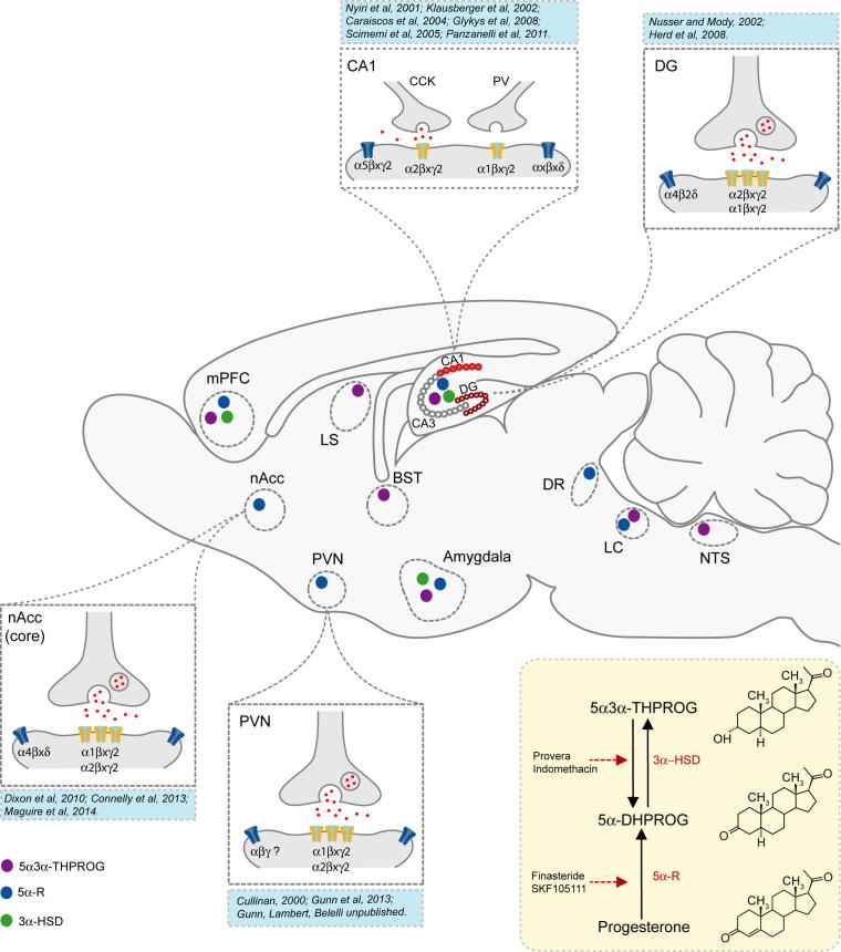
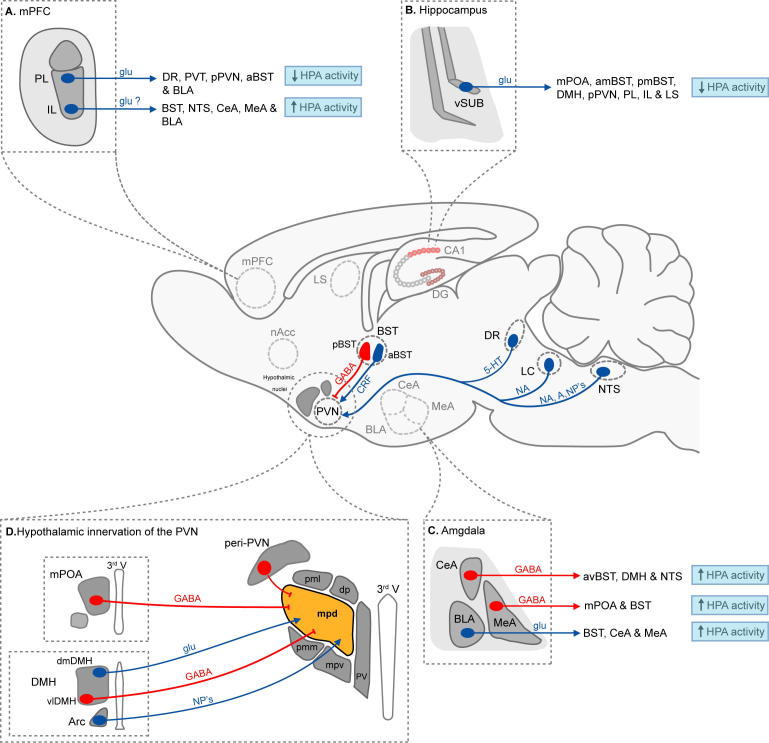
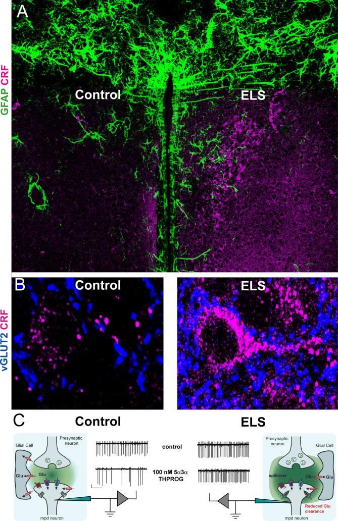
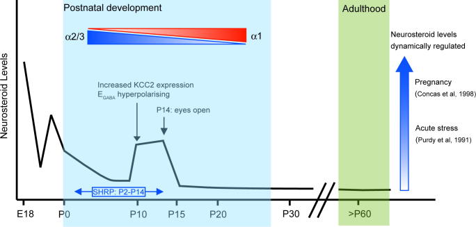
Verification
Claims the paper makes
The claims and statistics of this paper haven’t been checked yet.
Run the check — free, no account needed. It reads the open-access text and checks the sources it cites.
Claims other papers make about this one
Each is checked against this paper’s own text: does it say what the citing paper says it does?
The cited source says what the claim attributes to it.
In the citing paper
“and enhance the negative feedback on the HPA axis (21, 22).”
From Higher Circulating Cortisol in the Follicular vs. Luteal Phase of the Menstrual Cycle: A Meta-Analysis (2020) · cites this paper as [22]
Self-check
Percentages, sums and reported differences re-computed from the paper’s own numbers.
The self-check needs the paper’s full text.
Citations
Cites
Effects of stress throughout the lifespan on the brain, behaviour and cognition
Lupien, S. J., McEwen, B. S., Gunnar, M. R., & Heim, C. (2009) · Nature Reviews Neuroscience · doi:10.1038/nrn2639 · Cited by 6,256
Epigenetic programming by maternal behavior
Weaver, I. C. G., Cervoni, N., Champagne, F. A., D'Alessio, A. C., Sharma, S., Seckl, J. R., Dymov, S., Szyf, M., & Meaney, M. J. (2004) · Nature Neuroscience · doi:10.1038/nn1276 · Cited by 6,177
Stress and the brain: from adaptation to disease
de Kloet, E. R., Joëls, M., & Holsboer, F. (2005) · Nature Reviews Neuroscience · doi:10.1038/nrn1683 · Cited by 4,621
Requirement of hippocampal neurogenesis for the behavioral effects of antidepressants
Santarelli, L., Saxe, M., Gross, C., Surget, A., Battaglia, F., Dulawa, S., Weisstaub, N., Lee, J., Duman, R., Arancio, O., et al. (2003) · Science · doi:10.1126/science.1083328 · Cited by 4,321
Vale, W., Spiess, J., Rivier, C., & Rivier, J. (1981) · Science · doi:10.1126/science.6267699 · Cited by 3,965
Vale, W., Spiess, J., RIVIER, C. L., & Rivier, J. (1982) · Obstetrical & Gynecological Survey · doi:10.1097/00006254-198205000-00013 · Cited by 3,904
Neural regulation of endocrine and autonomic stress responses
Ulrich-Lai, Y. M., & Herman, J. P. (2009) · Nature Reviews Neuroscience · doi:10.1038/nrn2647 · Cited by 3,318
Liu, D., Diorio, J., Tannenbaum, B., Caldji, C., Francis, D., Freedman, A., Sharma, S., Pearson, D., Plotsky, P. M., & Meaney, M. J. (1997) · Science · doi:10.1126/science.277.5332.1659 · Cited by 3,170
Chronic antidepressant treatment increases neurogenesis in adult rat hippocampus
Malberg, J. E., Eisch, A. J., Nestler, E. J., & Duman, R. S. (2000) · The Journal of Neuroscience · doi:10.1523/jneurosci.20-24-09104.2000 · Cited by 3,111
Interneurons of the neocortical inhibitory system
Markram, H., Toledo-Rodriguez, M., Wang, Y., Gupta, A., Silberberg, G., & Wu, C. (2004) · Nature Reviews Neuroscience · doi:10.1038/nrn1519 · Cited by 2,954
Cited by
Sorted by citations
Singewald, N., Schmuckermair, C., Whittle, N., Holmes, A., & Ressler, K. J. (2015) · Pharmacology & Therapeutics · doi:10.1016/j.pharmthera.2014.12.004 · Cited by 390
“7 and (Gunn et al., 2014; Makkar et al., 2010)].”
Li, Z., Yu, J., Peng, Y., & Huang, B. (2016) · Physiologia Plantarum · doi:10.1111/ppl.12483 · Cited by 207
Porcu, P., Barron, A. M., Frye, C. A., Walf, A. A., Yang, S., He, X., Morrow, A. L., Panzica, G., & Melcangi, R. C. (2015) · Journal of Neuroendocrinology · doi:10.1111/jne.12351 · Cited by 186
Essential elements in depression and anxiety. Part I
Młyniec, K., Davies, C. L., Sánchez, I. G. D. A., Pytka, K., Budziszewska, B., & Nowak, G. (2014) · Pharmacological Reports · doi:10.1016/j.pharep.2014.03.001 · Cited by 174
GABA Receptors: Pharmacological Potential and Pitfalls
Jembrek, M. J., & Vlainić, J. (2015) · Current Pharmaceutical Design · doi:10.2174/1381612821666150914121624 · Cited by 171
GABAergic Control of Depression-Related Brain States
Luscher, B., & Fuchs, T. (2015) · Advances in Pharmacology · doi:10.1016/bs.apha.2014.11.003 · Cited by 149
Sex differences in the outcome of juvenile social isolation on HPA axis function in rats
Pisu, M. G., Garau, A., Boero, G., Biggio, F., Pibiri, V., Dore, R., Locci, V., Paci, E., Porcu, P., & Serra, M. (2016) · Neuroscience · doi:10.1016/j.neuroscience.2016.02.009 · Cited by 112
Imamura, Y., Shinozaki, T., Okada‐Ogawa, A., Noma, N., Shinoda, M., Iwata, K., Wada, A., Abe, O., Wang, K., & Svensson, P. J. (2019) · Journal of Oral Rehabilitation · doi:10.1111/joor.12795 · Cited by 101
Higashi, T., & Ogawa, S. (2015) · The Journal of Steroid Biochemistry and Molecular Biology · doi:10.1016/j.jsbmb.2015.10.003 · Cited by 101
Hamidovic, A., Karapetyan, K., Serdarevic, F., Choi, S. H., Eisenlohr-Moul, T., & Pinna, G. (2020) · Frontiers in Endocrinology · doi:10.3389/fendo.2020.00311 · Cited by 99
“Our hypothesis was based on the finding that the progesterone metabolite allopregnanolone positively modulates gamma-aminobutyric acid (GABA)A receptors via an allosteric binding site to potentiate inhibitory signaling (20) and enhance the negative feedback on the HPA axis (21, 22).”
Stress-Induced Chronic Visceral Pain of Gastrointestinal Origin
Meerveld, B. G., & Johnson, A. C. (2017) · Frontiers in Systems Neuroscience · doi:10.3389/fnsys.2017.00086 · Cited by 102
GABAA receptor subtype involvement in addictive behaviour
Stephens, D. N., King, S. L., Lambert, J. J., Belelli, D., & Duka, T. (2016) · Genes Brain & Behavior · doi:10.1111/gbb.12321 · Cited by 97
Self-citationEssential elements in depression and anxiety. Part II
Młyniec, K., Gaweł, M., Doboszewska, U., Starowicz, G., Pytka, K., Davies, C. L., & Budziszewska, B. (2014) · Pharmacological Reports · doi:10.1016/j.pharep.2014.09.009 · Cited by 94
Sze, Y., & Brunton, P. J. (2020) · European Journal of Neuroscience · doi:10.1111/ejn.14615 · Cited by 85
GABA Receptors and the Pharmacology of Sleep
Wisden, W., Yu, X., & Franks, N. P. (2017) · Handbook of experimental pharmacology · doi:10.1007/164_2017_56 · Cited by 92
Neural mechanisms underlying GABAergic regulation of adult hippocampal neurogenesis
Catavero, C. M., Bao, H., & Song, J. (2017) · Cell and Tissue Research · doi:10.1007/s00441-017-2668-y · Cited by 76
Zhu, X., Fréchou, M., Lière, P., Zhang, S., Pianos, A., Fernandez, N., Denier, C., Mattern, C., Schumacher, M., & Guennoun, R. (2017) · Journal of Neuroscience · doi:10.1523/jneurosci.3874-16.2017 · Cited by 79
Realising the therapeutic potential of neuroactive steroid modulators of the GABAA receptor
Belelli, D., Hogenkamp, D., Gee, K. W., & Lambert, J. J. (2020) · Neurobiology of Stress · doi:10.1016/j.ynstr.2019.100207 · Cited by 67
Self-citationZimmermann, T., Bartsch, J., Beer, A., Lomazzo, E., Guggenhuber, S., Lange, M. D., Bîndilă, L., Pape, H., & Lutz, B. (2018) · Neuropsychopharmacology · doi:10.1038/s41386-018-0274-7 · Cited by 68
Puga-Olguín, A., Rodríguez‐Landa, J. F., Rovirosa-Hernández, M. D. J., Germán-Ponciano, L. J., Caba, M., Meza, E., Guillén-Ruiz, G., & Olmos-Vázquez, O. J. (2018) · Behavioural Brain Research · doi:10.1016/j.bbr.2018.12.017 · Cited by 65
Neurobiological consequences of juvenile stress: A GABAergic perspective on risk and resilience
Albrecht, A., Müller, I., Ardi, Z., Çalışkan, G., Gruber, D., Ivens, S., Segal, M., Behr, J., Heinemann, U., Stork, O., et al. (2017) · Neuroscience & Biobehavioral Reviews · doi:10.1016/j.neubiorev.2017.01.005 · Cited by 65
Pleiotropic actions of allopregnanolone underlie therapeutic benefits in stress-related disease
Boero, G., Porcu, P., & Morrow, A. L. (2019) · Neurobiology of Stress · doi:10.1016/j.ynstr.2019.100203 · Cited by 63
Sze, Y., Gill, A. C., & Brunton, P. J. (2018) · Journal of Neuroendocrinology · doi:10.1111/jne.12644 · Cited by 69
The brain as a target of hormonal contraceptives: Evidence from animal studies
Porcu, P., Serra, M., & Concas, A. (2019) · Frontiers in Neuroendocrinology · doi:10.1016/j.yfrne.2019.100799 · Cited by 62
Perinatal exposure to bisphenol A at the intersection of stress, anxiety, and depression
Wiersielis, K., Samuels, B. A., & Roepke, T. A. (2020) · Neurotoxicology and Teratology · doi:10.1016/j.ntt.2020.106884 · Cited by 57
The critical role of microRNAs in stress response: Therapeutic prospect and limitation
Du, J., Li, M., Huang, Q., Liu, W., Li, W., Li, Y., & Gong, Z. (2018) · Pharmacological Research · doi:10.1016/j.phrs.2018.12.007 · Cited by 57
Stress-Induced Pain: A Target for the Development of Novel Therapeutics
Johnson, A. C., & Meerveld, B. G. (2014) · Journal of Pharmacology and Experimental Therapeutics · doi:10.1124/jpet.114.218065 · Cited by 57
Giatti, S., Foglio, B., Romano, S., Pesaresi, M., Panzica, G., García‐Segura, L. M., Caruso, D., & Melcangi, R. C. (2015) · Neuroendocrinology · doi:10.1159/000442982 · Cited by 52
A Rationale for Allopregnanolone Treatment of Alcohol Use Disorders: Basic and Clinical Studies
Morrow, A. L., Boero, G., & Porcu, P. (2019) · Alcoholism Clinical and Experimental Research · doi:10.1111/acer.14253 · Cited by 53
Hypothalamic-pituitary-adrenal axis targets for the treatment of epilepsy
Basu, T., Maguire, J. L., & Salpekar, J. A. (2021) · Neuroscience Letters · doi:10.1016/j.neulet.2020.135618 · Cited by 50
Neuroactive steroids and stress axis regulation: Pregnancy and beyond
Brunton, P. J. (2016) · The Journal of Steroid Biochemistry and Molecular Biology · doi:10.1016/j.jsbmb.2015.08.003 · Cited by 48
GABA-A Receptors Mediate Tonic Inhibition and Neurosteroid Sensitivity in the Brain
Reddy, D. S. (2018) · Vitamins and hormones · doi:10.1016/bs.vh.2017.12.001 · Cited by 46
Γ-Aminobutyric acid in adult brain: an update
Siucińska, E. (2019) · Behavioural Brain Research · doi:10.1016/j.bbr.2019.112224 · Cited by 43
Abuaish, S., Spinieli, R. L., & McGowan, P. O. (2018) · Psychoneuroendocrinology · doi:10.1016/j.psyneuen.2018.08.003 · Cited by 39
Steroids in Stroke with Special Reference to Progesterone
Guennoun, R., Zhu, X., Fréchou, M., Gaignard, P., Slama, A., Lière, P., & Schumacher, M. (2018) · Cellular and Molecular Neurobiology · doi:10.1007/s10571-018-0627-0 · Cited by 41
Puerarin ameliorated the behavioral deficits induced by chronic stress in rats
Qiu, Z.-K., Zhang, G.-H., Zhong, D.-S., He, J.-L., Liu, X., Chen, J., & Wei, D. (2017) · Scientific Reports · doi:10.1038/s41598-017-06552-x · Cited by 41
Steroids, steroid precursors, and neuroactive steroids in critically ill equine neonates
Dembek, K. A., Timko, K. J., Johnson, L. M., Hart, K. A., Barr, B. S., David, B., Burns, T. A., & Toribio, R. E. (2017) · The Veterinary Journal · doi:10.1016/j.tvjl.2017.05.009 · Cited by 41
Ecdysteroids: Isolation, Chemical Transformations, and Biological Activity
Savchenko, R. G., Veskina, N. A., Odinokov, V. N., Benkovskaya, G. V., & Parfenova, L. V. (2022) · Phytochemistry Reviews · doi:10.1007/s11101-021-09792-y · Cited by 39
α6-Containing GABAA Receptors: Functional Roles and Therapeutic Potentials
Sieghart, W., Chiou, L., Ernst, M. C., Fabjan, J., Savić, M. M., & Lee, M. T. (2022) · Pharmacological Reviews · doi:10.1124/pharmrev.121.000293 · Cited by 40
Tomaselli, G., & Vallée, M. (2019) · Frontiers in Neuroendocrinology · doi:10.1016/j.yfrne.2019.100789 · Cited by 41
Cueto‐Escobedo, J., Andrade-Soto, J., Lima, M. G., Maximino, C., Hernández-López, F., & Rodríguez‐Landa, J. F. (2020) · Behavioural Brain Research · doi:10.1016/j.bbr.2020.112590 · Cited by 33
Relating neurosteroid modulation of inhibitory neurotransmission to behaviour
Belelli, D., Phillips, G., Atack, J. R., & Lambert, J. J. (2021) · Journal of Neuroendocrinology · doi:10.1111/jne.13045 · Cited by 32
Self-citationLiu, X., Li, X., Teng, T., Jiang, Y., Xiang, Y., Fan, L., Yu, Y., Zhou, X., & Xie, P. (2022) · Journal of Affective Disorders · doi:10.1016/j.jad.2022.06.088 · Cited by 33
Neonatal blockade of GABA‐A receptors alters behavioral and physiological phenotypes in adult mice
Salari, A., & Amani, M. (2017) · International Journal of Developmental Neuroscience · doi:10.1016/j.ijdevneu.2017.01.007 · Cited by 31
Neurosteroids and neuropathic pain management: Basic evidence and therapeutic perspectives
Meyer, L., Taleb, O., Patte-Mensah, C., & Mensah-Nyagan, A.-G. (2019) · Frontiers in Neuroendocrinology · doi:10.1016/j.yfrne.2019.100795 · Cited by 30
Pre-pubertal stress and brain development in rodents
Brydges, N. M. (2015) · Current Opinion in Behavioral Sciences · doi:10.1016/j.cobeha.2015.08.003 · Cited by 29
Gatta, E., Camussi, D., Auta, J., Guidotti, A., & Pandey, S. C. (2022) · Pharmacology & Therapeutics · doi:10.1016/j.pharmthera.2022.108299 · Cited by 28
Franco‐Pérez, J., Montes, S., Sánchez-Hernández, J., & Ballesteros‐Zebadúa, P. (2020) · Radiation Oncology · doi:10.1186/s13014-020-01716-y · Cited by 27
Higashi, T., Aiba, N., Tanaka, T., Yoshizawa, K., & Ogawa, S. (2015) · Journal of Pharmaceutical and Biomedical Analysis · doi:10.1016/j.jpba.2015.08.040 · Cited by 27
Chen, C., Zhong, Z., Xin, Z., Hong, L.-H., Su, Y., & Yu, C. (2017) · Journal of Natural Medicines · doi:10.1007/s11418-017-1070-0 · Cited by 27
Khawaja, S. N., Alaswaiti, O. F., & Scrivani, S. J. (2022) · Dental Clinics of North America · doi:10.1016/j.cden.2022.07.004 · Cited by 26
Stamper, C. E., Hassell, J. E., Kapitz, A. J., Renner, K. J., Orchinik, M., & Lowry, C. A. (2017) · Stress · doi:10.1080/10253890.2017.1301426 · Cited by 26
Biochemical mechanisms in pathogenesis of infantile epileptic spasm syndrome
Riikonen, R. (2023) · Seizure · doi:10.1016/j.seizure.2023.01.004 · Cited by 26
Frau, R., Traccis, F., & Bortolato, M. (2019) · Journal of Neuroendocrinology · doi:10.1111/jne.12792 · Cited by 27
Brown, A. R., Herd, M. B., Belelli, D., & Lambert, J. J. (2014) · The Journal of Physiology · doi:10.1113/jphysiol.2014.280263 · Cited by 24
Self-citationMaternally derived hormones, neurosteroids and the development of behaviour
Mouton, J. C., & Duckworth, R. A. (2021) · Proceedings of the Royal Society B Biological Sciences · doi:10.1098/rspb.2020.2467 · Cited by 24
Giving a good start to a new life via maternal brain allostatic adaptations in pregnancy
Russell, J. A., & Brunton, P. J. (2019) · Frontiers in Neuroendocrinology · doi:10.1016/j.yfrne.2019.02.003 · Cited by 24
Bossé, G. D., Cadeddu, R., Floris, G., Farero, R. D., Vigato, E., Lee, S. J., Zhang, T., Gaikwad, N. W., Keefe, K. A., Phillips, P. E. M., et al. (2021) · Journal of Clinical Investigation · doi:10.1172/jci143990 · Cited by 22
Cruz, D. A., Glantz, L. A., McGaughey, K. D., Parke, G. J., Shampine, L. J., Kilts, J. D., Naylor, J. C., Marx, C. E., & Williamson, D. E. (2019) · Chronic Stress · doi:10.1177/2470547019838570 · Cited by 22
Flores-Ramos, M., Alcauter, S., López-Titla, M., Bernal-Santamaría, N., Calva-Coraza, E., & Edden, R. A. E. (2019) · Journal of Affective Disorders · doi:10.1016/j.jad.2018.08.033 · Cited by 21
How hormonal contraceptives shape brain and behavior: A review of preclinical studies
Concas, A., Serra, M., & Porcu, P. (2022) · Frontiers in Neuroendocrinology · doi:10.1016/j.yfrne.2022.101017 · Cited by 26
Cadeddu, R., Zandt, M. A. V., Santovito, L. S., Odeh, K., Anderson, C. J., Flanagan, D., Nordkild, P., Pinna, G., Pittenger, C., & Bortolato, M. (2023) · Neuropsychopharmacology · doi:10.1038/s41386-023-01603-6 · Cited by 21
miR-212 mediates counter-regulation on CRH expression and HPA axis activity in male mice
Tang, Y., Cai, X., Zhang, H., Shen, H., Wang, W., Shen, Z., Gu, W., Ling, C., & Li, M. (2017) · Journal of Molecular Endocrinology · doi:10.1530/jme-17-0124 · Cited by 21
Hasirci, A. S., Maldonado‐Devincci, A. M., Beattie, M. C., O’Buckley, T. K., & Morrow, A. L. (2017) · Alcoholism Clinical and Experimental Research · doi:10.1111/acer.13300 · Cited by 19
Wang, S., Yao, Z., Zhao, C.-B.-W., Ding, Y.-S., JingLUO, Bian, L.-H., Li, Q., Wang, X., Shi, J., Guo, J., et al. (2020) · Journal of Ethnopharmacology · doi:10.1016/j.jep.2020.113452 · Cited by 19
Diviccaro, S., Falvo, E., Piazza, R., Cioffi, L., Herian, M., Brivio, P., Calabrese, F., Giatti, S., Caruso, D., & Melcangi, R. C. (2022) · Neuropharmacology · doi:10.1016/j.neuropharm.2022.109405 · Cited by 19
Kuenzel, K., Friedrich, O., & Gilbert, D. F. (2016) · Frontiers in Molecular Neuroscience · doi:10.3389/fnmol.2016.00051 · Cited by 18
Mukerji, S. S., Misra, V., Lorenz, D. R., Chettimada, S., Keller, K., Letendre, S. L., Ellis, R. J., Morgello, S., Parker, R. A., & Gabuzda, D. H. (2020) · The Journal of Infectious Diseases · doi:10.1093/infdis/jiaa104 · Cited by 21
Zaman, S., Bilal, M., Du, H., & Che, S. (2020) · BioMed Research International · doi:10.1155/2020/4827045 · Cited by 18
Sze, Y., & Brunton, P. J. (2020) · Journal of Neuroendocrinology · doi:10.1111/jne.12916 · Cited by 18
Wang, S., Ding, Y.-S., Ma, X.-J., Zhao, C.-B.-W., Lin, M.-X., Luo, J., Jiang, Y., He, S., Guo, J., & Shi, J. (2018) · Molecules · doi:10.3390/molecules23092329 · Cited by 18
Salgado, R., Pereiro, N., López-Doval, S., & Lafuente, A. (2015) · Food and Chemical Toxicology · doi:10.1016/j.fct.2015.05.013 · Cited by 18
Human Microglia Synthesize Neurosteroids to Cope with Rotenone-Induced Oxidative Stress
Lucchi, C., Codeluppi, A., Filaferro, M., Vitale, G., Rustichelli, C., Avallone, R., Mandrioli, J., & Biagini, G. (2023) · Antioxidants · doi:10.3390/antiox12040963 · Cited by 17
Taleb, O., Patte‐Mensah, C., Meyer, L., Kemmel, V., Geoffroy, P., Miesch, M., & Mensah‐Nyagan, A. G. (2017) · Journal of Neuroendocrinology · doi:10.1111/jne.12568 · Cited by 18
Nejatbakhsh, M., Saboory, E., & Bagheri, M. (2018) · International Journal of Developmental Neuroscience · doi:10.1016/j.ijdevneu.2018.05.003 · Cited by 17
Kamli, H. M., Shaikh, A. A., Bappi, M. H., Raposo, A., Ahmad, M. F., Sonia, F. A., Akbor, M. S., Prottay, A. A. S., Gonçalves, S. A., Araújo, I. M., et al. (2023) · Biomedicine & Pharmacotherapy · doi:10.1016/j.biopha.2023.115768 · Cited by 16
Neurosteroid Receptor Modulators for Treating Traumatic Brain Injury
Verdoorn, T. A., Parry, T. J., Pinna, G., & Lifshitz, J. (2023) · Neurotherapeutics · doi:10.1007/s13311-023-01428-7 · Cited by 15
Yuan, X., Chai, J., Xu, W., & Zhao, Y. (2024) · Probiotics and Antimicrobial Proteins · doi:10.1007/s12602-024-10326-z · Cited by 15
Endogenous neurosteroids influence synaptic GABAAreceptors during postnatal development
Belelli, D., Brown, A. R., Mitchell, S. J., Gunn, B. G., Herd, M. B., Phillips, G. D., Seifi, M., Swinny, J. D., & Lambert, J. J. (2017) · Journal of Neuroendocrinology · doi:10.1111/jne.12537 · Cited by 15
Self-citationGuillén-Ruiz, G., Bernal‐Morales, B., Limón-Vázquez, A. K., Olmos-Vázquez, O. J., & Rodríguez‐Landa, J. F. (2024) · Journal of Integrative Neuroscience · doi:10.31083/j.jin2303051 · Cited by 14
Frau, R., & Bortolato, M. (2018) · Neuropharmacology · doi:10.1016/j.neuropharm.2018.05.013 · Cited by 13
Neurosteroids; potential underpinning roles in maintaining homeostasis
Rahmani, B., Ghasemi, R., Dargahi, L., Ahmadiani, A., & Haeri, A. (2015) · General and Comparative Endocrinology · doi:10.1016/j.ygcen.2015.09.030 · Cited by 14
Mohammed, S., Mahdi, F., Sulochana, S. P., & Paris, J. J. (2021) · Viruses · doi:10.3390/v13050813 · Cited by 14
Diviccaro, S., Giatti, S., Cioffi, L., Falvo, E., Herian, M., Caruso, D., & Melcangi, R. C. (2022) · Biomolecules · doi:10.3390/biom12111567 · Cited by 16
Isotope Dilution-Based Targeted and Nontargeted Carbonyl Neurosteroid/Steroid Profiling
Sharp, S., Mitchell, S. J., Vallée, M., Kuzmanova, E., Cooper, M. A., Belelli, D., Lambert, J. J., & Huang, J. T.-J. (2018) · Analytical Chemistry · doi:10.1021/acs.analchem.8b00055 · Cited by 13
Self-citationWang, Q., Deng, Z., Lan, J., Li, D., Fan, K., Chang, J., & Ma, Y. (2022) · International Journal of Molecular Sciences · doi:10.3390/ijms231911210 · Cited by 13
Reddy, D. S., & Bakshi, K. (2019) · Elsevier eBooks · doi:10.1016/b978-0-12-813814-4.00004-3 · Cited by 15
Dembek, K. A., Johnson, L. M., Timko, K. J., Minuto, J. S., Hart, K. A., Barr, B. S., & Toribio, R. E. (2019) · Journal of Veterinary Internal Medicine · doi:10.1111/jvim.15527 · Cited by 11
Exercise as a therapeutic strategy for insomnia: Current mechanisms and clinical relevance
Saadh, M. J., Saleh, A. Y., Kareem, R. A., Sharma, R. K., Roopashree, R., Sharma, G. C., Verma, R., Juneja, B., Sameer, H. N., Yaseen, A., et al. (2025) · Sleep Medicine · doi:10.1016/j.sleep.2025.108681 · Cited by 14
Porcu, P., Lallai, V., Locci, A., Catzeddu, S., Serra, V., Pisu, M. G., Serra, M., Dazzi, L., & Concas, A. (2016) · Psychopharmacology · doi:10.1007/s00213-016-4511-7 · Cited by 10
McCallie, K. R., Gaikwad, N. W., Cuadrado, M. E. C., Aleman, M. R., Madigan, J. E., Stevenson, D. K., & Bhutani, V. K. (2017) · Journal of Perinatology · doi:10.1038/jp.2016.268 · Cited by 10
Prefrontal 5α-reductase 2 mediates male-specific acute stress response
Cadeddu, R., Braccagni, G., Floris, G., Branca, C., Corridori, E., Salviati, S., Sánchez, P., Santovito, L. S., Torres, J. M., Ortega, E., et al. (2025) · Science Advances · doi:10.1126/sciadv.adr0563 · Cited by 9
Шушпанова, Т. В., & Bokhan, N. A. (2016) · Journal of Addiction Research & Therapy · doi:10.4172/2155-6105.1000271 · Cited by 9
Effects of γ-aminobutyric Acid on Mortality in Laying Hens During Summer Time
Choi, Y. (2019) · Journal of Agriculture & Life Science · doi:10.14397/jals.2019.53.2.131 · Cited by 9
Cadeddu, R., Mosher, L. J., Nordkild, P., Gaikwad, N. W., Ratto, G. M., Scheggi, S., & Bortolato, M. (2022) · Neurobiology of Stress · doi:10.1016/j.ynstr.2022.100489 · Cited by 10
Wotton, C. A., Quon, E. F., Palmer, A. C., & Bekar, L. K. (2018) · Journal of Neuroendocrinology · doi:10.1111/jne.12592 · Cited by 10
Rodríguez‐Landa, J. F., Olmos-Vázquez, O. J., Costa, B. P. D., Lima, M. G., Maximino, C., & Guillén-Ruiz, G. (2020) · Salud Mental · doi:10.17711/sm.0185-3325.2020.007 · Cited by 9
Ogun, A. O., Kim, H., Yoon, S., Lee, S., Jeon, H., Aulia, D., Hur, J. W., & Lee, S. (2025) · Animals · doi:10.3390/ani15060809 · Cited by 8
Lucchi, C., Marcucci, M., Aledresi, K. A. M. S., Costa, A., Cannazza, G., & Biagini, G. (2024) · Pharmaceuticals · doi:10.3390/ph17091187 · Cited by 7
Managing Burning Mouth Syndrome: Current and Future Directions
Sangalli, L., Mirfarsi, S., Kramer, J. M., Eisa, E., & Miller, C. S. (2025) · Drugs · doi:10.1007/s40265-025-02220-x · Cited by 7
Metadata
- Authors
- Gunn, B. G., Cunningham, L., Mitchell, S. G., Swinny, J. D., Lambert, J. J., & Belelli, D.
- Affiliations
- University of Dundee (GB)
- University of Portsmouth (GB)
- Corresponding author
- Belelli, Delia
- Venue
- Frontiers in Neuroendocrinology
- Publisher
- Elsevier BV
- Published
- 2015-01
- Volume
- 36
- Pages
- 28-48
- ISSN
- 0091-3022 (ISSN-L)
- PMC
- PMC4349499
- OpenAlex
- W2113668852
- Cited by
- 149
- References
- 317
- Citations by year
- 2026: 2 · 2025: 13 · 2024: 8 · 2023: 10 · 2022: 18 · 2021: 8 · 2020: 16 · 2019: 20 · 2018: 14 · 2017: 16 · 2016: 7 · 2015: 11
- FWCI
- 1.07
- Type
- journal article
- Language
- en
- Open access
- hybrid
- doi.org
- Licence
- Locations
- Frontiers in Neuroendocrinology (publishedVersion, cc-by)
- Europe PMC (PubMed Central) (publishedVersion, cc-by)
- PubMed Central (publishedVersion, cc-by)
- PubMed (publishedVersion)
- Discovery Research Portal (University of Dundee) (publishedVersion)
- Funders
- Wellbeing of Women (RG1265)
- Medical Research Council
- Topics
- Life Sciences › Neuroscience › Behavioral Neuroscience › Stress Responses and Cortisol
- Life Sciences › Neuroscience › Cellular and Molecular Neuroscience › Neuroscience and Neuropharmacology Research
- Life Sciences › Neuroscience › Cellular and Molecular Neuroscience › Neuropeptides and Animal Physiology
- Keywords
- GABAA receptor, neurosteroids, hypothalamic-pituitary-adrenal axis, stress response, childhood adversity, maternal care, chronic stress, stress-protective effect, mood disorders, intergenerational transmission
- MeSH terms
- Humans, Hypothalamo-Hypophyseal System, Pituitary-Adrenal System, Receptors, GABA-A, Stress, Psychological, Neurotransmitter Agents
About these checks
Registry, retraction, Cites and statistics results are computed, not judged, with one exception: a printed reference that no index lists is read once by a model before it counts as a citation error. Claims are extracted by a model and kept only when their quote appears verbatim in the text; each cited source is then checked, with a model’s help, for whether it says what the claim relies on. Those verdicts can be wrong — read the claim’s quote and the cited source before relying on one.
Statistics and arithmetic are recomputed, not judged.
Confidence bands are shown only for fields calibrated on our annotated test set. None is calibrated yet, so no band is shown.
What counts as a claim ›How we measure confidence ›How we check this ›
Are you an author, or do you see a problem with this page? Request a review
