

The DOI matches its Crossref record; 301 of the 301 references it cites were found in a registry or an index.
Targeting Small GTPases and Their Prenylation in Diabetes Mellitus
Open accessAbstract
A fundamental role of pancreatic β-cells to maintain proper blood glucose level is controlled by the Ras superfamily of small GTPases that undergo post-translational modifications, including prenylation. This covalent attachment with either a farnesyl or a geranylgeranyl group controls their localization, activity, and protein-protein interactions. Small GTPases are critical in maintaining glucose homeostasis acting in the pancreas and metabolically active tissues such as skeletal muscles, liver, or adipocytes. Hyperglycemia-induced upregulation of small GTPases suggests that inhibition of these pathways deserves to be considered as a potential therapeutic approach in treating T2D. This Perspective presents how inhibition of various points in the mevalonate pathway might affect protein prenylation and functioning of diabetes-affected tissues and contribute to chronic inflammation involved in diabetes mellitus (T2D) development. We also demonstrate the currently available molecular tools to decipher the mechanisms linking the mevalonate pathway's enzymes and GTPases with diabetes.
Overview
The claims and statistics of this paper haven’t been checked yet.
Run the check — free, no account needed. It reads the open-access text and checks the sources it cites.
Integrity signals
Registry
Cites
Statistics
Figures
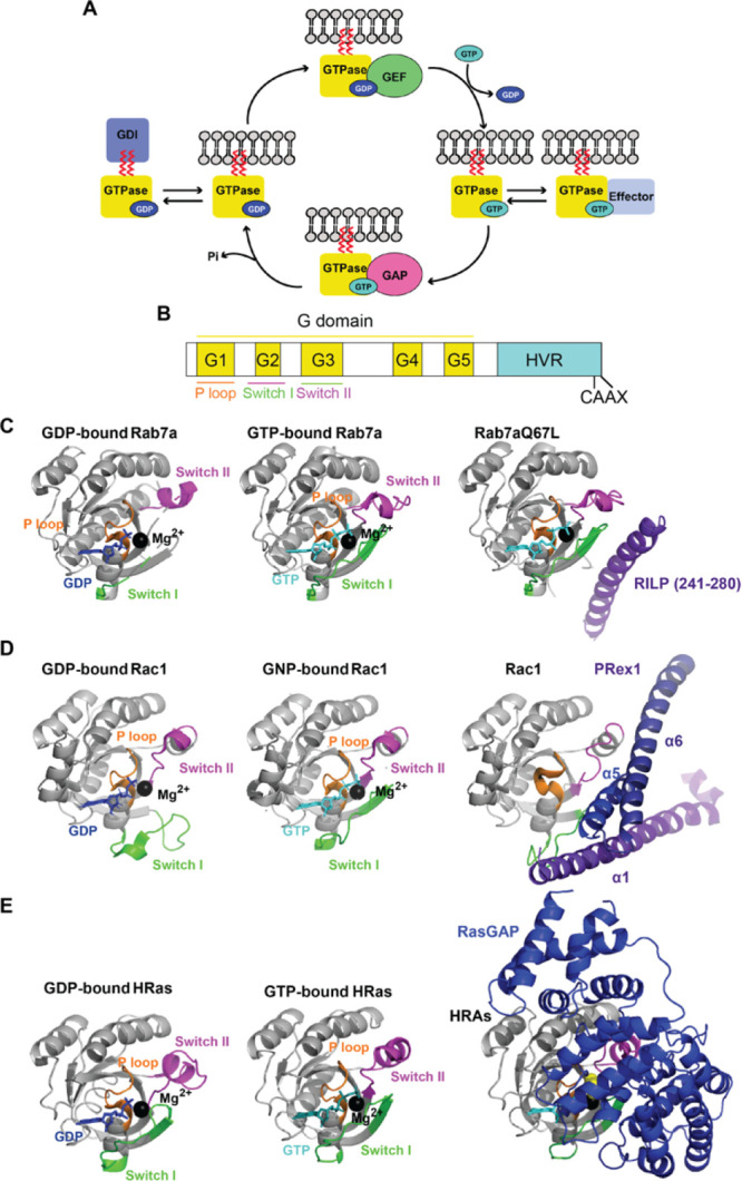
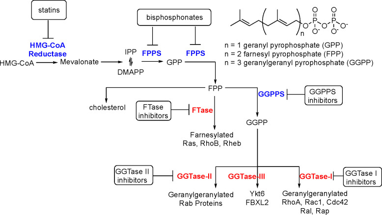
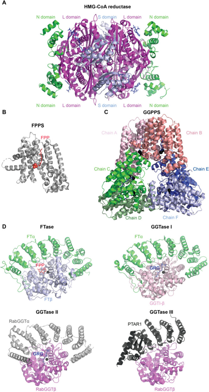
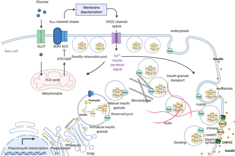
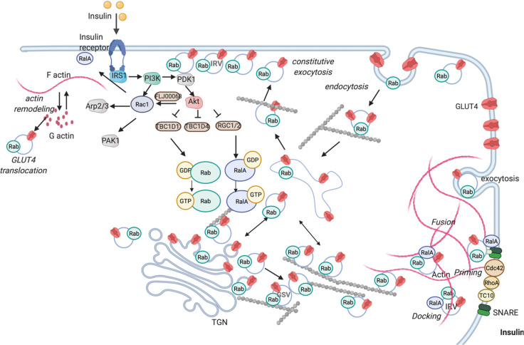
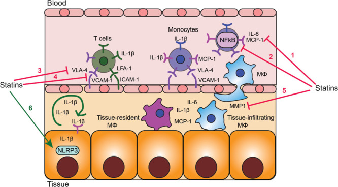
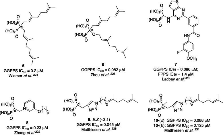
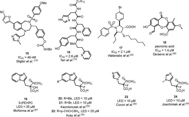
Verification
Claims the paper makes
The claims and statistics of this paper haven’t been checked yet.
Run the check — free, no account needed. It reads the open-access text and checks the sources it cites.
Claims other papers make about this one
Each is checked against this paper’s own text: does it say what the citing paper says it does?
The source backs part of the claim, not all of it — or backs it for a different population, condition or comparison.
In the citing paper
“All these small GTPase need post-translational modification to target them to cellular membranes and be active [16,17].”
From Brain Cholesterol Biosynthetic Pathway Is Altered in a Preclinical Model of Fragile X Syndrome (2022) · cites this paper as [17]
Experimental — accuracy not yet measured
Self-check
Percentages, sums and reported differences re-computed from the paper’s own numbers.
The self-check needs the paper’s full text.
Citations
Cites
Saeedi, P., Petersohn, I., Salpea, P., Malanda, B., Karuranga, S., Unwin, N., Colagiuri, S., Guariguata, L., Motala, A. A., Ogurtsova, K., et al. (2019) · Diabetes Research and Clinical Practice · doi:10.1016/j.diabres.2019.107843 · Cited by 10,792
Genomic analyses identify molecular subtypes of pancreatic cancer
Bailey, P., Chang, D. K., Nones, K., Johns, A. L., Patch, A.-M., Gingras, M.-C., Miller, D. K., Christ, A. N., Bruxner, T. J. C., Quinn, M. C., et al. (2016) · Nature · doi:10.1038/nature16965 · Cited by 3,621
Exercise, GLUT4, and skeletal muscle glucose uptake
Richter, E. A., & Hargreaves, M. (2013) · Physiological Reviews · doi:10.1152/physrev.00038.2012 · Cited by 1,407
The integrative biology of type 2 diabetes
Roden, M., & Shulman, G. I. (2019) · Nature · doi:10.1038/s41586-019-1797-8 · Cited by 1,167
RAS-targeted therapies: is the undruggable drugged?
Moore, A. R., Rosenberg, S. C., McCormick, F., & Malek, S. (2020) · Nature Reviews Drug Discovery · doi:10.1038/s41573-020-0068-6 · Cited by 1,103
Rational design and characterization of a Rac GTPase-specific small molecule inhibitor
Gao, Y., Dickerson, J. B., Guo, F., Zheng, J., & Zheng, Y. (2004) · Proceedings of the National Academy of Sciences · doi:10.1073/pnas.0307512101 · Cited by 1,091
Pharmacological Properties of Y-27632, a Specific Inhibitor of Rho-Associated Kinases
Ishizaki, T., Uehata, M., Tamechika, I., Keel, J., Nonomura, K., Maekawa, M., & Narumiya, S. (2000) · Molecular Pharmacology · doi:10.1016/s0026-895x(24)26508-1 · Cited by 940
Dunford, J. E., Thompson, K., Coxon, F. P., Luckman, S. P., Hahn, F. M., Poulter, C. D., Ebetino, F. H., & Rogers, M. J. (2001) · Journal of Pharmacology and Experimental Therapeutics · doi:10.1016/s0022-3565(24)38786-5 · Cited by 900
The promise and peril of chemical probes
Arrowsmith, C. H., Audia, J. E., Austin, C., Baell, J., Bennett, J., Blagg, J., Bountra, C., Brennan, P. E., Brown, P. J., Bunnage, M. E., et al. (2015) · Nature Chemical Biology · doi:10.1038/nchembio.1867 · Cited by 839
The expanding role of prodrugs in contemporary drug design and development
Rautio, J., Meanwell, N. A., Di, L., & Hageman, M. J. (2018) · Nature Reviews Drug Discovery · doi:10.1038/nrd.2018.46 · Cited by 781
Targeting inflammation in the treatment of type 2 diabetes: time to start
Donath, M. Y. (2014) · Nature Reviews Drug Discovery · doi:10.1038/nrd4275 · Cited by 690
The molecular mechanism of nitrogen-containing bisphosphonates as antiosteoporosis drugs
Kavanagh, K. L., Guo, K., Dunford, J. E., Wu, X., Knapp, S., Ebetino, F. H., Rogers, M. J., Russell, R. G. G., & Oppermann, U. C. T. (2006) · Proceedings of the National Academy of Sciences · doi:10.1073/pnas.0601643103 · Cited by 549
Rab family of small GTPases: an updated view on their regulation and functions
Homma, Y., Hiragi, S., & Fukuda, M. (2020) · FEBS Journal · doi:10.1111/febs.15453 · Cited by 482
10. Cardiovascular Disease and Risk Management: Standards of Medical Care in Diabetes—2019
American Diabetes Association (2019) · Diabetes Care · doi:10.2337/dc19-s010 · Cited by 471
Targeted Degradation of Oncogenic KRAS G12C by VHL-Recruiting PROTACs
Bond, M. J., Chu, L., Nalawansha, D. A., Li, K., & Crews, C. M. (2020) · ACS Central Science · doi:10.1021/acscentsci.0c00411 · Cited by 450
Mîinea, C. P., Sano, H., Kane, S., Sano, E., Fukuda, M., Peränen, J., Lane, W. S., & Lienhard, G. E. (2005) · Biochemical Journal · doi:10.1042/bj20050887 · Cited by 449
Essential role of Epac2/Rap1 signaling in regulation of insulin granule dynamics by cAMP
Shibasaki, T., Takahashi, H., Miki, T., Sunaga, Y., Matsumura, K., Yamanaka, M., Zhang, C., Tamamoto, A., Satoh, T., Miyazaki, J.-I., et al. (2007) · Proceedings of the National Academy of Sciences · doi:10.1073/pnas.0707054104 · Cited by 425
Alendronate Is a Specific, Nanomolar Inhibitor of Farnesyl Diphosphate Synthase
Bergstrom, J. D., Bostedor, R. G., Masarachia, P. J., Reszka, A. A., & Rodan, G. A. (2000) · Archives of Biochemistry and Biophysics · doi:10.1006/abbi.1999.1502 · Cited by 411
Liu, M., Bryant, M., Chen, J., Lee, S., Yaremko, B., Lipari, P., Malkowski, M. G., Ferrari, E., Nielsen, L. L., Prioli, N. A., et al. (1998) · PubMed · Cited by 309
Rad: a Member of the Ras Family Overexpressed in Muscle of Type II Diabetic Humans
Reynet, C., & Kahn, C. R. (1993) · Science · doi:10.1126/science.8248782 · Cited by 300
Update on GLUT4 Vesicle Traffic: A Cornerstone of Insulin Action
Jaldín‐Fincati, J. R., Pavarotti, M. A., Frendo‐Cumbo, S., Bilan, P. J., & Klip, A. (2017) · Trends in Endocrinology and Metabolism · doi:10.1016/j.tem.2017.05.002 · Cited by 287
Protein Prenylation: Enzymes, Therapeutics, and Biotechnology Applications
Palsuledesai, C. C., & Distefano, M. D. (2014) · ACS Chemical Biology · doi:10.1021/cb500791f · Cited by 263
The Mevalonate Pathway Is a Druggable Target for Vaccine Adjuvant Discovery
Xia, Y., Xie, Y., Yu, Z., Xiao, H., Jiang, G., Zhou, X., Yang, Y., Li, X., Zhao, M., Li, L., et al. (2018) · Cell · doi:10.1016/j.cell.2018.08.070 · Cited by 259
Rational Design of Small Molecule Inhibitors Targeting RhoA Subfamily Rho GTPases
Shang, X., Marchioni, F., Sipes, N. S., Evelyn, C. R., Jerabek‐Willemsen, M., Duhr, S., Seibel, W. L., Wortman, M. D., & Zheng, Y. (2012) · Chemistry & Biology · doi:10.1016/j.chembiol.2012.05.009 · Cited by 242
Molecular control of Rab activity by GEFs, GAPs and GDI
Müller, M. P., & Goody, R. S. (2017) · Small GTPases · doi:10.1080/21541248.2016.1276999 · Cited by 242
Targeting Rac and Cdc42 GTPases in Cancer
Maldonado, M. D. M., & Dharmawardhane, S. (2018) · Cancer Research · doi:10.1158/0008-5472.can-18-0619 · Cited by 242
The Rab27a/Granuphilin Complex Regulates the Exocytosis of Insulin-Containing Dense-Core Granules
Yi, Z., Yokota, H., Torii, S., Aoki, T., Hosaka, M., Zhao, S., Takata, K., Takeuchi, T., & Izumi, T. (2002) · Molecular and Cellular Biology · doi:10.1128/mcb.22.6.1858-1867.2002 · Cited by 228
Control of the innate immune response by the mevalonate pathway
Akula, M. K., Shi, M., Jiang, Z., Foster, C. E., Miao, D., Li, A. S., Zhang, X., Gavin, R. M., Forde, S., Germain, G., et al. (2016) · Nature Immunology · doi:10.1038/ni.3487 · Cited by 215
JeBailey, L., Wanono, O., Niu, W., Roessler, J., Rudich, A., & Klip, A. (2007) · Diabetes · doi:10.2337/db06-0823 · Cited by 211
Sun, J., Blaskovich, M. A., Knowles, D. B., Qian, Y., Ohkanda, J., Bailey, R. D., Hamilton, A. D., & Sebti, S. M. (1999) · PubMed · Cited by 210
Chen, X., Leto, D. E., Chiang, S.-H., Wang, Q., & Saltiel, A. R. (2007) · Developmental Cell · doi:10.1016/j.devcel.2007.07.007 · Cited by 203
Usharani, P., Mateen, A., Naidu, M. U. R., Raju, Y. S. N., & Chandra, N. (2008) · Drugs in R&D · doi:10.2165/00126839-200809040-00004 · Cited by 200
Rab8A and Rab13 are activated by insulin and regulate GLUT4 translocation in muscle cells
Sun, Y., Bilan, P. J., Liu, Z., & Klip, A. (2010) · Proceedings of the National Academy of Sciences · doi:10.1073/pnas.1009523107 · Cited by 197
Rogers, M. J., Mönkkönen, J., & Munoz, M. A. (2020) · Bone · doi:10.1016/j.bone.2020.115493 · Cited by 194
GLUT4 Retention in Adipocytes Requires Two Intracellular Insulin-regulated Transport Steps
Zeigerer, A., Lampson, M. A., Karylowski, O., Sabatini, D. D., Adesnik, M., Ren, M., & McGraw, T. E. (2002) · Molecular Biology of the Cell · doi:10.1091/mbc.e02-02-0071 · Cited by 183
Liver governs adipose remodelling via extracellular vesicles in response to lipid overload
Zhao, Y., Zhao, M., Jiang, S., Wu, J., Liu, J., Yuan, X., Shen, D., Zhang, J., Zhou, N., He, J., et al. (2020) · Nature Communications · doi:10.1038/s41467-020-14450-6 · Cited by 181
Zhang, Y., Cao, R., Yin, F., Hudock, M. P., Guo, R., Krysiak, K., Mukherjee, S., Gao, Y., Robinson, H., Song, Y., et al. (2009) · Journal of the American Chemical Society · doi:10.1021/ja808285e · Cited by 179
Development of specific Rho-kinase inhibitors and their clinical application
Tamura, M., Nakao, H., Yoshizaki, H., Shiratsuchi, M., Shigyo, H., Yamada, H., Ozawa, T., Totsuka, J., & Hidaka, H. (2005) · Biochimica et Biophysica Acta (BBA) - Proteins and Proteomics · doi:10.1016/j.bbapap.2005.06.015 · Cited by 178
Rab10 and myosin-Va mediate insulin-stimulated GLUT4 storage vesicle translocation in adipocytes
Chen, Y., Wang, Y., Zhang, J., Deng, Y., Jiang, L., Song, E., Wu, X., Hammer, J. A., Xu, T., & Lippincott‐Schwartz, J. (2012) · The Journal of Cell Biology · doi:10.1083/jcb.201111091 · Cited by 177
Jiang, S.-Y., Li, H., Tang, J.-J., Wang, J., Luo, J., Liu, B., Wang, J.-K., Shi, X.-J., Cui, H.-W., Tang, J., et al. (2018) · Nature Communications · doi:10.1038/s41467-018-07590-3 · Cited by 173
Pleiotropic effects of statins: A focus on cancer
Ahmadi, M., Amiri, S., Pecic, S., Machaj, F., Rosik, J., Łoś, M., Alizadeh, J., Mahdian, R., Rosa, S. C. D. S., Schaafsma, D., et al. (2020) · Biochimica et Biophysica Acta (BBA) - Molecular Basis of Disease · doi:10.1016/j.bbadis.2020.165968 · Cited by 167
Galicia-García, U., Jebari‐Benslaiman, S., Larrea, A., Uribe, K. B., Siddiqi, H. F., Ostolaza, H., Benito‐Vicente, A., & Martín, C. (2020) · International Journal of Molecular Sciences · doi:10.3390/ijms21134725 · Cited by 165
Vogt, A., Qian, Y., McGuire, T. F., Hamilton, A. D., & Sebti, S. M. (1996) · PubMed · Cited by 164
Glucose-stimulated Cdc42 Signaling Is Essential for the Second Phase of Insulin Secretion
Wang, Z., Oh, E., & Thurmond, D. C. (2007) · Journal of Biological Chemistry · doi:10.1074/jbc.m610553200 · Cited by 159
Ishikura, S., Bilan, P. J., & Klip, A. (2006) · Biochemical and Biophysical Research Communications · doi:10.1016/j.bbrc.2006.12.140 · Cited by 158
Jeong, A., Suazo, K. F., Wood, W. G., Distefano, M. D., & Li, L. (2018) · Critical Reviews in Biochemistry and Molecular Biology · doi:10.1080/10409238.2018.1458070 · Cited by 152
Kowluru, R. A., Kowluru, A., Veluthakal, R., Mohammad, G., Syed, I., dos Santos, J. M., & Mishra, M. (2014) · Diabetologia · doi:10.1007/s00125-014-3194-z · Cited by 151
Jiang, Z. Y., Chawla, A., Bose, A., Way, M., & Czech, M. (2002) · Journal of Biological Chemistry · doi:10.1074/jbc.m108280200 · Cited by 150
HDL and LDL cholesterol significantly influence β-cell function in type 2 diabetes mellitus
Kruit, J. K., Brunham, L. R., Verchere, C. B., & Hayden, M. R. (2010) · Current Opinion in Lipidology · doi:10.1097/mol.0b013e328339387b · Cited by 148
Targeting the Small GTPase Superfamily through Their Regulatory Proteins
Gray, J. L., von Delft, F., & Brennan, P. E. (2019) · Angewandte Chemie International Edition · doi:10.1002/anie.201900585 · Cited by 148
Sylow, L., Kleinert, M., Pehmøller, C. K., Prats, C., Chiu, T. T., Klip, A., Richter, E. A., & Jensen, T. E. (2013) · Cellular Signalling · doi:10.1016/j.cellsig.2013.11.007 · Cited by 147
Muscle cells engage Rab8A and myosin Vb in insulin-dependent GLUT4 translocation
Ishikura, S., & Klip, A. (2008) · American Journal of Physiology-Cell Physiology · doi:10.1152/ajpcell.00277.2008 · Cited by 139
Insulin granule biogenesis and exocytosis
Omar‐Hmeadi, M., & Idevall‐Hagren, O. (2020) · Cellular and Molecular Life Sciences · doi:10.1007/s00018-020-03688-4 · Cited by 137
Hunt, J. T., Ding, C. Z., Batorsky, R. S., Bednarz, M. S., Bhide, R. S., Cho, Y. H., Chong, S., Chao, S. T., Gullo-Brown, J. L., Guo, P., et al. (2000) · Journal of Medicinal Chemistry · doi:10.1021/jm000248z · Cited by 135
Wang, Z., Oh, E., Clapp, D. W., Chernoff, J., & Thurmond, D. C. (2011) · Journal of Biological Chemistry · doi:10.1074/jbc.m111.291500 · Cited by 134
Rab18 Dynamics in Adipocytes in Relation to Lipogenesis, Lipolysis and Obesity
Pulido, M. R., Díaz‐Ruiz, A., Jiménez, Y., García‐Navarro, S., Gracia‐Navarro, F., Tinahones, F. J., López‐Miranda, J., Frühbeck, G., Vázquez‐Martínez, R., & Malagón, M. M. (2011) · PLoS ONE · doi:10.1371/journal.pone.0022931 · Cited by 133
Glucose transporters in pancreatic islets
Berger, C., & Zdzieblo, D. (2020) · Pflügers Archiv - European Journal of Physiology · doi:10.1007/s00424-020-02383-4 · Cited by 133
Allosteric non-bisphosphonate FPPS inhibitors identified by fragment-based discovery
Jahnke, W., Rondeau, J., Cotesta, S., Marzinzik, A. L., Pellé, X., Geiser, M., Strauss, A., Götte, M., Bitsch, F., Hemmig, R., et al. (2010) · Nature Chemical Biology · doi:10.1038/nchembio.421 · Cited by 129
Pancreatic β-Cell Protein Granuphilin Binds Rab3 and Munc-18 and Controls Exocytosis
Coppola, T., Frantz, C., Perret-Menoud, V., Gattesco, S., Hirling, H., & Regazzi, R. (2002) · Molecular Biology of the Cell · doi:10.1091/mbc.02-02-0025 · Cited by 127
The Effect of Sustained Inflammation on Hepatic Mevalonate Pathway Results in Hyperglycemia
Okin, D. A., & Medzhitov, R. M. (2016) · Cell · doi:10.1016/j.cell.2016.02.023 · Cited by 126
Lobell, R. B., Liu, D., Buser, C. A., Davide, J. P., DePuy, E., Hamilton, K., Koblan, K. S., Lee, Y., Mosser, S. D., Motzel, S. L., et al. (2002) · PubMed · Cited by 121
A Not-So-Ancient Grease History: Click Chemistry and Protein Lipid Modifications
Suazo, K. F., Park, K., & Distefano, M. D. (2021) · Chemical Reviews · doi:10.1021/acs.chemrev.0c01108 · Cited by 120
Overexpression of Rad Inhibits Glucose Uptake in Cultured Muscle and Fat Cells
Moyers, J. S., Bilan, P. J., Reynet, C., & Kahn, C. R. (1996) · Journal of Biological Chemistry · doi:10.1074/jbc.271.38.23111 · Cited by 119
Sylow, L., Nielsen, I. L., Kleinert, M., Møller, L. L. V., Ploug, T., Schjerling, P., Bilan, P. J., Klip, A., Jensen, T. E., & Richter, E. A. (2016) · The Journal of Physiology · doi:10.1113/jp272039 · Cited by 114
Rim2α Determines Docking and Priming States in Insulin Granule Exocytosis
Yasuda, T., Shibasaki, T., Minami, K., Takahashi, H., Mizoguchi, A., Uriu, Y., Numata, T., Mori, Y., Miyazaki, J.-I., Miki, T., et al. (2010) · Cell Metabolism · doi:10.1016/j.cmet.2010.05.017 · Cited by 110
GGTase3 is a newly identified geranylgeranyltransferase targeting a ubiquitin ligase
Kuchay, S., Wang, H., Marzio, A., Jain, K., Homer, H., Fehrenbacher, N., Philips, M. R., Zheng, N., & Pagano, M. (2019) · Nature Structural & Molecular Biology · doi:10.1038/s41594-019-0249-3 · Cited by 109
Nevins, A. K., & Thurmond, D. C. (2006) · Journal of Biological Chemistry · doi:10.1074/jbc.m603604200 · Cited by 108
A Ral GAP complex links PI 3-kinase/Akt signaling to RalA activation in insulin action
Chen, X.-W., Leto, D., Xiong, T., Yu, G., Cheng, A., Decker, S., & Saltiel, A. R. (2011) · Molecular Biology of the Cell · doi:10.1091/mbc.e10-08-0665 · Cited by 108
Rho‐kinase as a molecular target for insulin resistance and hypertension
Kanda, T., Wakino, S., Homma, K., Yoshioka, K., Tatematsu, S., Hasegawa, K., Takamatsu, I., Sugano, N., Hayashi, K., & Saruta, T. (2005) · The FASEB Journal · doi:10.1096/fj.05-4197fje · Cited by 107
Chang, L. C., Adams, R. D., & Saltiel, A. R. (2002) · Proceedings of the National Academy of Sciences · doi:10.1073/pnas.202495599 · Cited by 107
Kikuchi, Y., Yamada, M., Imakiire, T., Kushiyama, T., Higashi, K., Hyodo, N., Yamamoto, K., Oda, T., Suzuki, S., & Miura, S. (2007) · Journal of Endocrinology · doi:10.1677/joe-06-0045 · Cited by 106
Chiu, T. T., Patel, N., Shaw, A. E., Bamburg, J. R., & Klip, A. (2010) · Molecular Biology of the Cell · doi:10.1091/mbc.e10-04-0316 · Cited by 105
Characterization of a Dual Rac/Cdc42 Inhibitor MBQ-167 in Metastatic Cancer
Humphries‐Bickley, T., Castillo‐Pichardo, L., O'Farril, E. H., Borrero-García, L. D., Forestier-Román, I., Gerena, Y., Blanco, M., Rivera-Robles, M. J., Rodríguez-Medina, J. R., Cubano, L. A., et al. (2017) · Molecular Cancer Therapeutics · doi:10.1158/1535-7163.mct-16-0442 · Cited by 105
Asahara, S., Shibutani, Y., Teruyama, K., Inoue, H. Y., Kawada, Y., Etoh, H., Matsuda, T., Kimura-Koyanagi, M., Hashimoto, N., Sakahara, M., et al. (2013) · Diabetologia · doi:10.1007/s00125-013-2849-5 · Cited by 103
Nevins, A. K., & Thurmond, D. (2005) · Journal of Biological Chemistry · doi:10.1074/jbc.m409528200 · Cited by 101
Virtual Screening Approach for the Identification of New Rac1 Inhibitors
Ferri, N., Corsini, A., Bottino, P., Clerici, F., & Contini, A. (2009) · Journal of Medicinal Chemistry · doi:10.1021/jm8015987 · Cited by 101
Effect of acute physiological hyperinsulinemia on gene expression in human skeletal muscle in vivo
Coletta, D. K., Balas, B., Chávez, A. O., Baig, M. R., Abdul‐Ghani, M. A., Kashyap, S., Folli, F. B., Tripathy, D., Mandarino, L. J., Cornell, J. E., et al. (2008) · American Journal of Physiology-Endocrinology and Metabolism · doi:10.1152/ajpendo.00607.2007 · Cited by 97
Structure-guided discovery of a single-domain antibody agonist against human apelin receptor
Ma, Y., Ding, Y., Song, X., Ma, X., Li, X., Zhang, N., Song, Y., Sun, Y., Shen, Y., Zhong, W., et al. (2020) · Science Advances · doi:10.1126/sciadv.aax7379 · Cited by 94
Granuphilin Modulates the Exocytosis of Secretory Granules through Interaction with Syntaxin 1a
Torii, S., Zhao, S., Yi, Z., Takeuchi, T., & Izumi, T. (2002) · Molecular and Cellular Biology · doi:10.1128/mcb.22.15.5518-5526.2002 · Cited by 94
Shen, N., Yu, X., Pan, F., Gao, X., Xue, B., & Li, C. (2011) · Journal of Biological Chemistry · doi:10.1074/jbc.m110.190165 · Cited by 91
Direct inhibition of RAS: Quest for the Holy Grail?
Spencer-Smith, R., & O’Bryan, J. P. (2017) · Seminars in Cancer Biology · doi:10.1016/j.semcancer.2017.12.005 · Cited by 90
Usui, I., Imamura, T., Huang, J., Satoh, H., & Olefsky, J. M. (2003) · Journal of Biological Chemistry · doi:10.1074/jbc.m208904200 · Cited by 89
ROCKII inhibition promotes the maturation of human pancreatic beta-like cells
Ghazizadeh, Z., Kao, D.-I., Amin, S., Cook, B. D., Rao, S. D., Zhou, T., Zhang, T., Xiang, Z.-Y., Kenyon, R., Kaymakcalan, O. E., et al. (2017) · Nature Communications · doi:10.1038/s41467-017-00129-y · Cited by 88
Chen, M., Liu, A., Ouyang, Y., Huang, Y., Chao, X., & Pi, R. (2013) · Expert Opinion on Investigational Drugs · doi:10.1517/13543784.2013.778242 · Cited by 87
Takaguri, A., Satoh, K., Itagaki, M., Tokumitsu, Y., & Ichihara, K. (2008) · Journal of Pharmacological Sciences · doi:10.1254/jphs.fp0072403 · Cited by 87
Mitogenic action of insulin: friend, foe or ‘frenemy’?
Draznin, B. (2009) · Diabetologia · doi:10.1007/s00125-009-1558-6 · Cited by 87
Watanabe, M., Fiji, H. D. G., Guo, L., Chan, L. N., Kinderman, S. S., Slamon, D. J., Kwon, O., & Tamanoi, F. (2008) · Journal of Biological Chemistry · doi:10.1074/jbc.m706229200 · Cited by 86
Waller, D. D., Park, J., & Tsantrizos, Y. S. (2019) · Critical Reviews in Biochemistry and Molecular Biology · doi:10.1080/10409238.2019.1568964 · Cited by 86
Sakamoto, K., Masutani, T., & Hirokawa, T. (2020) · Scientific Reports · doi:10.1038/s41598-020-78712-5 · Cited by 85
Cardama, G. A., Comin, M. J., Hornos, L., González, N., Defelipe, L. A., Turjanski, A. G., Alonso, D. F., Gomez, D. E., & Menna, P. L. (2013) · Anti-Cancer Agents in Medicinal Chemistry · doi:10.2174/18715206113136660334 · Cited by 85
Zhu, L.-H., Wang, L., Wang, D., Jiang, H., Tang, Q., Yan, L., Bian, Z., Wang, X.-A., & Li, H. (2009) · Free Radical Biology and Medicine · doi:10.1016/j.freeradbiomed.2009.10.040 · Cited by 85
Ueda, S., Kataoka, T., & Satoh, T. (2008) · Biology of the Cell · doi:10.1042/bc20070160 · Cited by 84
Protease-Resistant and Cell-Permeable Double-Stapled Peptides Targeting the Rab8a GTPase
Cromm, P. M., Spiegel, J., Küchler, P., Dietrich, L., Kriegesmann, J., Wendt, M., Goody, R. S., Waldmann, H., & Grossmann, T. N. (2016) · ACS Chemical Biology · doi:10.1021/acschembio.6b00386 · Cited by 84
The Ras Superfamily of Small GTPases in Non-neoplastic Cerebral Diseases
Qu, L., Pan, C., He, S., Lang, B., Gao, G.-D., Wang, X., & Wang, Y. (2019) · Frontiers in Molecular Neuroscience · doi:10.3389/fnmol.2019.00121 · Cited by 84
Hudock, M. P., Zhang, Y., Guo, R., Cao, R., No, J. H., Liang, P., Ko, T., Chang, T.-H., Chang, S.-C., Song, Y., et al. (2008) · Journal of Medicinal Chemistry · doi:10.1021/jm800325y · Cited by 83
A SNARE geranylgeranyltransferase essential for the organization of the Golgi apparatus
Shirakawa, R., Goto‐Ito, S., Goto, K., Wakayama, S., Kubo, H., Sakata, N., Trinh, D. A., Yamagata, A., Sato, Y., Masumoto, H., et al. (2020) · The EMBO Journal · doi:10.15252/embj.2019104120 · Cited by 83
Rho GTPases—Emerging Regulators of Glucose Homeostasis and Metabolic Health
Møller, L. L. V., Klip, A., & Sylow, L. (2019) · Cells · doi:10.3390/cells8050434 · Cited by 82
Kazi, A., Xiang, S., Yang, H., Chen, L., Kennedy, P., Ayaz, M., Fletcher, S., Cummings, C., Lawrence, H. R., Beato, F., et al. (2019) · Clinical Cancer Research · doi:10.1158/1078-0432.ccr-18-3399 · Cited by 81
Sano, H., Peck, G. R., Kettenbach, A. N., Gerber, S. A., & Lienhard, G. E. (2011) · Journal of Biological Chemistry · doi:10.1074/jbc.c111.228908 · Cited by 81
Chun, K., Araki, K., Jee, Y., Lee, D.-H., Oh, B., Hu, H., Park, K. S., Lee, S. W., Zabolotny, J. M., & Kim, Y. (2012) · Endocrinology · doi:10.1210/en.2011-1036 · Cited by 81
Myosin Va mediates Rab8A-regulated GLUT4 vesicle exocytosis in insulin-stimulated muscle cells
Sun, Y., Chiu, T. T., Foley, K. P., Bilan, P. J., & Klip, A. (2014) · Molecular Biology of the Cell · doi:10.1091/mbc.e13-08-0493 · Cited by 81
Therapeutic medications against diabetes: What we have and what we expect
Hu, C., & Jia, W. (2018) · Advanced Drug Delivery Reviews · doi:10.1016/j.addr.2018.11.008 · Cited by 81
Digeranyl bisphosphonate inhibits geranylgeranyl pyrophosphate synthase
Wiemer, A. J., Tong, H., Swanson, K. M., & Hohl, R. J. (2006) · Biochemical and Biophysical Research Communications · doi:10.1016/j.bbrc.2006.12.094 · Cited by 81
Balamatsias, D., Kong, A. M., Waters, J. E., Sriratana, A., Gurung, R., Bailey, C. G., Rasko, J. E. J., Tiganis, T., Macaulay, S. L., & Mitchell, C. A. (2011) · Journal of Biological Chemistry · doi:10.1074/jbc.m111.306621 · Cited by 80
Noc2 is essential in normal regulation of exocytosis in endocrine and exocrine cells
Matsumoto, M., Miki, T., Shibasaki, T., Kawaguchi, M., Shinozaki, H., Nio‐Kobayashi, J., Saraya, A., Koseki, H., Miyazaki, M., Iwanaga, T., et al. (2004) · Proceedings of the National Academy of Sciences · doi:10.1073/pnas.0306709101 · Cited by 79
Protein prenylation and human diseases: a balance of protein farnesylation and geranylgeranylation
Xu, N., Shen, N., Wang, X., Jiang, S., Xue, B., & Li, C. (2015) · Science China Life Sciences · doi:10.1007/s11427-015-4836-1 · Cited by 77
Syed, I., Jayaram, B., Subasinghe, W., & Kowluru, A. (2010) · Biochemical Pharmacology · doi:10.1016/j.bcp.2010.05.006 · Cited by 77
Goalstone, M. L., Carel, K. D., Leitner, J. W., & Draznin, B. (1997) · Endocrinology · doi:10.1210/endo.138.12.5621 · Cited by 76
Exocyst function regulated by effector phosphorylation
Chen, X.-W., Leto, D., Xiao, J., Goss, J., Wang, Q., Shavit, J. A., Xiong, T., Yu, G., Ginsburg, D., Toomre, D., et al. (2011) · Nature Cell Biology · doi:10.1038/ncb2226 · Cited by 76
Peterson, Y. K., Kelly, P., Weinbaum, C., & Casey, P. J. (2006) · Journal of Biological Chemistry · doi:10.1074/jbc.m600168200 · Cited by 74
A role for Rab14 in the endocytic trafficking of GLUT4 in 3T3-L1 adipocytes
Reed, S. E., Hodgson, L. R., Song, S., May, M., Mastick, C. C., Verkade, P., & Tavaré, J. M. (2013) · Journal of Cell Science · doi:10.1242/jcs.104307 · Cited by 73
Liu, J., Jiang, S., Zhao, Y., Sun, Q., Zhang, J., Shen, D., Wu, J., Shen, N., Fu, X., Sun, X., et al. (2018) · The Journal of Pathology · doi:10.1002/path.5131 · Cited by 72
Explaining statin inhibition effectiveness of HMG-CoA reductase by quantum biochemistry computations
da COSTA, R. F., Freire, V. N., BEZERRA, E. M., Cavada, B. S., Caetano, E. W. S., Filho, J. L. D. L., & Albuquerque, E. L. (2011) · Physical Chemistry Chemical Physics · doi:10.1039/c1cp22824b · Cited by 72
Malmgren, S., Spégel, P., Danielsson, A. P. H., Nagorny, C. L., Andersson, L. E., Nitert, M. D., Ridderstråle, M., Mulder, H., & Ling, C. (2013) · Journal of Biological Chemistry · doi:10.1074/jbc.m112.422527 · Cited by 71
Kalwat, M. A., Yoder, S. M., Wang, Z.-X., & Thurmond, D. (2012) · Biochemical Pharmacology · doi:10.1016/j.bcp.2012.12.003 · Cited by 71
Sadacca, L. A., Bruno, J., Wen, J., Xiong, W., & McGraw, T. E. (2013) · Molecular Biology of the Cell · doi:10.1091/mbc.e13-02-0103 · Cited by 71
Krysiak, R. G., Gdula‐Dymek, A., & Okopień, B. (2011) · The American Journal of Cardiology · doi:10.1016/j.amjcard.2010.11.023 · Cited by 70
ICER induced by hyperglycemia represses the expression of genes essential for insulin exocytosis
Abderrahmani, A., Cheviet, S., Ferdaoussi, M., Coppola, T., Waeber, G., & Regazzi, R. (2006) · The EMBO Journal · doi:10.1038/sj.emboj.7601008 · Cited by 70
Rab11 and its effector Rip11 participate in regulation of insulin granule exocytosis
Sugawara, K., Shibasaki, T., Mizoguchi, A., Saito, T., & Seino, S. (2009) · Genes to Cells · doi:10.1111/j.1365-2443.2009.01285.x · Cited by 69
Subasinghe, W., Syed, I., & Kowluru, A. (2010) · American Journal of Physiology-Regulatory, Integrative and Comparative Physiology · doi:10.1152/ajpregu.00421.2010 · Cited by 69
RABIF/MSS4 is a Rab-stabilizing holdase chaperone required for GLUT4 exocytosis
Gulbranson, D. R., Davis, E. M., Demmitt, B. A., Ouyang, Y., Ye, Y., Yu, H., & Shen, J. (2017) · Proceedings of the National Academy of Sciences · doi:10.1073/pnas.1712176114 · Cited by 67
Effect of Insulin on Farnesyltransferase Activity in 3T3-L1 Adipocytes
Goalstone, M. L., & Draznin, B. (1996) · Journal of Biological Chemistry · doi:10.1074/jbc.271.44.27585 · Cited by 67
A Link between Cdc42 and Syntaxin Is Involved in Mastoparan-Stimulated Insulin Release
Daniel, S., Noda, M., Cerione, R. A., & Sharp, G. W. G. (2002) · Biochemistry · doi:10.1021/bi025604p · Cited by 66
SEC16A is a RAB10 effector required for insulin-stimulated GLUT4 trafficking in adipocytes
Bruno, J., Brumfield, A., Chaudhary, N., Iaea, D. B., & McGraw, T. E. (2016) · The Journal of Cell Biology · doi:10.1083/jcb.201509052 · Cited by 65
Novel Activities of Select NSAID R-Enantiomers against Rac1 and Cdc42 GTPases
Oprea, T. I., Sklar, L. A., Agola, J. O., Guo, Y., Silberberg, M., Roxby, J., Vestling, A., Romero, E., Surviladze, Z., Murray-Krezan, C. M., et al. (2015) · PLoS ONE · doi:10.1371/journal.pone.0142182 · Cited by 63
Veluthakal, R., Suresh, M. V., McDonald, P., Olson, L. K., & Kowluru, A. (2008) · Biochemical Pharmacology · doi:10.1016/j.bcp.2008.09.021 · Cited by 62
Li, H., Peng, W., Jian, W., Li, Y., Li, Q., Li, W., & Xu, Y. (2012) · Cardiovascular Diabetology · doi:10.1186/1475-2840-11-65 · Cited by 62
A complex of Rab13 with MICAL-L2 and α-actinin-4 is essential for insulin-dependent GLUT4 exocytosis
Sun, Y., Jaldín‐Fincati, J. R., Liu, Z., Bilan, P. J., & Klip, A. (2015) · Molecular Biology of the Cell · doi:10.1091/mbc.e15-05-0319 · Cited by 61
RhoA/Rho kinase pathway contributes to the pathogenesis of thermal hyperalgesia in diabetic mice
Ohsawa, M., Aasato, M., Hayashi, S.-S., & Kamei, J. (2011) · Pain · doi:10.1016/j.pain.2010.10.005 · Cited by 61
Different effects of statins on induction of diabetes mellitus: an experimental study
Zhao, S., & Wang, Z. (2015) · Drug Design Development and Therapy · doi:10.2147/dddt.s87979 · Cited by 60
Stegman, B., Puri, R., Cho, L., Shao, M., Ballantyne, C. M., Barter, P. J., Chapman, M. J., Erbel, R. M., Libby, P., Raichlen, J. S., et al. (2014) · Diabetes Care · doi:10.2337/dc14-1121 · Cited by 59
Jiang, S., Shen, D., Jia, W., Han, X., Shen, N., Tao, W., Gao, X., Xue, B., & Li, C. (2015) · The Journal of Pathology · doi:10.1002/path.4652 · Cited by 57
Deraeve, C., Guo, Z., Bon, R. S., Blankenfeldt, W., DiLucrezia, R., Wolf, A., Menninger, S., Stigter, E. A., Wetzel, S., Choidas, A., et al. (2012) · Journal of the American Chemical Society · doi:10.1021/ja211305j · Cited by 56
McKenna, C. E., Kashemirov, B. A., Błażewska, K. M., Mallard, I., Stewart, C. A., Rojas, J., Lundy, M. W., Ebetino, F. H., Baron, R., Dunford, J. E., et al. (2010) · Journal of Medicinal Chemistry · doi:10.1021/jm900232u · Cited by 56
Self-citationEfficient drug and gene delivery to liver fibrosis: rationale, recent advances, and perspectives
Mahdinloo, S., Kiaie, S. H., Amiri, A., Hemmati, S., Valizadeh, H., & Zakeri‐Milani, P. (2020) · Acta Pharmaceutica Sinica B · doi:10.1016/j.apsb.2020.03.007 · Cited by 54
Uenishi, E., Shibasaki, T., Takahashi, H., Seki, C., Hamaguchi, H., Yasuda, T., Tatebe, M., Oiso, Y., Takenawa, T., & Seino, S. (2013) · Journal of Biological Chemistry · doi:10.1074/jbc.m113.464420 · Cited by 54
Guzmán‐Ruiz, R., Tercero‐Alcázar, C., Rabanal‐Ruiz, Y., Díaz‐Ruiz, A., Bekay, R. E., Rangel-Zúñiga, O. A., Navarro‐Ruiz, M. C., Molero, L., Membrives, A., Ruiz-Rabelo, J. F., et al. (2020) · The FASEB Journal · doi:10.1096/fj.201902703r · Cited by 54
Hammar, E. B., Tomás, A., Bosco, D., & Halban, P. A. (2008) · Endocrinology · doi:10.1210/en.2008-1135 · Cited by 54
Zverina, E. A., Lamphear, C. L., Wright, E. N., & Fierke, C. A. (2012) · Current Opinion in Chemical Biology · doi:10.1016/j.cbpa.2012.10.015 · Cited by 53
TBC1D13 is a RAB35 Specific GAP that Plays an Important Role in GLUT4 Trafficking in Adipocytes
Davey, J. R., Humphrey, S. J., Junutula, J. R., Mishra, A. K., Lambright, D. G., James, D. E., & Stöckli, J. (2012) · Traffic · doi:10.1111/j.1600-0854.2012.01397.x · Cited by 53
In vivo activation of ROCK1 by insulin is impaired in skeletal muscle of humans with type 2 diabetes
Chun, K., Choi, K., Lee, D.-H., Jung, Y., Henry, R. R., Ciaraldi, T. P., & Kim, Y. (2010) · American Journal of Physiology-Endocrinology and Metabolism · doi:10.1152/ajpendo.00538.2010 · Cited by 53
Riad, A., Du, J., Stiehl, S., Westermann, D., Mohr, Z., Sobirey, M., Doehner, W., Adams, V., Pauschinger, M., Schultheiß, H., et al. (2007) · European Journal of Pharmacology · doi:10.1016/j.ejphar.2007.04.065 · Cited by 53
RalA controls glucose homeostasis by regulating glucose uptake in brown fat
Skorobogatko, Y., Dragan, M., Cordon, C., Reilly, S. M., Hung, C.-W., Xia, W., Zhao, P., Wallace, M., Lackey, D. E., Chen, X.-W., et al. (2018) · Proceedings of the National Academy of Sciences · doi:10.1073/pnas.1801050115 · Cited by 52
Kaddai, V., Gonzalez, T., Keslair, F., Grémeaux, T., Bonnafous, S., Gugenheim, J., Tran, A., Gual, P., MARCHAND‐BRUSTEL, Y. L., & Cormont, M. (2009) · PLoS ONE · doi:10.1371/journal.pone.0005257 · Cited by 52
Syed, I., Kyathanahalli, C., & Kowluru, A. (2011) · American Journal of Physiology-Regulatory, Integrative and Comparative Physiology · doi:10.1152/ajpregu.00786.2010 · Cited by 52
A mitochondrial proteome profile indicative of type 2 diabetes mellitus in skeletal muscles
Chae, S., Kim, S., Koo, Y. D., Lee, J. H., Kim, H., Ahn, B. Y., Ha, Y., Kim, Y., Jang, M. G., Koo, K., et al. (2018) · Experimental & Molecular Medicine · doi:10.1038/s12276-018-0154-6 · Cited by 51
The RalA GTPase Is a Central Regulator of Insulin Exocytosis from Pancreatic Islet Beta Cells
Lopez, J. A., Kwan, E. P., Xie, L., He, Y., James, D. E., & Gaisano, H. Y. (2008) · Journal of Biological Chemistry · doi:10.1074/jbc.m800321200 · Cited by 51
Molecular imaging of β-cells: diabetes and beyond
Wei, W., Ehlerding, E. B., Lan, X., Luo, Q., & Cai, W. (2018) · Advanced Drug Delivery Reviews · doi:10.1016/j.addr.2018.06.022 · Cited by 51
Mishra, M., Duraisamy, A. J., Bhattacharjee, S., & Kowluru, R. A. (2018) · Antioxidants and Redox Signaling · doi:10.1089/ars.2018.7542 · Cited by 51
Bisphosphonates and Glucose Homeostasis: A Population-Based, Retrospective Cohort Study
Toulis, K. A., Nirantharakumar, K., Ryan, R. P., Marshall, T. P., & Hemming, K. (2015) · The Journal of Clinical Endocrinology & Metabolism · doi:10.1210/jc.2014-3481 · Cited by 51
Jialal, I., Miguelino, E., Griffen, S. C., & Devaraj, S. (2007) · The Journal of Clinical Endocrinology & Metabolism · doi:10.1210/jc.2007-0453 · Cited by 50
Stapled peptide inhibitors of RAB25 target context-specific phenotypes in cancer
Mitra, S., Montgomery, J. E., Kolar, M. J., Li, G., Jeong, K. J., Peng, B., Verdine, G. L., Mills, G. B., & Moellering, R. E. (2017) · Nature Communications · doi:10.1038/s41467-017-00888-8 · Cited by 50
Human isoprenoid synthase enzymes as therapeutic targets
Park, J., Matralis, A. N., Berghuis, A. M., & Tsantrizos, Y. S. (2014) · Frontiers in Chemistry · doi:10.3389/fchem.2014.00050 · Cited by 50
Role of RalA downstream of Rac1 in insulin-dependent glucose uptake in muscle cells
Nozaki, S., Ueda, S., Takenaka, N., Kataoka, T., & Satoh, T. (2012) · Cellular Signalling · doi:10.1016/j.cellsig.2012.07.013 · Cited by 49
Techniques for measuring cellular zinc
Carpenter, M. C., Lo, M. N., & Palmer, A. E. (2016) · Archives of Biochemistry and Biophysics · doi:10.1016/j.abb.2016.08.018 · Cited by 49
Direct Interaction of Rab4 with Syntaxin 4
Lü, L., Omata, W., Kojima, I., & Shibata, H. (2001) · Journal of Biological Chemistry · doi:10.1074/jbc.m003883200 · Cited by 49
Kowluru, R. A., Kowluru, A., Chakrabarti, S., & Khan, Z. A. (2004) · Diabetes · doi:10.2337/diabetes.53.3.775 · Cited by 48
Karimifard, M., Aminorroaya, A., Kachuei, A., Salamat, M. R., Alijanvand, M. H., Yamini, S. A., Karimifar, M., Feizi, A., & Amini, M. (2018) · Journal of Diabetes Investigation · doi:10.1111/jdi.12944 · Cited by 48
Henriksbo, B. D., Tamrakar, A. K., Xu, J., Duggan, B. M., Cavallari, J. F., Phulka, J. S., Stämpfli, M. R., Ashkar, A. A., & Schertzer, J. D. (2019) · Diabetes · doi:10.2337/db18-0999 · Cited by 48
Lacbay, C. M., Waller, D. D., Park, J., Palou, M. G., Vincent, F., Huang, X. F., Ta, V., Berghuis, A. M., Sébag, M., & Tsantrizos, Y. S. (2018) · Journal of Medicinal Chemistry · doi:10.1021/acs.jmedchem.8b00886 · Cited by 47
Functional Regulation of an Oxidative Stress Mediator, Rac1, in Diabetic Retinopathy
Mohammad, G., Duraisamy, A. J., Kowluru, A., & Kowluru, R. A. (2019) · Molecular Neurobiology · doi:10.1007/s12035-019-01696-5 · Cited by 46
Implications of farnesyltransferase and its inhibitors as a promising strategy for cancer therapy
Klochkov, S. G., Евгеньевна, Н. М., Yarla, N. S., Parvathaneni, M., Sharma, B., Tarasov, V. V., Barreto, G. E., Бачурин, С. О., Ashraf, G. M., & Aliev, G. M. (2017) · Seminars in Cancer Biology · doi:10.1016/j.semcancer.2017.10.010 · Cited by 46
Petersen, N., Frimurer, T. M., Pedersen, M. T., Egerod, K. L., Albrechtsen, N. J. W., Holst, J. J., Grapin‐Botton, A., Jensen, K. B., & Schwartz, T. W. (2018) · Gastroenterology · doi:10.1053/j.gastro.2018.06.039 · Cited by 46
Hepatic Rab24 controls blood glucose homeostasis via improving mitochondrial plasticity
Seitz, S., Kwon, Y., Hartleben, G., Jülg, J., Sekar, R., Krahmer, N., Najafi, B., Loft, A., Gancheva, S., Stemmer, K., et al. (2019) · Nature Metabolism · doi:10.1038/s42255-019-0124-x · Cited by 46
Welsh, G. I., Leney, S. E., Lloyd‐Lewis, B., Wherlock, M. D., Lindsay, A. J., McCaffrey, M., & Tavaré, J. M. (2007) · Journal of Cell Science · doi:10.1242/jcs.007310 · Cited by 45
Metabolic regulation through the endosomal system
Gilleron, J., Gerdes, J. M., & Zeigerer, A. (2019) · Traffic · doi:10.1111/tra.12670 · Cited by 44
Ilany, J., Bilan, P. J., Kapur, S., Caldwell, J. S., Patti, M., Marette, A., & Kahn, C. R. (2006) · Proceedings of the National Academy of Sciences · doi:10.1073/pnas.0511246103 · Cited by 44
Omi, H., Okayama, N., Shimizu, M., Fukutomi, T., Imaeda, K., Okouchi, M., & Itoh, M. (2003) · Microvascular Research · doi:10.1016/s0026-2862(02)00033-x · Cited by 44
Bhuin, T., & Roy, J. K. (2015) · Europe PMC (PubMed Central) · Cited by 43
Effects of Simvastatin on Glucose Metabolism in Mouse MIN6 Cells
Zhou, J., Li, W., Xie, Q., Hou, Y., Zhan, S., Yang, X., Xu, X., Cai, J., & Huang, Z. (2014) · Journal of Diabetes Research · doi:10.1155/2014/376570 · Cited by 43
Sidarala, V., Veluthakal, R., Syeda, K., Vlaar, C. P., Newsholme, P., & Kowluru, A. (2015) · Biochemical Pharmacology · doi:10.1016/j.bcp.2015.04.001 · Cited by 43
Elumalai, S., Karunakaran, U., Lee, I. K., Moon, J. S., & Won, K. C. (2016) · Redox Biology · doi:10.1016/j.redox.2016.11.009 · Cited by 43
Zoledronate inhibits the proliferation, adhesion and migration of vascular smooth muscle cells
Wu, L., Zhu, L., Shi, W., Zhang, J., Ma, D., & Yu, B. (2008) · European Journal of Pharmacology · doi:10.1016/j.ejphar.2008.10.043 · Cited by 43
Ling-jia, G., Gao, Q., Ni, L.-S., Wang, M., & Shen, F. (2013) · Chemical and Pharmaceutical Bulletin · doi:10.1248/cpb.c13-00066 · Cited by 43
A Rab10:RalA G protein cascade regulates insulin-stimulated glucose uptake in adipocytes
Karunanithi, S., Xiong, T., Uhm, M., Leto, D. E., Sun, J., Chen, X., & Saltiel, A. R. (2014) · Molecular Biology of the Cell · doi:10.1091/mbc.e14-06-1060 · Cited by 42
Zhao, Y., Wu, T., Zhao, M., & Li, C. (2020) · Journal of Biological Chemistry · doi:10.1074/jbc.rev119.008897 · Cited by 42
RILP Restricts Insulin Secretion Through Mediating Lysosomal Degradation of Proinsulin
Zhou, Y., Liu, Z., Zhang, S., Zhuang, R., Liu, H., Liu, X., Qiu, X., Zhang, M., Zheng, Y., Li, L., et al. (2019) · Diabetes · doi:10.2337/db19-0086 · Cited by 42
Matsunaga, K., Taoka, M., Isobe, T., & Izumi, T. (2016) · Journal of Cell Science · doi:10.1242/jcs.195479 · Cited by 41
von Eynatten, M., Schneider, J. G., Hadziselimovic, S., Hamann, A., Bierhaus, A., Nawroth, P. P., & Dugi, K. A. (2005) · Diabetes Care · doi:10.2337/diacare.28.3.754 · Cited by 41
Veluthakal, R., Tunduguru, R., Arora, D. K., Sidarala, V., Syeda, K., Vlaar, C. P., Thurmond, D. C., & Kowluru, A. (2015) · Diabetologia · doi:10.1007/s00125-015-3707-4 · Cited by 41
Phosphorylation of Rab GTPases in the regulation of membrane trafficking
Waschbüsch, D., & Khan, A. R. (2020) · Traffic · doi:10.1111/tra.12765 · Cited by 40
Kadoglou, N. P. E., Sailer, N., Kapelouzou, A., Lampropoulos, S., Vitta, I., Kostakis, A. I., & Liapis, C. D. (2011) · Acta Diabetologica · doi:10.1007/s00592-011-0310-0 · Cited by 40
The GTPase Rab1 Is Required for NLRP3 Inflammasome Activation and Inflammatory Lung Injury
Zhang, Y., Wang, L., Lv, Y., Jiang, C., Wu, G., Dull, R. O., Minshall, R. D., Malik, A. B., & Hu, G. (2018) · The Journal of Immunology · doi:10.4049/jimmunol.1800777 · Cited by 40
Regulation of RabGAPs involved in insulin action
Mafakheri, S., Chadt, A., & Al‐Hasani, H. (2018) · Biochemical Society Transactions · doi:10.1042/bst20170479 · Cited by 39
Wang, H., Ishizaki, R., Xu, J. C., Kasai, K., Kobayashi, E., Gomi, H., & Izumi, T. (2012) · Molecular Biology of the Cell · doi:10.1091/mbc.e12-04-0265 · Cited by 39
Matthiesen, R. A., Varney, M. L., Xu, P. C., Rier, A. S., Wiemer, D. F., & Holstein, S. A. (2017) · Bioorganic & Medicinal Chemistry · doi:10.1016/j.bmc.2017.10.023 · Cited by 39
Takata, M., Urakaze, M., Temaru, R., Yamazaki, K., Nakamura, N., Nobata, Y., Kishida, M., Sato, A., & Kobayashi, M. (2001) · British Journal of Pharmacology · doi:10.1038/sj.bjp.0704305 · Cited by 39
Extra-skeletal effects of bisphosphonates
Panagiotakou, A., Yavropoulou, M. P., Ansari, N. N., Makras, P., Basdra, E. K., Papavassiliou, A. G., & Kassi, E. N. (2020) · Metabolism · doi:10.1016/j.metabol.2020.154264 · Cited by 38
Increased amounts of farnesylated p21Ras in tissues of hyperinsulinaemic animals
Goalstone, M. L., Wall, K., Leitner, J. W., Kurowski, T. G., Ruderman, N. B., Pan, S., Ivy, J. L., Moller, D. E., & Draznin, B. (1999) · Diabetologia · doi:10.1007/s001250051156 · Cited by 38
Distinct Actions of Rab3 and Rab27 GTPases on Late Stages of Exocytosis of Insulin
Cazares, V. A., Subramani, A., Saldate, J. J., Hoerauf, W. W., & Stuenkel, E. L. (2014) · Traffic · doi:10.1111/tra.12182 · Cited by 38
Statins activate the NLRP3 inflammasome and impair insulin signaling via p38 and mTOR
Henriksbo, B. D., Tamrakar, A. K., Phulka, J. S., Barra, N. G., & Schertzer, J. D. (2020) · American Journal of Physiology-Endocrinology and Metabolism · doi:10.1152/ajpendo.00125.2020 · Cited by 38
Alsaggar, M., & Liu, D. (2018) · Journal of drug targeting · doi:10.1080/1061186x.2018.1437919 · Cited by 38
Diabetic retinopathy and transcriptional regulation of a small molecular weight G-Protein, Rac1
Kowluru, R. A., Mishra, M., & Kumar, B. (2016) · Experimental Eye Research · doi:10.1016/j.exer.2016.04.014 · Cited by 38
Tao, W., Wu, J., Xie, B., Zhao, Y., Shen, N., Jiang, S., Wang, X., Xu, N., Chen, J., Chen, S., et al. (2015) · Journal of Biological Chemistry · doi:10.1074/jbc.m115.657742 · Cited by 37
Haney, S. L., Varney, M. L., Chhonker, Y. S., Shin, S. C., Mehla, K., Crawford, A. J., Smith, H. J., Smith, L. M., Murry, D. J., Hollingsworth, M. A., et al. (2019) · Oncogene · doi:10.1038/s41388-019-0794-6 · Cited by 37
The GTPase Rab37 Participates in the Control of Insulin Exocytosis
Ljubicic, S., Bezzi, P., Brajkovic, S., Nesca, V., Guay, C., Ohbayashi, N., Fukuda, M., Abderrhamani, A., & Regazzi, R. (2013) · PLoS ONE · doi:10.1371/journal.pone.0068255 · Cited by 37
Rac1 Modulates Endothelial Function and Platelet Aggregation in Diabetes Mellitus
Schiattarella, G. G., Carrizzo, A., Ilardi, F., Damato, A., Ambrosio, M., Madonna, M., Trimarco, V., Marino, M., Angelis, E. D., Settembrini, S., et al. (2018) · Journal of the American Heart Association · doi:10.1161/jaha.117.007322 · Cited by 37
Development of Selective, Potent RabGGTase Inhibitors
Stigter, E. A., Guo, Z., Bon, R. S., Wu, Y., Choidas, A., Wolf, A., Menninger, S., Waldmann, H., Blankenfeldt, W., & Goody, R. S. (2012) · Journal of Medicinal Chemistry · doi:10.1021/jm300624s · Cited by 37
Kong, X., Yan, D., Sun, J., Wu, X., Mulder, H., Hua, X., & Ma, X. (2014) · Endocrinology · doi:10.1210/en.2014-1314 · Cited by 37
Chen, J., Dai, M., & Wang, Y. (2014) · Evidence-based Complementary and Alternative Medicine · doi:10.1155/2014/484269 · Cited by 36
Research works printed but not linked: 10
- Chen F., 2017, Int. J. Clin. Exp. Med., 10, 13224
- Li J., 2012, Mol. Vision, 18, 1354
- Štulc T., 2012, Neuro Endocrinol. Lett., 33, 73
- Dunford J. E., 2001, J. Pharmacol. Exp. Ther., 296, 235
- Vogt A., 1996, Oncogene, 13, 1991
- Sun J., 1999, Cancer Res., 59, 4919
- Liu M., 1998, Cancer Res., 58, 4947
- Lobell R. B., 2002, Mol. Cancer Ther., 1, 747
- Ishizaki T., 2000, Mol. Pharmacol., 57, 976
- Bhuin T., 2015, Int. J. Mol. Cell. Med., 4, 1
Cited by
Sorted by citations
Brain Cholesterol Biosynthetic Pathway Is Altered in a Preclinical Model of Fragile X Syndrome
Parente, M., Tonini, C., Buzzelli, V., Carbone, E., Trezza, V., & Pallottini, V. (2022) · International Journal of Molecular Sciences · doi:10.3390/ijms23063408 · Cited by 11
“All these small GTPase need posttranslational modification to target them to cellular membranes and be active [16,17].”
Background
Metadata
- Authors
- Gendaszewska-Darmach, E., Garstka, M. A., & Błażewska, K. M.
- Affiliations
- Institute of Molecular and Industrial Biotechnology, Faculty of Biotechnology and Food Sciences, Lodz University of Technology, Stefanowskiego Street 4/10, 90-924 Łódź, Poland
- Second Affiliated Hospital of Xi'an Jiaotong University (CN)
- Lodz University of Technology (PL)
- Corresponding author
- Garstka, Malgorzata A.
- Błażewska, Katarzyna M.
- Venue
- Journal of Medicinal Chemistry
- Publisher
- American Chemical Society (ACS)
- Published
- 2021-07-08
- Volume
- 64
- Issue
- 14
- Pages
- 9677-9710
- ISSN
- 0022-2623 (ISSN-L)
- 1520-4804
- PMC
- PMC8389838
- OpenAlex
- W3181493345
- Cited by
- 35
- References
- 301
- Citations by year
- 2026: 6 · 2025: 4 · 2024: 5 · 2023: 6 · 2022: 12 · 2021: 2
- FWCI
- 0.66
- Type
- journal article
- Language
- en
- Open access
- hybrid
- doi.org
- Locations
- Journal of Medicinal Chemistry (publishedVersion, cc-by)
- PubMed Central (publishedVersion, cc-by)
- PubMed (publishedVersion)
- Funders
- Narodowe Centrum Nauki (2014/14/E/ST5/00491, 2018/31/B/NZ9/02433)
- Second Affiliated Hospital, Xiâ??an Jiaotong University (82668330, 82668428)
- Topics
- Life Sciences › Biochemistry, Genetics and Molecular Biology › Molecular Biology › Protein Kinase Regulation and GTPase Signaling
- Health Sciences › Medicine › Endocrinology, Diabetes and Metabolism › Pancreatic function and diabetes
- Life Sciences › Biochemistry, Genetics and Molecular Biology › Molecular Biology › Sphingolipid Metabolism and Signaling
- Keywords
- small GTPases, Ras superfamily, protein prenylation, farnesyltransferase, geranylgeranyltransferase, mevalonate pathway, type 2 diabetes, pancreatic beta cells, glucose homeostasis, hyperglycemia, chronic inflammation, GTPase inhibitors, therapeutic target
- MeSH terms
- Animals, Diabetes Mellitus, Type 2, Dose-Response Relationship, Drug, Enzyme Inhibitors, Humans, Hypoglycemic Agents, Structure-Activity Relationship, Molecular Structure, Monomeric GTP-Binding Proteins
About these checks
Registry, retraction, Cites and statistics results are computed, not judged, with one exception: a printed reference that no index lists is read once by a model before it counts as a citation error. Claims are extracted by a model and kept only when their quote appears verbatim in the text; each cited source is then checked, with a model’s help, for whether it says what the claim relies on. Those verdicts can be wrong — read the claim’s quote and the cited source before relying on one.
Statistics and arithmetic are recomputed, not judged.
Confidence bands are shown only for fields calibrated on our annotated test set. None is calibrated yet, so no band is shown.
What counts as a claim ›How we measure confidence ›How we check this ›
Are you an author, or do you see a problem with this page? Request a review
