Cited by 1,574Cell death mechanisms and regulationMitochondrial Function and PathologyCancer-related Molecular Pathways

The DOI matches its Crossref record; 120 of the 120 references it cites were found in a registry or an index.

BCL-2 family proteins: changing partners in the dance towards death

Open access
Kale, Justin, Osterlund, Elizabeth J, Andrews, David W·2018·Cell Death & Differentiation

Abstract

The BCL-2 family of proteins controls cell death primarily by direct binding interactions that regulate mitochondrial outer membrane permeabilization (MOMP) leading to the irreversible release of intermembrane space proteins, subsequent caspase activation and apoptosis. The affinities and relative abundance of the BCL-2 family proteins dictate the predominate interactions between anti-apoptotic and pro-apoptotic BCL-2 family proteins that regulate MOMP. We highlight the core mechanisms of BCL-2 family regulation of MOMP with an emphasis on how the interactions between the BCL-2 family proteins govern cell fate. We address the critical importance of both the concentration and affinities of BCL-2 family proteins and show how differences in either can greatly change the outcome. Further, we explain the importance of using full-length BCL-2 family proteins (versus truncated versions or peptides) to parse out the core mechanisms of MOMP regulation by the BCL-2 family. Finally, we discuss how post-translational modifications and differing intracellular localizations alter the mechanisms of apoptosis regulation by BCL-2 family proteins. Successful therapeutic intervention of MOMP regulation in human disease requires an understanding of the factors that mediate the major binding interactions between BCL-2 family proteins in cells.

Overview

The claims and statistics of this paper haven’t been checked yet.

Run the check — free, no account needed. It reads the open-access text and checks the sources it cites.

A few free checks a day · papers already verified open instantly · results are public

Integrity signals

Registry

Crossref · 10.1038/cdd.2017.186

Cites

120 of 120 references verified0 citation errors · 0 not checkable

Statistics

Figures

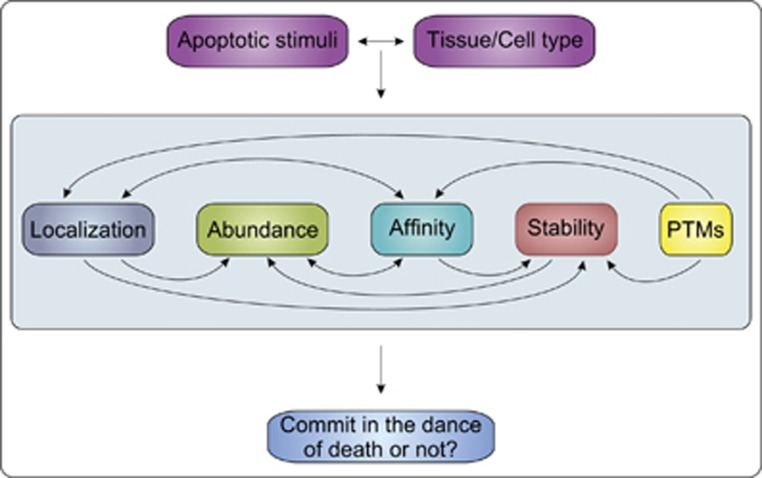
Figure 1 Cellular factors regulating commitment to the apoptotic dance of death by the BCL-2 family proteins. BCL-2 family proteins function by direct binding interactions that lead to mutual sequestration and/or membrane permeabilization. These direct protein–protein binding interactions are functionally regulated in cells by a number of interrelated processes. Differences in apoptotic stimuli and cell types lead to different responses due to the integration of the effects of localization, abundance, affinity, stability and post-translational modifications (PTMs). Related interactions are connected by arrows. For example, PTMs affect protein stability, localization and binding affinities. Affinities determine which interactions dominate but also affect localization and stability (heterodimerization with BH3-only proteins stabilizes MCL-1). The affinities of BCL-2 family proteins for intracellular membranes and other binding partners at these membranes dictate BCL-2 family localization. Co-localization increases local concentrations (abundance). Protein stability also impacts abundance of the BCL-2 family proteins. The relative abundance and affinity ultimately determine which binding interactions dominate and whether or not the cell undergoes MOMP committing it to apoptosis
Figure 1 Cellular factors regulating commitment to the apoptotic dance of death by the BCL-2 family proteins. BCL-2 family proteins function by direct binding interactions that lead to mutual sequestration and/or membrane permeabilization. These direct protein–protein binding interactions are functionally regulated in cells by a number of interrelated processes. Differences in apoptotic stimuli and cell types lead to different responses due to the integration of the effects of localization, abundance, affinity, stability and post-translational modifications (PTMs). Related interactions are connected by arrows. For example, PTMs affect protein stability, localization and binding affinities. Affinities determine which interactions dominate but also affect localization and stability (heterodimerization with BH3-only proteins stabilizes MCL-1). The affinities of BCL-2 family proteins for intracellular membranes and other binding partners at these membranes dictate BCL-2 family localization. Co-localization increases local concentrations (abundance). Protein stability also impacts abundance of the BCL-2 family proteins. The relative abundance and affinity ultimately determine which binding interactions dominate and whether or not the cell undergoes MOMP committing it to apoptosisCC BY-NC-SA · PubMed Central
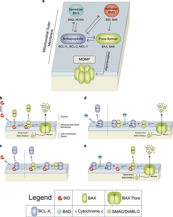
Figure 2 The dance of the BCL-2 family within the bilayer regulates mitochondrial outer membrane permeabilization (MOMP) and apoptosis (a) Schematic of the embedded together model. All binding interactions are reversible and equilibria are governed by local affinities. Interactions with the lipid bilayer change the affinities of the interactions and therefore have an active role in the functions of the proteins. Binding of the activator BH3-only proteins (e.g., BID, BIM) to membranes increases their affinity for the pore-formers (e.g. BAX, BAK), which are activated (arrows) to permeabilize the mitochondrial outer membrane. The anti-apoptotic proteins (e.g., BCL-XL, BCL-2, MCL-1) inhibit both the activator BH3-only proteins and the pore-forming proteins by mutual sequestration (T’d arrows). The sensitizer BH3-only proteins (e.g., BAD, NOXA) bind to and inhibit the anti-apoptotic proteins also by mutual sequestration. Recruitment of the complexes to the membrane by constitutive interactions (e.g., BAK) and dynamic interactions (e.g., BAX, BID, BIM) increases the affinities and local concentrations and reduces the diffusion of the BCL-2 family proteins. Localization at different intracellular membranes also dictates the binding equilibria between each family member. The efficiency of inhibition by mutual sequestration of anti-apoptotic proteins depends on both affinities and off-rates of the interactions. Interaction of the BH4 region of the anti-apoptotic proteins with BAX shifts the BAX-membrane binding equilibrium to favor the unbound state (retrotranslocation, not shown). (b–d). Interactions of the BCL-2 family that promote or inhibit MOMP illustrated for cBID. BAX, BCL-XL and BAD as examples of different functional categories. (b) BID is activated by caspase-8 mediated cleavage to cBID (cleaved BID) a protein comprised of two fragments BID-P7 and BID-P15 held together by hydrophobic interactions. Rapid high-affinity binding to membranes dissociates the p7 fragment to solution and favors insertion of the p15 fragment (tBID; truncated BID) into the membrane. Membrane-bound tBID recruits inactive BAX from the cytosol. Binding to tBID activates BAX to insert in the bilayer, oligomerize and permeabilize the mitochondrial outer membrane releasing intermembrane space proteins including cytochrome c and SMAC. (c) Active tBID and BAX can recruit BCL-XL to the membrane resulting in inhibition of both pro and anti-apoptotic proteins by mutual sequestration. BCL-XL prevents tBID from activating BAX and prevents BAX from oligomerizing resulting in the inhibition of MOMP. BAX bound to BCL-XL is in the active (oligomerization competent) conformation. (d, e) BAD inhibits unbound BCL-XL by mutual sequestration. The affinity of BCL-XL is higher for tBID than for active BAX (Tables 1A and 1B) therefore, in the absence of other regulatory interactions or PTMs if BCL-XL is bound to tBID and BAX then high concentrations of BAD will displace active BAX (d) and then tBID (e) from BCL-XL resulting in MOMP
Figure 2 The dance of the BCL-2 family within the bilayer regulates mitochondrial outer membrane permeabilization (MOMP) and apoptosis (a) Schematic of the embedded together model. All binding interactions are reversible and equilibria are governed by local affinities. Interactions with the lipid bilayer change the affinities of the interactions and therefore have an active role in the functions of the proteins. Binding of the activator BH3-only proteins (e.g., BID, BIM) to membranes increases their affinity for the pore-formers (e.g. BAX, BAK), which are activated (arrows) to permeabilize the mitochondrial outer membrane. The anti-apoptotic proteins (e.g., BCL-XL, BCL-2, MCL-1) inhibit both the activator BH3-only proteins and the pore-forming proteins by mutual sequestration (T’d arrows). The sensitizer BH3-only proteins (e.g., BAD, NOXA) bind to and inhibit the anti-apoptotic proteins also by mutual sequestration. Recruitment of the complexes to the membrane by constitutive interactions (e.g., BAK) and dynamic interactions (e.g., BAX, BID, BIM) increases the affinities and local concentrations and reduces the diffusion of the BCL-2 family proteins. Localization at different intracellular membranes also dictates the binding equilibria between each family member. The efficiency of inhibition by mutual sequestration of anti-apoptotic proteins depends on both affinities and off-rates of the interactions. Interaction of the BH4 region of the anti-apoptotic proteins with BAX shifts the BAX-membrane binding equilibrium to favor the unbound state (retrotranslocation, not shown). (b–d). Interactions of the BCL-2 family that promote or inhibit MOMP illustrated for cBID. BAX, BCL-XL and BAD as examples of different functional categories. (b) BID is activated by caspase-8 mediated cleavage to cBID (cleaved BID) a protein comprised of two fragments BID-P7 and BID-P15 held together by hydrophobic interactions. Rapid high-affinity binding to membranes dissociates the p7 fragment to solution and favors insertion of the p15 fragment (tBID; truncated BID) into the membrane. Membrane-bound tBID recruits inactive BAX from the cytosol. Binding to tBID activates BAX to insert in the bilayer, oligomerize and permeabilize the mitochondrial outer membrane releasing intermembrane space proteins including cytochrome c and SMAC. (c) Active tBID and BAX can recruit BCL-XL to the membrane resulting in inhibition of both pro and anti-apoptotic proteins by mutual sequestration. BCL-XL prevents tBID from activating BAX and prevents BAX from oligomerizing resulting in the inhibition of MOMP. BAX bound to BCL-XL is in the active (oligomerization competent) conformation. (d, e) BAD inhibits unbound BCL-XL by mutual sequestration. The affinity of BCL-XL is higher for tBID than for active BAX (Tables 1A and 1B) therefore, in the absence of other regulatory interactions or PTMs if BCL-XL is bound to tBID and BAX then high concentrations of BAD will displace active BAX (d) and then tBID (e) from BCL-XL resulting in MOMPCC BY-NC-SA · PubMed Central
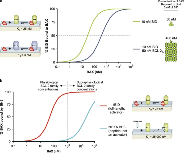
Figure 3 Affinities and concentrations dictate the predominate interactions of the BCL-2 family members (a) At membranes cBID binds BAX and BCL-XL with an affinity of 25 nM and 3 nM, respectively. Equations describing competitive binding of two different ‘ligands’ (BAX, BCL-XL) to one protein molecule (cBID)21 were used to model the Interaction of 10 nM cBID with increasing concentrations of BAX in the absence (green) and presence (purple) of 50 nM BCL-XL. In the absence of BCL-XL, 50% of 10 nM cBID is bound to BAX at a BAX concentration of 30 nM. Upon the addition of BCL-XL, ~13 times more BAX (408 nM) is required to bind 50% of the cBID. In this case, BCL-XL is binding the majority of the cBID, preventing BAX activation. The functional consequence of the differing affinities results in BCL-XL out-competing BAX for binding to cBID, effectively inhibiting apoptosis at physiologically relevant concentrations of BAX. (b) The affinities between BH3-only proteins and BAX determine whether a BH3-only protein functions as an activator or sensitizer. Equations describing the binding of two proteins21 were used to model the interaction of 50 nM BAX with increasing concentrations of the indicated BH3-only protein/peptide; full-length tBID (red) and NOXA BH3 peptide (cyan).The affinities of full-length tBID or NOXA BH3 peptide for BAX are 25 nM20 and 25 000 nM (estimated from ref. 35) respectively. Typical in vitro BAX activation assays use peptide concentrations in the micromolar range, well above the nanomolar concentrations predicted for BCL-2 family proteins in cells. At supraphysiological concentrations (>1000 nM) of peptides (e.g., NOXA, cyan) some BH3 sequences that typically function as sensitizers can bind to and activate BAX
Figure 3 Affinities and concentrations dictate the predominate interactions of the BCL-2 family members (a) At membranes cBID binds BAX and BCL-XL with an affinity of 25 nM and 3 nM, respectively. Equations describing competitive binding of two different ‘ligands’ (BAX, BCL-XL) to one protein molecule (cBID)21 were used to model the Interaction of 10 nM cBID with increasing concentrations of BAX in the absence (green) and presence (purple) of 50 nM BCL-XL. In the absence of BCL-XL, 50% of 10 nM cBID is bound to BAX at a BAX concentration of 30 nM. Upon the addition of BCL-XL, ~13 times more BAX (408 nM) is required to bind 50% of the cBID. In this case, BCL-XL is binding the majority of the cBID, preventing BAX activation. The functional consequence of the differing affinities results in BCL-XL out-competing BAX for binding to cBID, effectively inhibiting apoptosis at physiologically relevant concentrations of BAX. (b) The affinities between BH3-only proteins and BAX determine whether a BH3-only protein functions as an activator or sensitizer. Equations describing the binding of two proteins21 were used to model the interaction of 50 nM BAX with increasing concentrations of the indicated BH3-only protein/peptide; full-length tBID (red) and NOXA BH3 peptide (cyan).The affinities of full-length tBID or NOXA BH3 peptide for BAX are 25 nM20 and 25 000 nM (estimated from ref. 35) respectively. Typical in vitro BAX activation assays use peptide concentrations in the micromolar range, well above the nanomolar concentrations predicted for BCL-2 family proteins in cells. At supraphysiological concentrations (>1000 nM) of peptides (e.g., NOXA, cyan) some BH3 sequences that typically function as sensitizers can bind to and activate BAXCC BY-NC-SA · PubMed Central
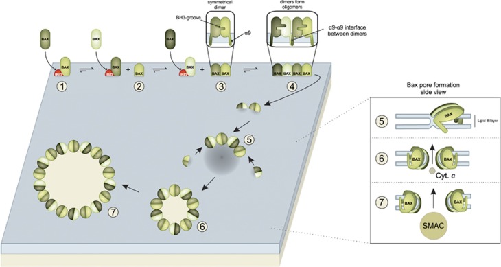
Figure 4 BAX activation is a multi-step process characterized by hetero-and homotypic interactions that result in MOMP. Full-length activator BH3-only proteins, like tBID, have a high affinity for membranes and bind them rapidly before interacting with the pore-formers, BAX and BAK. The following steps are shown for BAX activation; however, the BAK activation mechanism appears to be very similar. (Step 1) BAX interacts with tBID at the membrane. (Step 2) BAX then undergoes multiple conformation changes, inserting into the bilayer with α9 spanning the membrane. Insertion into the bilayer is the rate-limiting step in the BAX activation mechanism. (Step 3) The transition from the BAX and BH3-only protein heterodimer to BAX homodimers is not well understood but would occur spontaneously if the homotypic interaction is of higher affinity than that of the heterotypic interaction between BAX and activator BH3-only proteins. The end result is that the membrane-embedded active BAX monomers dimerize via reciprocal interactions between their BH3-grooves. (Step 4) BAX dimers interact with each other via multiple lower affinity interactions of which parallel α9–α9 interactions between dimers appear particularly important for the stabilization of large pores. (Step 5) BAX oligomers composed of symmetrical dimer subunits start to destabilize and thin the bilayer. Weak affinity interactions between dimers allow additional dimer subunits to add to the oligomer at any point. (Step 6) BAX oligomers form small pores in the bilayer that can initially release smaller intermembrane space (IMS) proteins like cytochrome c (12 kDa). (Step 7) Continuing activation of BAX results in a higher concentration of dimers in the bilayer that add to the oligomer, resulting in pore expansion and the release of larger IMS proteins like SMAC (54 kDa dimer)
Figure 4 BAX activation is a multi-step process characterized by hetero-and homotypic interactions that result in MOMP. Full-length activator BH3-only proteins, like tBID, have a high affinity for membranes and bind them rapidly before interacting with the pore-formers, BAX and BAK. The following steps are shown for BAX activation; however, the BAK activation mechanism appears to be very similar. (Step 1) BAX interacts with tBID at the membrane. (Step 2) BAX then undergoes multiple conformation changes, inserting into the bilayer with α9 spanning the membrane. Insertion into the bilayer is the rate-limiting step in the BAX activation mechanism. (Step 3) The transition from the BAX and BH3-only protein heterodimer to BAX homodimers is not well understood but would occur spontaneously if the homotypic interaction is of higher affinity than that of the heterotypic interaction between BAX and activator BH3-only proteins. The end result is that the membrane-embedded active BAX monomers dimerize via reciprocal interactions between their BH3-grooves. (Step 4) BAX dimers interact with each other via multiple lower affinity interactions of which parallel α9–α9 interactions between dimers appear particularly important for the stabilization of large pores. (Step 5) BAX oligomers composed of symmetrical dimer subunits start to destabilize and thin the bilayer. Weak affinity interactions between dimers allow additional dimer subunits to add to the oligomer at any point. (Step 6) BAX oligomers form small pores in the bilayer that can initially release smaller intermembrane space (IMS) proteins like cytochrome c (12 kDa). (Step 7) Continuing activation of BAX results in a higher concentration of dimers in the bilayer that add to the oligomer, resulting in pore expansion and the release of larger IMS proteins like SMAC (54 kDa dimer)CC BY-NC-SA · PubMed Central
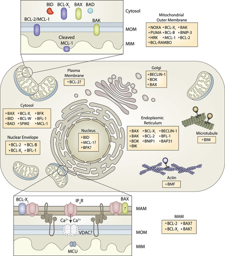
Figure 5 BCL-2 family protein subcellular localization in non-apoptotic cells BCL-2 family proteins reported at each location are listed in the corresponding box. Uncertainty or data for which there are conflicting reports are indicated by a ‘?’. Localization that changes during apoptosis is summarized in Tables 3A and 3B. Interactions at the mitochondria are shown in the enlargement top left. Interactions at the mitochondrial associated membrane (MAM), a subdomain of the endoplasmic reticulum in close contact with mitochondria are enlarged below left. Among other proteins, mitofusin proteins (shown as dimer linking the two membranes) bring a specialized subdomain of ER membranes in contact with the mitochondrial outer membrane forming the MAM
Figure 5 BCL-2 family protein subcellular localization in non-apoptotic cells BCL-2 family proteins reported at each location are listed in the corresponding box. Uncertainty or data for which there are conflicting reports are indicated by a ‘?’. Localization that changes during apoptosis is summarized in Tables 3A and 3B. Interactions at the mitochondria are shown in the enlargement top left. Interactions at the mitochondrial associated membrane (MAM), a subdomain of the endoplasmic reticulum in close contact with mitochondria are enlarged below left. Among other proteins, mitofusin proteins (shown as dimer linking the two membranes) bring a specialized subdomain of ER membranes in contact with the mitochondrial outer membrane forming the MAMCC BY-NC-SA · PubMed Central

Verification

Claims the paper makes

The claims and statistics of this paper haven’t been checked yet.

Run the check — free, no account needed. It reads the open-access text and checks the sources it cites.

A few free checks a day · papers already verified open instantly · results are public

Claims other papers make about this one

Each is checked against this paper’s own text: does it say what the citing paper says it does?

Self-check

Percentages, sums and reported differences re-computed from the paper’s own numbers.

The self-check needs the paper’s full text.

Citations

Cites

Show all 120 references

Cited by

Sorted by citations

See all 1,574

Metadata

Authors
Kale, J., Osterlund, E. J., & Andrews, D. W.
Affiliations
  • Sunnybrook Hospital (CA)
  • University of Toronto (CA)
Corresponding author
Andrews, David W
Venue
Cell Death & Differentiation
Published
2017-11-17
Volume
25
Issue
1
Pages
65-80
ISSN
  • 1350-9047 (ISSN-L)
  • 1476-5403
OpenAlex
W2769631482
Cited by
1,574
References
120
Citations by year
2026: 123 · 2025: 189 · 2024: 225 · 2023: 231 · 2022: 242 · 2021: 228 · 2020: 155 · 2019: 123 · 2018: 52 · 2017: 3
FWCI
8.52
Type
journal article
Language
en
Open access
Locations
  • Cell Death and Differentiation (publishedVersion, cc-by-nc-sa)
  • PubMed Central (publishedVersion, cc-by-nc-sa)
  • PubMed (publishedVersion)
Funders
Canadian Institutes of Health Research ((FDN143312))
Topics
  • Life Sciences › Biochemistry, Genetics and Molecular Biology › Molecular Biology › Cell death mechanisms and regulation
  • Life Sciences › Biochemistry, Genetics and Molecular Biology › Molecular Biology › Mitochondrial Function and Pathology
  • Health Sciences › Medicine › Oncology › Cancer-related Molecular Pathways
Keywords
Bcl-2 family proteins, mitochondrial outer membrane permeabilization, apoptosis, anti-apoptotic proteins, pro-apoptotic proteins, protein-protein interaction, binding affinity, protein concentration, post-translational modification, subcellular localization, caspase activation, cell fate regulation
MeSH terms
Cell Membrane Permeability, Protein Processing, Post-Translational, Apoptosis, Proto-Oncogene Proteins c-bcl-2, bcl-2 Homologous Antagonist-Killer Protein, Apoptosis Regulatory Proteins, bcl-2-Associated X Protein, Mitochondrial Membranes

About these checks

Registry, retraction, Cites and statistics results are computed, not judged, with one exception: a printed reference that no index lists is read once by a model before it counts as a citation error. Claims are extracted by a model and kept only when their quote appears verbatim in the text; each cited source is then checked, with a model’s help, for whether it says what the claim relies on. Those verdicts can be wrong — read the claim’s quote and the cited source before relying on one.

Statistics and arithmetic are recomputed, not judged.

Confidence bands are shown only for fields calibrated on our annotated test set. None is calibrated yet, so no band is shown.

What counts as a claim ›How we measure confidence ›How we check this ›

Are you an author, or do you see a problem with this page? Request a review