

The DOI matches its Crossref record; 4 of the 4 references it cites were found in a registry or an index.
Mature neurons: equipped for survival
Overview
The claims and statistics of this paper haven’t been checked yet.
Run the check — free, no account needed. It reads the open-access text and checks the sources it cites.
Integrity signals
Registry
Cites
Statistics
Figures
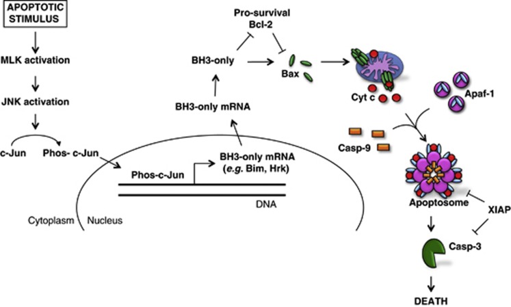
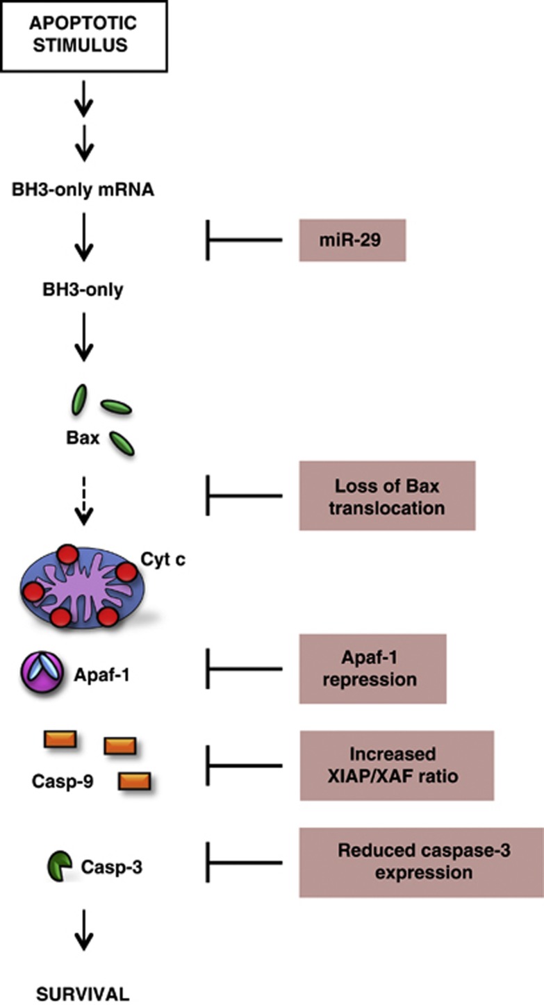
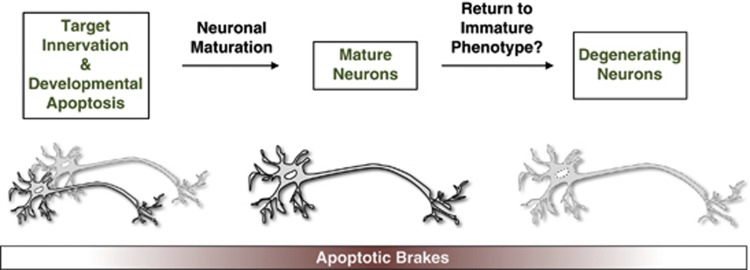
Verification
Claims the paper makes
The claims and statistics of this paper haven’t been checked yet.
Run the check — free, no account needed. It reads the open-access text and checks the sources it cites.
Claims other papers make about this one
Each is checked against this paper’s own text: does it say what the citing paper says it does?
In the citing paper
“Some of these targets—including Mst-1, BIM, and MLK3—contribute to the cell death processes of secondary injury [85,86].”
From MicroRNA-138-5p Targets Pro-Apoptotic Factors and Favors Neural Cell Survival: Analysis in the Injured Spinal Cord (2022) · cites this paper as [86]
Self-check
Percentages, sums and reported differences re-computed from the paper’s own numbers.
The self-check needs the paper’s full text.
Citations
Cites
Neurotrophin signal transduction in the nervous system
Kaplan, D. R., & Miller, F. D. (2000) · Current Opinion in Neurobiology · doi:10.1016/s0959-4388(00)00092-1 · Cited by 1,944
Apoptosis in neurodegenerative disorders
Mattson, M. P. (2000) · Nature Reviews Molecular Cell Biology · doi:10.1038/35040009 · Cited by 1,605
Mitochondrial membrane permeabilization in neuronal injury
Galluzzi, L., Blomgren, K., & Kroemer, G. (2009) · Nature Reviews Neuroscience · doi:10.1038/nrn2665 · Cited by 418
Fas ligand/Fas system in the brain: regulator of immune and apoptotic responses
Choi, C., & Benveniste, E. N. (2004) · Brain Research Reviews · doi:10.1016/j.brainresrev.2003.08.007 · Cited by 270
Research works printed but not linked: 78
- Oppenheim RW . Cell death during development of the nervous system. Annu Rev Neurosci 1991; 14: 453–501. · DOI · Paper page
- Kim WR, Sun W . Programmed cell death during postnatal development of the rodent nervous system. Dev Growth Differ 2011; 53: 225–235. · DOI
- Davies AM . Regulation of neuronal survival and death by extracellular signals during development. EMBO J 2003; 22: 2537–2545. · DOI
- Buss RR, Sun W, Oppenheim RW . Adaptive roles of programmed cell death during nervous system development. Annu Rev Neurosci 2006; 29: 1–35. · DOI
- Benn SC, Woolf CJ . Adult neuron survival strategies--slamming on the brakes. Nat Rev Neurosci 2004; 5: 686–700. · DOI
- Yuan J, Yankner BA . Apoptosis in the nervous system. Nature 2000; 407: 802–809. · DOI
- Ernst M . Ueber Untergang von Zellen wahrend der normalen Entwicklung bei Wirbeltieren. Zeitschr Anat u Entw gesch 1926; 79: 228–262. · DOI
- Glucksmann A . Ueber die Bedeutung von Zellvorgangen fur die Formbildung epithelialer Organe. Zeitschr Anat u Entw gesch 1930; 93: 35–92. · DOI
- Hamburger V, Levi-Montalcini R . Proliferation, differentiation and degeneration in the spinal ganglia of the chick embryo under normal and experimental conditions. J Exp Zool 1949; 111: 457–501. · DOI
- Levi-Montalcini R, Booker B . Destruction of the sympathetic ganglia in mammals by an antiserum to a nerve-growth protein. Proc Natl Acad Sci USA 1960; 46: 384–391. · DOI
- Angeletti PU, Levi-Montalcini R, Caramia F . Analysis of the effects of the antiserum to the nerve growth factor in adult mice. Brain Res 1971; 27: 343–355. · DOI
- Goedert M, Otten U, Thoenen H . Biochemical effects of antibodies against nerve growth factor on developing and differentiated sympathetic ganglia. Brain Res 1978; 148: 264–268. · DOI
- Otten U, Goedert M, Schwab M, Thibault J . Immunization of adult rats against 2.5 S NGF: effects on the peripheral sympathetic nervous system. Brain Res 1979; 176: 79–90. · DOI
- Gorin PD, Johnson EM Jr . Effects of long-term nerve growth factor deprivation on the nervous system of the adult rat: an experimental autoimmune approach. Brain Res 1980; 198: 27–42. · DOI
- Lazarus KJ, Bradshaw RA, West NR, Bunge P . Adaptive survival or rat sympathetic neurons cultured without supporting cells or exogenous nerve growth factor. Brain Res 1976; 113: 159–164. · DOI
- Chun LL, Patterson PH . Role of nerve growth factor in the development of rat sympathetic neurons in vitro. II. Developmental studies. J Cell Biol 1977; 75: 705–711. · DOI
- Easton RM, Deckwerth TL, Sh PA, Johnson EM Jr . Analysis of the mechanism of loss of trophic factor dependence associated with neuronal maturation: A phenotype indistinguishable from BAX deletion. J Neurosci 1997; 17: 9656–9666. · DOI
- Deshmukh M, Johnson EM Jr . Programmed cell death in neurons: focus on the pathway of nerve growth factor deprivation-induced death of sympathetic neurons. Mol Pharm 1997; 51: 897–906. · DOI
- Freeman RS, Burch RL, Crowder RJ, Lomb DJ, Schoell MC, Straub JA et al. NGF deprivation-induced gene expression: after ten years, where do we stand? Prog Brain Res 2004; 146: 111–126. · DOI
- Vogelbaum MA, Tong JX, Rich KM . Developmental regulation of apoptosis in dorsal root ganglion neurons. J Neurosci 1998; 18: 8928–8935. · DOI
- Kimpinski K, Campenot RB, Mearow K . Effects of the neurotrophins nerve growth factor, neurotrophin-3, and brain-derived neurotrophic factor (BDNF) on neurite growth from adult sensory neurons in compartmented cultures. J Neurobiol 1997; 33: 395–410. · DOI
- Lindsay RM . Nerve growth factors (NGF, BDNF) enhance axonal regeneration but are not required for survival of adult sensory neurons. J Neurosci 1988; 8: 2394–2405. · DOI
- Tong JX, Eichler ME, Rich KM . Intracellular calcium levels influence apoptosis in mature sensory neurons after trophic factor deprivation. Exp Neurol 1996; 138: 45–52. · DOI
- Harris JA, Rubel EW . Afferent regulation of neuron number in the cochlear nucleus: cellular and molecular analyses of a critical period. Hear Res 2006; 216-217: 127–137. · DOI
- Mostafapour SP, Del Puerto NM, Rubel EW . bcl-2 Overexpression eliminates deprivation-induced cell death of brainstem auditory neurons. J Neurosci 2002; 22: 4670–4674. · DOI
- Tierney TS, Russell FA, Moore DR . Susceptibility of developing cochlear nucleus neurons to deafferentation-induced death abruptly ends just before the onset of hearing. J Comp Neurol 1997; 378: 295–306. · DOI
- Mostafapour SP, Cochran SL, Del Puerto NM, Rubel EW . Patterns of cell death in mouse anteroventral cochlear nucleus neurons after unilateral cochlea removal. J Comp Neurol 2000; 426: 561–571. · DOI
- Snider WD, Elliott JL, Yan Q . Axotomy-induced neuronal death during development. J Neurobiol 1992; 23: 1231–1246. · DOI
- Snider WD, Thanedar S . Target dependence of hypoglossal motor neurons during development in maturity. J Comp Neurol 1989; 279: 489–498. · DOI
- Yan Q, Snider WD, Pinzone JJ, Johnson EM Jr . Retrograde transport of nerve growth factor (NGF) in motoneurons of developing rats: assessment of potential neurotrophic effects. Neuron 1988; 1: 335–343. · DOI
- Sendtner M, Kreutzberg GW, Thoenen H . Ciliary neurotrophic factor prevents the degeneration of motor neurons after axotomy. Nature 1990; 345: 440–441. · DOI
- Yu WH . Sex difference in neuronal loss induced by axotomy in the rat brain stem motor nuclei. Exp Neurol 1988; 102: 230–235. · DOI
- Kuzis K, Coffin JD, Eckenstein FP . Time course and age dependence of motor neuron death following facial nerve crush injury: role of fibroblast growth factor. Exp Neurol 1999; 157: 77–87. · DOI
- Rich KM, Luszczynski JR, Osborne PA, Johnson EM Jr . Nerve growth factor protects adult sensory neurons from cell death and atrophy caused by nerve injury. J Neurocytol 1987; 16: 261–268. · DOI
- McKernan DP, Caplis C, Donovan M, O’Brien CJ, Cotter TG . Age-dependent susceptibility of the retinal ganglion cell layer to cell death. Invest Ophthalmol Vis Sci 2006; 47: 807–814. · DOI
- Guerin MB, McKernan DP, O’Brien CJ, Cotter TG . Retinal ganglion cells: dying to survive. Int J Dev Biol 2006; 50: 665–674. · DOI
- Bittigau P, Sifringer M, Pohl D, Stadthaus D, Ishimaru M, Shimizu H et al. Apoptotic neurodegeneration following trauma is markedly enhanced in the immature brain. Ann Neurol 1999; 45: 724–735. · DOI
- Cheng Y, Deshmukh M, D’Costa A, DeMaro JA, Gidday JM, Shah A et al. Caspase inhibitor affords neuroprotection with delayed administration in a rat model of neonatal hypoxic-ischemic brain injury. J Clin Invest 1998; 101: 1992–1999. · DOI
- Hu BR, Liu CL, Ouyang Y, Blomgren K, Siesjo BK . Involvement of Caspase-3 in Cell Death After Hypoxia-Ischemia Declines During Brain Maturation. J Cereb Blood Flow Metab 2000; 20: 1294–1300. · DOI
- Liu CL, Siesjo BK, Hu BR . Pathogenesis of hippocampal neuronal death after hypoxia-ischemia changes during brain development. Neuroscience 2004; 127: 113–123. · DOI
- Blomgren K, Zhu C, Hallin U, Hagberg H . Mitochondria and ischemic reperfusion damage in the adult and in the developing brain. Biochem Biophys Res Commun 2003; 304: 551–559. · DOI
- Labrada L, Liang XH, Zheng W, Johnston C, Levine B . Age-dependent resistance to lethal alphavirus encephalitis in mice: analysis of gene expression in the central nervous system and identification of a novel interferon-inducible protective gene, mouse ISG12. J Virol 2002; 76: 11688–11703. · DOI
- Ahn J, Jee Y, Seo I, Yoon SY, Kim D, Kim YK et al. Primary neurons become less susceptible to coxsackievirus B5 following maturation: the correlation with the decreased level of CAR expression on cell surface. J Med Virol 2008; 80: 434–440. · DOI
- Hetman M, Kharebava G . Survival signaling pathways activated by NMDA receptors. Curr Top Med Chem 2006; 6: 787–799. · DOI
- Xiao L, Hu C, Feng C, Chen Y . Switching of N-methyl-D-aspartate (NMDA) receptor-favorite intracellular signal pathways from ERK1/2 protein to p38 mitogen-activated protein kinase leads to developmental changes in NMDA neurotoxicity. J Biol Chem 2011; 286: 20175–20193. · DOI
- Mota M, Reeder M, Chernoff J, Bazenet CE . Evidence for a role of mixed lineage kinases in neuronal apoptosis. J Neurosci 2001; 21: 4949–4957. · DOI
- Xu Z, Maroney AC, Dobrzanski P, Kukekov NV, Greene LA . The MLK family mediates c-Jun N-terminal kinase activation in neuronal apoptosis. Mol Cell Biol 2001; 21: 4713–4724. · DOI
- Imaizumi K, Tsuda M, Imai Y, Wanaka A, Takagi T, Tohyama M . Molecular cloning of a novel polypeptide, DP5, induced during programmed neuronal death. J Biol Chem 1997; 272: 18842–18848. · DOI
- Harris CA, Johnson EM Jr. . BH3-only Bcl-2 family members are coordinately regulated by the JNK pathway and require Bax to induce apoptosis in neurons. J Biol Chem 2001; 276: 37754–37760. · DOI
- Whitfield J, Neame SJ, Paquet L, Bernard O, Ham J . Dominant-negative c-Jun promotes neuronal survival by reducing BIM expression and inhibiting mitochondrial cytochrome c release. Neuron 2001; 29: 629–643. · DOI
- Deckwerth TL, Elliott JL, Knudson CM, Johnson EM Jr, Snider WD, Korsmeyer SJ . Bax is required for neuronal death after trophic factor deprivation and during development. Neuron 1996; 17: 401–411. · DOI
- Wright KM, Vaughn AE, Deshmukh M . Apoptosome dependent caspase-3 activation pathway is non-redundant and necessary for apoptosis in sympathetic neurons. Cell Death Differ 2007; 14: 625–633. · DOI
- Snider WD . Functions of the neurotrophins during nervous system development: what the knockouts are teaching us. Cell 1994; 77: 627–638. · DOI
- Tsui-Pierchala BA, Ginty DD . Characterization of an NGF-P-TrkA retrograde-signaling complex and age-dependent regulation of TrkA phosphorylation in sympathetic neurons. J Neurosci 1999; 19: 8207–8218. · DOI
- Putcha GV, Deshmukh M, Johnson EM Jr . Inhibition of apoptotic signaling cascades causes loss of trophic factor dependence during neuronal maturation. J Cell Biol 2000; 149: 1011–1018. · DOI
- Polster BM, Robertson CL, Bucci CJ, Suzuki M, Fiskum G . Postnatal brain development and neural cell differentiation modulate mitochondrial Bax and BH3 peptide-induced cytochrome c release. Cell Death Differ 2003; 10: 365–370. · DOI
- Walsh GS, Orike N, Kaplan DR, Miller FD . The invulnerability of adult neurons: a critical role for p73. J Neurosci 2004; 24: 9638–9647. · DOI
- Kole AJ, Swahari V, Hammond SM, Deshmukh M . miR-29b is activated during neuronal maturation and targets BH3-only genes to restrict apoptosis. Genes Dev 2011; 25: 125–130. · DOI
- Giam M, Huang DC, Bouillet P . BH3-only proteins and their roles in programmed cell death. Oncogene 2008; 27 (Suppl 1): S128–S136. · DOI
- Lomonosova E, Chinnadurai G . BH3-only proteins in apoptosis and beyond: an overview. Oncogene 2008; 27 (Suppl 1): S2–S19. · DOI
- Khanna S, Rink C, Ghoorkhanian R, Gnyawali S, Heigel M, Wijesinghe DS et al. Loss of miR-29b following acute ischemic stroke contributes to neural cell death and infarct size. J Cereb Blood Flow Metab 2013 e-pub ahead of print 1 May 2013 doi:10.1038/jcbfm.2013.68. · DOI
- Wright KM, Smith MI, Farrag L, Deshmukh M . Chromatin modification of Apaf-1 restricts the apoptotic pathway in mature neurons. J Cell Biol 2007; 179: 825–832. · DOI
- Yakovlev AG, Ota K, Wang G, Movsesyan V, Bao W-L, Yoshihara K et al. Differential expression of apoptotic protease-activating factor-1 and caspase-3 genes and susceptibility to apoptosis during brain development and after traumatic brain injury. J Neurosci 2001; 21: 7439–7446. · DOI
- Johnson CE, Huang YY, Parrish AB, Smith MI, Vaughn AE, Zhang Q et al. Differential Apaf-1 levels allow cytochrome c to induce apoptosis in brain tumors but not in normal neural tissues. Proc Natl Acad Sci USA 2007; 104: 20820–20825. · DOI · Paper page
- Donovan M, Cotter TG . Caspase-independent photoreceptor apoptosis in vivo and differential expression of apoptotic protease activating factor-1 and caspase-3 during retinal development. Cell Death Differ 2002; 9: 1220–1231. · DOI
- Ota K, Yakovlev AG, Itaya A, Kameoka M, Tanaka Y, Yoshihara K . Alteration of apoptotic protease-activating factor-1 (APAF-1)-dependent apoptotic pathway during development of rat brain and liver. J Biochem 2002; 131: 131–135. · DOI
- Blomgren K, Zhu C, Wang X, Karlsson JO, Leverin AL, Bahr BA et al. Synergistic activation of caspase-3 by m-calpain after neonatal hypoxia-ischemia: a mechanism of ‘pathological apoptosis’? J Biol Chem 2001; 276: 10191–10198. · DOI
- Harris JA, Hardie NA, Bermingham-McDonogh O, Rubel EW . Gene expression differences over a critical period of afferent-dependent neuron survival in the mouse auditory brainstem. J Comp Neurol 2005; 493: 460–474. · DOI
- Perrelet D, Perrin FE, Liston P, Korneluk RG, MacKenzie A, Ferrer-Alcon M et al. Motoneuron resistance to apoptotic cell death in vivo correlates with the ratio between X-linked inhibitor of apoptosis proteins (XIAPs) and its inhibitor, XIAP-associated factor 1. J Neurosci 2004; 24: 3777–3785. · DOI
- Chan PH . Mitochondria and neuronal death/survival signaling pathways in cerebral ischemia. Neurochem Res 2004; 29: 1943–1949. · DOI
- Martin LJ, Al-Abdulla NA, Brambrink AM, Kirsch JR, Sieber FE, Portera-Cailliau C . Neurodegeneration in excitotoxicity, global cerebral ischemia, and target deprivation: a perspective on the contributions of apoptosis and necrosis. Brain Res Bull 1998; 46: 281–309. · DOI
- Choi DW . Ischemia-induced neuronal apoptosis. Curr Opin Neurobiol 1996; 6: 667–672. · DOI
- Guegan C, Vila M, Rosoklija G, Hays AP, Przedborski S . Recruitment of the mitochondrial-dependent apoptotic pathway in amyotrophic lateral sclerosis. J Neurosci 2001; 21: 6569–6576. · DOI
- Kiechle T, Dedeoglu A, Kubilus J, Kowall NW, Beal MF, Friedlander RM et al. Cytochrome C and caspase-9 expression in Huntington’s disease. Neuromolecular Med 2002; 1: 183–195. · DOI
- Hebert SS, Horre K, Nicolai L, Papadopoulou AS, Mandemakers W, Silahtaroglu AN et al. Loss of microRNA cluster miR-29a/b-1 in sporadic Alzheimer's disease correlates with increased BACE1/beta-secretase expression. Proc Natl Acad Sci USA 2008; 105: 6415–6420. · DOI · Paper page
- Lee ST, Chu K, Im WS, Yoon HJ, Im JY, Park JE et al. Altered microRNA regulation in Huntington's disease models. Exp Neurol 2011; 227: 172–179. · DOI · Paper page
- Shioya M, Obayashi S, Tabunoki H, Arima K, Saito Y, Ishida T et al. Aberrant microRNA expression in the brains of neurodegenerative diseases: miR-29a decreased in Alzheimer disease brains targets neurone navigator 3. Neuropathol Appl Neurobiol 2010; 36: 320–330. · DOI · Paper page
- Haase G, Pettmann B, Raoul C, Henderson CE . Signaling by death receptors in the nervous system. Curr Opin Neurobiol 2008; 18: 284–291. · DOI
Cited by
Sorted by citations
Regulation of apoptosis in health and disease: the balancing act of BCL-2 family proteins
Singh, R., Letai, A., & Sarosiek, K. (2019) · Nature Reviews Molecular Cell Biology · doi:10.1038/s41580-018-0089-8 · Cited by 2,111
Necroptosis and RIPK1-mediated neuroinflammation in CNS diseases
Yuan, J., Amin, P., & Ofengeim, D. (2019) · Nature Reviews Neuroscience · doi:10.1038/s41583-018-0093-1 · Cited by 967
Neuronal Cell Death Mechanisms in Major Neurodegenerative Diseases
Chi, H., Chang, H.-Y., & Sang, T.-K. (2018) · International Journal of Molecular Sciences · doi:10.3390/ijms19103082 · Cited by 412
Age-dependent instability of mature neuronal fate in induced neurons from Alzheimer’s patients
Mertens, J., Herdy, J. R., Traxler, L., Schafer, S. T., Schlachetzki, J. C. M., Böhnke, L., Reid, D. A., Lee, H., Zangwill, D., Fernandes, D. P., et al. (2021) · Cell Stem Cell · doi:10.1016/j.stem.2021.04.004 · Cited by 240
Traxler, L., Herdy, J. R., Stefanoni, D., Eichhorner, S., Pelucchi, S., Szücs, A., Santagostino, A., Kim, Y., Agarwal, R. K., Schlachetzki, J. C. M., et al. (2022) · Cell Metabolism · doi:10.1016/j.cmet.2022.07.014 · Cited by 179
Regulation of Adult Neurogenesis in Mammalian Brain
Niklison-Chirou, M. V., Agostini, M., Amelio, I., & Melino, G. (2020) · International Journal of Molecular Sciences · doi:10.3390/ijms21144869 · Cited by 143
Schwartz, L. M. (2019) · Frontiers in Physiology · doi:10.3389/fphys.2018.01887 · Cited by 65
Mechanisms of Neuron Loss in Alzheimer’s Disease
Mukhin, V. N., Pavlov, K. I., & Klimenko, V. M. (2017) · Neuroscience and Behavioral Physiology · doi:10.1007/s11055-017-0427-x · Cited by 27
Maza, R. M., Barreda-Manso, M. A., Reigada, D., Silván, Á., Muñoz-Galdeano, T., Soto, A., del Águila, Á., & Nieto-Díaz, M. (2022) · Biomedicines · doi:10.3390/biomedicines10071559 · Cited by 12
Metadata
- Authors
- Kole, A. J., Annis, R. P., & Deshmukh, M.
- Venue
- Cell Death & Disease
- Published
- 2013-06-27
- Volume
- 4
- Issue
- 6
- Pages
- e689-e689
- ISSN
- 2041-4889
- PMC
- PMC3702294
- Cited by
- 170
- References
- 4
- Language
- en
About these checks
Registry, retraction, Cites and statistics results are computed, not judged, with one exception: a printed reference that no index lists is read once by a model before it counts as a citation error. Claims are extracted by a model and kept only when their quote appears verbatim in the text; each cited source is then checked, with a model’s help, for whether it says what the claim relies on. Those verdicts can be wrong — read the claim’s quote and the cited source before relying on one.
Statistics and arithmetic are recomputed, not judged.
Confidence bands are shown only for fields calibrated on our annotated test set. None is calibrated yet, so no band is shown.
What counts as a claim ›How we measure confidence ›How we check this ›
Are you an author, or do you see a problem with this page? Request a review
