

The DOI matches its Crossref record; 55 of the 56 references it cites were found in a registry or an index.
Regulation of DNA‐damage responses and cell‐cycle progression by the chromatin remodelling factor CHD4
Open accessAbstract
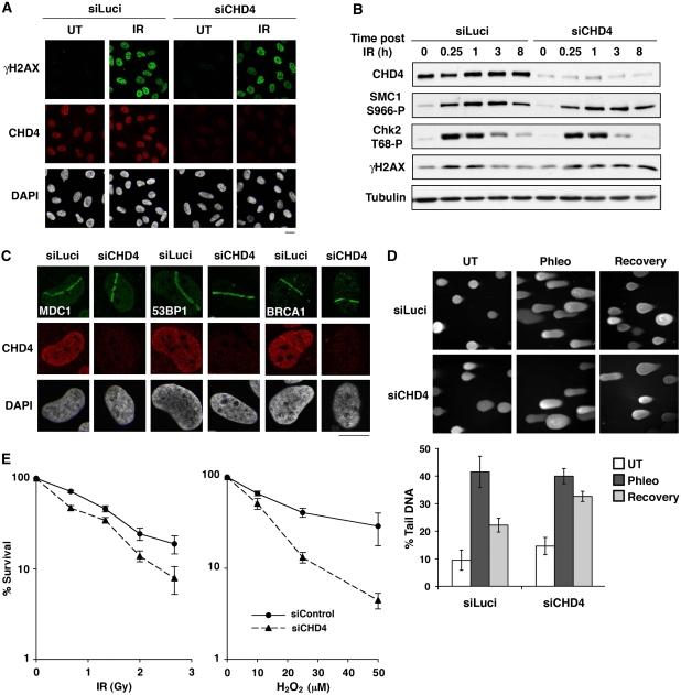
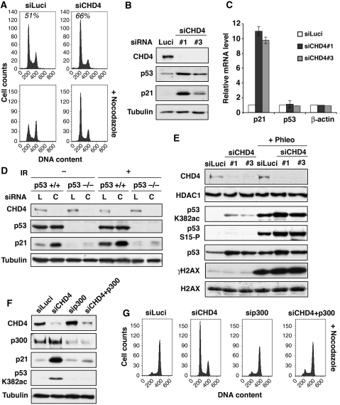
The chromatin remodelling factor chromodomain helicase DNA‐binding protein 4 (CHD4) is a catalytic subunit of the NuRD transcriptional repressor complex. Here, we reveal novel functions for CHD4 in the DNA‐damage response (DDR) and cell‐cycle control. We show that CHD4 mediates rapid poly(ADP‐ribose)‐dependent recruitment of the NuRD complex to DNA‐damage sites, and we identify CHD4 as a phosphorylation target for the apical DDR kinase ataxia‐telangiectasia mutated. Functionally, we show that CHD4 promotes repair of DNA double‐strand breaks and cell survival after DNA damage. In addition, we show that CHD4 acts as an important regulator of the G1/S cell‐cycle transition by controlling p53 deacetylation. These results provide new insights into how the chromatin remodelling complex NuRD contributes to maintaining genome stability. This paper adds to the emerging concept that chromatin remodelling contributes to DNA‐damage responses. CHD4, a known NuRD component, is identified as ATM target, localizes to DNA breaks and contributes to repair while associating with PARPylated factors.
Overview
The claims and statistics of this paper haven’t been checked yet.
Run the check — free, no account needed. It reads the open-access text and checks the sources it cites.
Integrity signals
Registry
Cites
Statistics
Figures
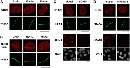
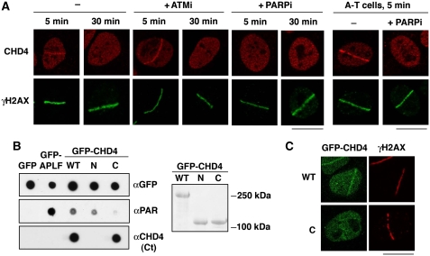
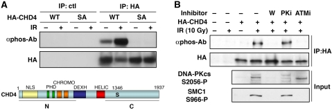
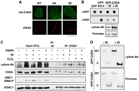
Verification
Claims the paper makes
The claims and statistics of this paper haven’t been checked yet.
Run the check — free, no account needed. It reads the open-access text and checks the sources it cites.
Claims other papers make about this one
Each is checked against this paper’s own text: does it say what the citing paper says it does?
The source backs part of the claim, not all of it — or backs it for a different population, condition or comparison.
In the citing paper
“Other repressive complexes, such as NuRD and histone deacetylases (HDACs) (43)(44)(45)(46), also transiently accumulate at DSBs, supporting this idea.”
From DNA double-strand breaks promote methylation of histone H3 on lysine 9 and transient formation of repressive chromatin (2014) · cites this paper as [43]
Experimental — accuracy not yet measuredThe source backs part of the claim, not all of it — or backs it for a different population, condition or comparison.
In the citing paper
“Thus, immediately after DNA damage, both euchromatin and heterochromatin domains have similar structural organization, including high density of H3K9me3 and the presence of repressive complexes, such as kap-1, HP1α, methyltransferases, HDACs, and CHD3/CHD4 remodeling ATPases (31,32,(43)(44)(45)(46)(47).”
From DNA double-strand breaks promote methylation of histone H3 on lysine 9 and transient formation of repressive chromatin (2014) · cites this paper as [43]
Experimental — accuracy not yet measured
Self-check
Percentages, sums and reported differences re-computed from the paper’s own numbers.
The self-check needs the paper’s full text.
Citations
Cites
Targeting the DNA repair defect in BRCA mutant cells as a therapeutic strategy
Farmer, H., McCabe, N., Lord, C. J., Tutt, A. N. J., Johnson, D. A., Richardson, T. B., Santarosa, M., Dillon, K. J., Hickson, I., Knights, C., et al. (2005) · Nature · doi:10.1038/nature03445 · Cited by 6,683
Self-citationThe DNA-damage response in human biology and disease
Jackson, S. P., & Bartek, J. (2009) · Nature · doi:10.1038/nature08467 · Cited by 6,092 · cited 3×
“In response to DNA damage, cells initiate a coordinated programme of events, termed the DNA-damage response (DDR), which is critical for maintenance of genome integrity and the prevention of ageing and tumourigenesis (Harper and Elledge, 2007; Jackson and Bartek, 2009).”
BackgroundSelf-citationDNA double-stranded breaks induce histone H2AX phosphorylation on serine 139
Rogakou, E. P., Pilch, D. R., Orr, A. H., Ivanova, V. S., & Bonner, W. M. (1998) · Journal of Biological Chemistry · doi:10.1074/jbc.273.10.5858 · Cited by 5,438 · cited 2×
“The work must be attributed back to the original author and commercial use is not permitted without specific permission.”
BackgroundBlinded by the Light: The Growing Complexity of p53
Vousden, K. H., & Prives, C. (2009) · Cell · doi:10.1016/j.cell.2009.04.037 · Cited by 3,163 · cited 2×
“The work must be attributed back to the original author and commercial use is not permitted without specific permission.”
BackgroundATM and ATR Substrate Analysis Reveals Extensive Protein Networks Responsive to DNA Damage
Matsuoka, S., Ballif, B. A., Smogorzewska, A., McDonald, E. R., Hurov, K. E., Luo, J., Bakalarski, C. E., Zhao, Z., Solimini, N., Lerenthal, Y., et al. (2007) · Science · doi:10.1126/science.1140321 · Cited by 3,075 · cited 5×
“Interestingly, proteomic screens in human cells have identified CHD4 as a target for DDR kinases (Matsuoka et al, 2007; Mu et al, 2007; Stokes et al, 2007).”
BackgroundMethodInfluentialThe biology of chromatin remodeling complexes.
Clapier, C. R., & Cairns, B. (2009) · Annual Review of Biochemistry · doi:10.1146/annurev.biochem.77.062706.153223 · Cited by 2,384 · cited 3×
“It is, therefore, possible that such factors will display synergistic and/or antagonistic functions in the regulation of chromatin compaction at DNA-damage sites, as happens in the context of transcription (Ramirez-Carrozzi et al, 2006; Clapier and Cairns, 2009).”
BackgroundAdvances in autism genetics: on the threshold of a new neurobiology
Abrahams, B. S., & Geschwind, D. H. (2008) · Nature Reviews Genetics · doi:10.1038/nrg2346 · Cited by 1,866
The DNA Damage Response: Ten Years After
Harper, J. W., & Elledge, S. J. (2007) · Molecular Cell · doi:10.1016/j.molcel.2007.11.015 · Cited by 1,705 · cited 3×
“In response to DNA damage, cells initiate a coordinated programme of events, termed the DNA-damage response (DDR), which is critical for maintenance of genome integrity and the prevention of ageing and tumourigenesis (Harper and Elledge, 2007; Jackson and Bartek, 2009).”
BackgroundKruse, J.-P., & Gu, W. (2009) · Cell · doi:10.1016/j.cell.2009.04.050 · Cited by 1,610 · cited 3×
“The work must be attributed back to the original author and commercial use is not permitted without specific permission.”
BackgroundPARP inhibition: PARP1 and beyond
Rouleau, M., Patel, A., Hendzel, M. J., Kaufmann, S. H., & Poirier, G. G. (2010) · Nature Reviews Cancer · doi:10.1038/nrc2812 · Cited by 1,298 · cited 1×
“…DNA nicks also activate poly(ADP-ribose) poly- merase (PARP) enzymes that modify target proteins with poly(ADP-ribose) chains (PAR chains) at DNA-damage sites, thereby stimulating the recruitment and/or activity of repair factors (Malanga and Althaus, 2005; Hakme et al, 2008; Rouleau et al, 2010).”
Background
Cited by
Sorted by citations
Items 201–300 of 374
CHD4 mutations promote endometrial cancer stemness by activating TGF-beta signaling.
Li, Y., Liu, Q., McGrail, D. J., Dai, H., Li, K., & Lin, S. (2018) · Europe PMC (PubMed Central) · Cited by 29
Methylomic Signatures of High Grade Serous Ovarian Cancer
Cárdenas, H., Fang, F., Jiang, G., Perkins, S. M., Zhang, C., Emerson, R. E., Hutchins, G., Keer, H. N., Liu, Y., Matei, D. E., et al. (2020) · Epigenetics · doi:10.1080/15592294.2020.1853402 · Cited by 29
Pal, D., Raj, K., Nandi, S. S., Sinha, S., Mishra, A., Mondal, A., Lagoa, R., Burcher, J. T., & Bishayee, A. (2023) · Cancers · doi:10.3390/cancers15102808 · Cited by 29
Hou, M., Luo, C., Chang, T., Hung, W., Chen, T.-Y., Tsai, Y.-L., Chai, C., & Pan, M. (2017) · Experimental Cell Research · doi:10.1016/j.yexcr.2017.08.029 · Cited by 28
Tu, Z., Wang, C., Davis, A. K., Hu, M., Zhao, C., Xin, M., Lu, Q. R., & Zheng, Y. (2021) · Blood · doi:10.1182/blood.2020009997 · Cited by 32
Wang, S., Li, L., Liang, Q., Ye, Y., Lan, Z., Dong, Q., Chen, A., Fu, M., Li, Y., Liu, X., et al. (2022) · Journal of Molecular and Cellular Cardiology · doi:10.1016/j.yjmcc.2022.10.009 · Cited by 26
The nucleosome: orchestrating DNA damage signaling and repair within chromatin
Agarwal, P., & Miller, K. M. (2016) · Biochemistry and Cell Biology · doi:10.1139/bcb-2016-0017 · Cited by 28
HECTD1 promotes base excision repair in nucleosomes through chromatin remodelling
Bennett, L. G., Madders, E. C. E. T., & Parsons, J. L. (2019) · Nucleic Acids Research · doi:10.1093/nar/gkz1129 · Cited by 27
Damage site chromatin: open or closed?
Ball, A. R., & Yokomori, K. (2011) · Current Opinion in Cell Biology · doi:10.1016/j.ceb.2011.03.012 · Cited by 28
“Both Jackson’s and Elledge’s groups showed that CHD4 directly interacts with PAR and that the NuRD complex is recruited in a PAR-dependent manner to the damage sites [40 ,42].”
BackgroundEpigenetic modifications in acute myeloid leukemia: The emerging role of circular RNAs (Review)
Issah, M. A., Wu, D., Zhang, F., Zheng, W., Liu, Y., Fu, H., Zhou, H., Chen, R., & Shen, J. (2021) · International Journal of Oncology · doi:10.3892/ijo.2021.5287 · Cited by 26
Cruz, G. M. S., Kong, X., Silva, B. A., Khatibzadeh, N., Thai, R., Berns, M. W., & Yokomori, K. (2015) · Nucleic Acids Research · doi:10.1093/nar/gkv976 · Cited by 29
Multiple functions of PARP1 in the repair of DNA double strand breaks
Ortega, R., Bitler, B. G., & Arnoult, N. (2025) · DNA repair · doi:10.1016/j.dnarep.2025.103873 · Cited by 27
SHPRH regulates rRNA transcription by recognizing the histone code in an mTOR-dependent manner
Lee, D., An, J., Park, Y.-U., Liaw, H., Woodgate, R., Park, J. H., & Myung, K. (2017) · Proceedings of the National Academy of Sciences · doi:10.1073/pnas.1701978114 · Cited by 27
Profound synthetic lethality between SMARCAL1 and FANCM
Feng, S., Liu, K., Shang, J., Hoeg, L., Pastore, G., Yang, W. E., Roy, S., Sastre-Moreno, G., Young, J. T. F., Wu, W., et al. (2024) · Molecular Cell · doi:10.1016/j.molcel.2024.10.016 · Cited by 24
A di-acetyl-decorated chromatin signature couples liquid condensation to suppress DNA end synapsis
Bao, K., Ma, Y., Li, Y., Shen, X., Zhao, J., Tian, S., Zhang, C., Liang, C., Zhao, Z., Yang, Y., et al. (2024) · Molecular Cell · doi:10.1016/j.molcel.2024.02.002 · Cited by 24
Poly-ADP-ribosylation signaling during DNA damage repair
Timinszky, G. (2015) · Frontiers in bioscience · doi:10.2741/4318 · Cited by 26
“The Nucleosome Remodeling Deacetylase (NuRD) complex and several Polycomb Group (PcG) proteins are recruited to DNA lesions in a PARylation-dependent manner (49, 50).”
BackgroundMethodInfluentialMechanism of DNA replication fork breakage and PARP1 hyperactivation during replication catastrophe
Ortega, P., Bournique, E., Li, J., Sanchez, A., Santiago, G., Harris, B. R., Striepen, J., Maciejowski, J., Green, A. M., & Buisson, R. (2025) · Science Advances · doi:10.1126/sciadv.adu0437 · Cited by 22
“In addition, PARP1 can directly modify chromatin organization around the DNA breaks by facilitating the recruitment and regulation of chromatin remodelers ( 53 – 56 ).”
Guilty as CHARGED: p53's expanding role in disease
Nostrand, J. L. V., & Attardi, L. D. (2014) · Cell Cycle · doi:10.4161/15384101.2014.987627 · Cited by 25
“In addition, Chd4 regulates p53 at the post-translational level by deacetylating p53, such that Chd4 loss enhances p53 acetylation and activity.(56) In addition, embryos with duplication of 1p36, which contains the tumor suppressor gene Chd5, exhibit enhanced senescence and apoptosis as well as perinatal lethality, wh…”
BackgroundDNA-damage response in chromatin of ribosomal genes and the surrounding genome
Foltánková, V., Legartová, S., Kozubek, S., Hofer, M., & Bártová, E. (2013) · Gene · doi:10.1016/j.gene.2013.03.108 · Cited by 24
Knockdown of GATAD2A suppresses cell proliferation in thyroid cancer in vitro
Wang, Z., Kang, J., Deng, X., Guo, B., Wu, B., & Fan, Y. (2017) · Oncology Reports · doi:10.3892/or.2017.5436 · Cited by 23
“Moreover, activation of PARP is a response to DNA damaging compounds and acts as a target of caspase-3 for degradation.”
BackgroundInfluentialKlanert, G., Fernández, D. J., Weinguny, M., Eisenhut, P., Buehler, E. C., Melcher, M., Titus, S. A., Diendorfer, A. B., Gludovacz, E., Jadhav, V., et al. (2019) · Scientific Reports · doi:10.1038/s41598-019-45159-2 · Cited by 22
Yang, S. F., Nelson, C. B., Wells, J., Fernando, M., Lu, R., Allen, J. A. M., Malloy, L., Lamm, N., Murphy, V. J., Mackay, J. P., et al. (2024) · Nature Communications · doi:10.1038/s41467-024-46578-0 · Cited by 22
Hirota, A., Nakajima‐Koyama, M., Ashida, Y., & Nishida, E. (2018) · Journal of Biological Chemistry · doi:10.1074/jbc.ra118.004086 · Cited by 21
“6B), which is consistent with the previous report using human cancer cells (37).”
BackgroundResultMouse Chd4-NURD is required for neonatal spermatogonia survival and normal gonad development
de Castro, R. O., Carbajal, A., de Almeida, L. P., Goitea, V. E., Griffin, C. T., & Pezza, R. J. (2022) · Epigenetics & Chromatin · doi:10.1186/s13072-022-00448-5 · Cited by 21
Biology of the Mi-2/NuRD Complex in SLAC (Stemness, Longevity/Ageing, and Cancer)
Zhang, Y. (2011) · Gene Regulation and Systems Biology · doi:10.4137/grsb.s6510 · Cited by 22
New factors in mammalian DNA repair—the chromatin connection
Raschellà, G., Melino, G., & Malewicz, M. (2017) · Oncogene · doi:10.1038/onc.2017.60 · Cited by 21
Distinct Requirements of CHD4 during B Cell Development and Antibody Response
Yen, W.-F., Sharma, R., Cols, M., Lau, C. M., Chaudhry, A., Chowdhury, P., Yewdell, W. T., Vaidyanathan, B., Sun, A. L., Coffre, M., et al. (2019) · Cell Reports · doi:10.1016/j.celrep.2019.04.011 · Cited by 22
“The NuRD complex also contains the HDAC1/2 deacetylases, which could also influence p53 stability via regulating its acetylation (Hirota et al., 2019; Polo et al., 2010).”
Sweet Melody or Jazz? Transcription Around DNA Double-Strand Breaks
Long, Q., Liu, Z., & Gullerová, M. (2021) · Frontiers in Molecular Biosciences · doi:10.3389/fmolb.2021.655786 · Cited by 19
“Moreover, PARP1 recruits repressive chromatin modifiers, such as CHD4 (Polo et al., 2010) and NuRD (nucleosome remodeling and histone deacetylation complex) to DSBs (Chou et al.”
BackgroundSriram, C. S., Jangra, A., Kasala, E. R., Bodduluru, L. N., & Bezbaruah, B. K. (2014) · Neurochemistry International · doi:10.1016/j.neuint.2014.07.001 · Cited by 19
Kwintkiewicz, J., Padilla‐Banks, E., Jefferson, W. N., Jacobs, I. M., Wade, P. A., & Williams, C. J. (2011) · Biology of Reproduction · doi:10.1095/biolreprod.111.096032 · Cited by 19
“The NuRD complex also participates with cohesin in the cellular response to DNA damage, perhaps by affecting cell cycle checkpoints [20–24].”
BackgroundCHD4 as an important mediator in regulating the malignant behaviors of colorectal cancer
Chang, C., Huang, C., Chang, S., Wu, C., Chen, H., Luo, C., & Yip, H. (2021) · International Journal of Biological Sciences · doi:10.7150/ijbs.56976 · Cited by 18
“It is involved in the regulation of cell cycle and DNA repair [18-20].”
BackgroundMin, S., Lee, H., Ji, J., Heo, Y., Kim, Y., Chae, S., Choi, Y. W., Kang, H. C., Nakanishi, M., & Cho, H. (2021) · Nucleic Acids Research · doi:10.1093/nar/gkab1093 · Cited by 18
“Previous study reported that CHD4, a component of the NuRD complex, facilitates HDAC1 recruitment at DSB sites, and CHD4 is recruited in a PARP-dependent manner (42).”
CHD4 drives gastric cancer metastasis via MYH9/GSK3β/β-catenin axis and WNT/EMT pathway activation
Shi, Y., Zhao, Z., Zhou, S., Zhou, Z., Huang, Z., Zhou, Z., & Zhang, C. (2025) · Cancer Letters · doi:10.1016/j.canlet.2025.217813 · Cited by 17
Maintenance of Genome Integrity by Mi2 Homologs CHD-3 and LET-418 in Caenorhabditis elegans
Turcotte, C. A., Sloat, S. A., Rigothi, J. A., Rosenkranse, E., Northrup, A. L., Andrews, N. P., & Checchi, P. M. (2018) · Genetics · doi:10.1534/genetics.118.300686 · Cited by 17
“Mi2 facilitates repair of meiotic DSBs As several studies have reporteda role forNuRD in response to DNA damage in somatic tissue (Larsen et al. 2010; Polo et al. 2010; Smeenk et al. 2010; Gong et al. 2015), we questioned whether a similar scenario could occur in adult germ cells.”
BackgroundLiu, J., Li, B., Li, W., Pan, T., Diao, Y.-P., & Wang, F. (2022) · Frontiers in Molecular Biosciences · doi:10.3389/fmolb.2022.808162 · Cited by 17
“Their inactivation leads to hyperacetylation of p53 and then boosts p53 stability and transcriptional activity (Ito et al., 2002; Polo et al., 2010; Chen et al., 2015).”
BackgroundThe fast-growing business of Serine ADP-ribosylation
Longarini, E. J., & Matić, I. (2022) · DNA repair · doi:10.1016/j.dnarep.2022.103382 · Cited by 18
Zhang, Q., Zhu, F., Yin, T., Shi, D., & Zhang, J. (2024) · Scientific Reports · doi:10.1038/s41598-024-69233-6 · Cited by 17
Chromatin Remodeling Complexes
Clapier, C. R., & Cairns, B. R. (2013) · Springer eBooks · doi:10.1007/978-1-4614-8624-4_3 · Cited by 18
Kaufmann, T., Herbert, S., Hackl, B., Besold, J. M., Schramek, C., Gotzmann, J., Elsayad, K., & Slade, D. (2020) · Nucleic Acids Research · doi:10.1093/nar/gkaa859 · Cited by 17
“CHD4 recruitment to laser damage sites was reduced in the presence of the PARP1 catalytic dead mutant E988K, confirming that CHD4 recruitment is PAR dependent (Supplementary Figure S5C) (48,49).”
BackgroundMta2 promotes Tipin-dependent maintenance of replication fork integrity
Errico, A., Aze, A., & Costanzo, V. (2014) · Cell Cycle · doi:10.4161/cc.29157 · Cited by 16
Bauer, T. L., Collmar, K., Kaltofen, T., Loeffler, A.-K., Decker, L., Mueller, J., Pinter, S., Eisler, S. A., Mahner, S., Fraungruber, P., et al. (2021) · Cancers · doi:10.3390/cancers13153801 · Cited by 16
“The regulation of gene expression activity and DDR are two relevant functions of the NuRD complex which could lead to the observed anti-proliferative effect upon suppression of MBD3 in the presence of cPt.”
BackgroundInfluentialMarques, J. G., Pavlovic, B., Ngo, Q. A., Pedot, G., Roemmele, M., Volken, L., Kisele, S., Perbet, R., Wachtel, M., & Schäfer, B. W. (2023) · Cancer Research · doi:10.1158/0008-5472.can-22-3950 · Cited by 16
Liu, Z., Ajit, K., Wu, Y., Zhu, W., & Gullerová, M. (2024) · The EMBO Journal · doi:10.1038/s44318-024-00111-7 · Cited by 16
“Various speci fi c NuRD subunits are rapidly recruited to loci of DNA damage (Chou et al, 2010; Larsen et al, 2010; Polo et al, 2010; Smeenk et al, 2010).”
BackgroundKong, X., Cruz, G. M. S., Trinh, S., Zhu, X., Berns, M. W., & Yokomori, K. (2018) · Journal of Cell Science · doi:10.1242/jcs.219311 · Cited by 16
“Furthermore, PAR modification at DNA damage sites is critical for the recruitment of chromatin modifying enzymes that promote DNA repair (Ahel et al., 2009; Ayrapetov et al., 2014; Ball and Yokomori, 2011; Chou et al., 2010; Gottschalk et al., 2009; Izhar et al., 2015; Khoury-Haddad et al., 2014; Larsen et al., 2010;…”
BackgroundChromatin Remodeling in the Brain-a NuRDevelopmental Odyssey
Larrigan, S., Shah, S. J., Fernandes, J. A. L., & Mattar, P. A. (2021) · International Journal of Molecular Sciences · doi:10.3390/ijms22094768 · Cited by 18
“In cell lines, CHD4 has been shown to participate in the DNA damage response [78–80], and CHD4 deficiency has been repeatedly shown to trigger to ATM-dependent intra-S-phase checkpoint activation [31,79,80].”
BackgroundLaser Microirradiation to Study In Vivo Cellular Responses to Simple and Complex DNA Damage
Kong, X., Cruz, G. M. S., Silva, B. A., Wakida, N. M., Khatibzadeh, N., Berns, M. W., & Yokomori, K. (2018) · Journal of Visualized Experiments · doi:10.3791/56213 · Cited by 17
NEAT1 promotes genome stability via m 6 A methylation-dependent regulation of CHD4Preprint
Mamontova, V., Trifault, B., Gribling-Burrer, A.-S., Bohn, P. S., Boten, L., Preckwinkel, P., Gallant, P., Solvie, D., Ade, C. P., Papadopoulos, D., et al. (2024) · Genes & Development · doi:10.1101/gad.351913.124 · Cited by 16
“The chromodomain helicase DNA binding protein 4 (CHD4) histone deacetylase caught our attention, as it both promotes DSBR and binds several lncRNAs, including NEAT1 (Polo et al. 2010; Hendrickson et al. 2016; Zhao et al. 2018).”
BackgroundThe neuro-glial-vascular interrelations in genomic instability symptoms
Barzilai, A. (2011) · Mechanisms of Ageing and Development · doi:10.1016/j.mad.2011.06.001 · Cited by 15
Wang, J., Zhong, F., Li, J., Yue, H., Li, W., & Lü, X. (2023) · Journal of Translational Medicine · doi:10.1186/s12967-022-03854-1 · Cited by 15
MKP1 phosphatase mediates G1-specific dephosphorylation of H3Serine10P in response to DNA damage
Sharma, A. K., Khan, S. A., Sharda, A., Reddy, D., & Gupta, S. (2015) · Mutation research. Fundamental and molecular mechanisms of mutagenesis · doi:10.1016/j.mrfmmm.2015.06.001 · Cited by 15
Yamada, M., Sato, N., Ikeda, S., Arai, T., Sawabe, M., Mori, S., Yamada, Y., Muramatsu, M., & Tanaka, M. (2014) · Genes Chromosomes and Cancer · doi:10.1002/gcc.22227 · Cited by 14
“VC 2014 Wiley Periodicals, Inc. GENES, CHROMOSOMES & CANCER 54:122–128 (2015) et al., 2010; Li and Kumar, 2010; Polo et al., 2010; Stanley et al., 2013).”
BackgroundThe Expanding Mi-2/NuRD Complexes: A Schematic Glance
Zhang, Y., & Li, Y. (2010) · Proteomics Insights · doi:10.4137/pri.s6329 · Cited by 15
The Role of the Neuro-Astro-Vascular Unit in the Etiology of Ataxia Telangiectasia
Meshulam, L., Galron, R., Kanner, S., Pittà, M. D., Bonifazi, P., Goldin, M. M., Frenkel, D., Ben‐Jacob, E., & Barzilai, A. (2012) · Frontiers in Pharmacology · doi:10.3389/fphar.2012.00157 · Cited by 14
Dasgupta, A., Nandi, S., Gupta, S. D., Roy, S., & Das, C. (2024) · Biochimica et Biophysica Acta (BBA) - Gene Regulatory Mechanisms · doi:10.1016/j.bbagrm.2024.195033 · Cited by 14
Connecting Chromatin Modifying Factors to DNA Damage Response
Lai, W., Li, H., Liu, S., & Tao, Y. (2013) · International Journal of Molecular Sciences · doi:10.3390/ijms14022355 · Cited by 13
“Moreover, CHD4 functions as a key regulator of the G1/S cell cycle transition by p53 deacetylation [32].”
Herpesvirus DNA polymerase processivity factors: Not just for DNA synthesis
Cohan, B., & Frappier, L. (2021) · Virus Research · doi:10.1016/j.virusres.2021.198394 · Cited by 13
MTA family of proteins in DNA damage response: mechanistic insights and potential applications
Li, D., Yang, Y., & Kumar, R. (2014) · Cancer and Metastasis Reviews · doi:10.1007/s10555-014-9524-2 · Cited by 13
“Consequently, knockdown of CHD4 sensitizes cells to DNA damage agents and deregulates cell cycle progression [90, 92].”
The Nucleosome Remodeling and Deacetylase Complex in Development and Disease
Basta, J. M., & Rauchman, M. I. (2017) · Elsevier eBooks · doi:10.1016/b978-0-12-800802-7.00003-4 · Cited by 14
Serafim, R. B., Cardoso, C. C., Storti, C. B., da Silva, P., Qi, H., Parasuram, R., Navegante, G., Peron, J. P. S., Silva, W. A., Espreáfico, E. M., et al. (2024) · Oncogene · doi:10.1038/s41388-024-02937-1 · Cited by 13
Histone Displacement during Nucleotide Excision Repair
Dinant, C., Bártek, J., & Bekker‐Jensen, S. (2012) · International Journal of Molecular Sciences · doi:10.3390/ijms131013322 · Cited by 13
“chromatin relaxation is CHD4, which has been shown to be recruited to sites of DSBs in a PARP-dependent manner (see also below) [43].”
BackgroundCHD4 acts as a critical regulator in the survival of spermatogonial stem cells in mice
Li, P., Tang, J., Yu, Z., Jin, C., Wang, Z., Li, M., Zou, D., Mang, X., Liu, J., Lü, Y., et al. (2022) · Biology of Reproduction · doi:10.1093/biolre/ioac162 · Cited by 11
“At the same time, in Chd4MKO mice, the expression of cell cycle-related gene Trp53 was upregulated following Chd4 knockout, which is consistent with the established literature.”
BackgroundInfluentialSherrard, A., Bishop, P., Panagi, M., Villagomez, M. B., Alibhai, D., & Kaidi, A. (2018) · Biology Open · doi:10.1242/bio.031476 · Cited by 11
“S1B), indicative of their incorporation into chromatin (Polo et al., 2010).”
Self-citationBuddaseth, S., Göttmann, W., Blasczyk, R., & Huyton, T. (2013) · Biochimica et Biophysica Acta (BBA) - Molecular Cell Research · doi:10.1016/j.bbamcr.2013.02.001 · Cited by 11
“Knockdown studies and BrdU incorporation assays demonstrated a specific role of CHD4 in cell cycle progression, in particular as a critical regulator of the G1 to S (G1/S) transition through the control of p53 deacetylation [21,22].”
BackgroundRemodeler Catalyzed Nucleosome Repositioning: Influence of Structure and Stability
Morgan, A. M., LeGresley, S. E., & Fischer, C. J. (2020) · International Journal of Molecular Sciences · doi:10.3390/ijms22010076 · Cited by 11
Abad, C., Robayo, M. C., Moreno, M. D. M. M., Bernardi, M. T., Otero, M. G., Kosanovic, C., Griswold, A. J., Pierson, T. M., Walz, K., & Young, J. I. (2024) · Translational Psychiatry · doi:10.1038/s41398-023-02678-x · Cited by 9
“NuRD complexes have been implicated in regulating gene transcription, genome stability and DNA repair [11 – 14], as well as controlling events related to cell cycle progression, stem cell differentiation, and cerebral corticogenesis [15, 16].”
BackgroundVGLL3 modulates chemosensitivity through promoting DNA double-strand break repair
Wu, W. J., Fan, Z., Fu, H., Ma, X., Wang, D., Liu, H., Zhang, C., Zheng, H., Yang, Y., Wu, H., et al. (2024) · Science Advances · doi:10.1126/sciadv.adr2643 · Cited by 9
“HA-CHD4 and pcDNA4TO- GFP-CHD4 were provided by S. Jackson and J. Lukas, respectively ( 51 , 52 ).”
Morawska, M., Kiełbus, M., Paziewska, M., Szelest, M., Karczmarczyk, A., Zaleska, J., Własiuk, P., Giannopoulos, K., & Grząśko, N. (2024) · Cancer Genetics · doi:10.1016/j.cancergen.2024.11.001 · Cited by 11
ATM and ATR checkpoint kinase pathways: A concise review
Chauhan, N., Wagh, V., Joshi, P., & Jariyal, H. (2020) · Advances in Human Biology · doi:10.4103/aihb.aihb_78_19 · Cited by 9
The Chd4 subunit of the NuRD complex regulates Pdx1-controlled genes involved in β-cell function
Davidson, R. K., Weaver, S. A., CASEY, N., Kanojia, S., Hogarth, E., Aguirre, R. S., Sims, E. K., Evans‐Molina, C., & Spaeth, J. M. (2022) · Journal of Molecular Endocrinology · doi:10.1530/jme-22-0011 · Cited by 11
The Tale of CHD4 in DNA Damage Response and Chemotherapeutic Response
Lin, S.-Y., Zhang, J., & Shih, D. J. H. (2019) · Cancer Research and Cellular Therapeutics · doi:10.31579/2640-1053/052 · Cited by 9
“In addition, CHD4 can be phosphorylated by the DNA damage response kinases ATM [34, 35] and ATR [36], and in turn CHD4 can also regulate the Tip60dependent phosphorylation of ATM [37] in response to DNA damage.”
BackgroundAberrant chromatin remodeling in gynecological cancer (Review)
Okawa, R., Banno, K., Iida, M., Yanokura, M., Takeda, T., Iijima, M., Irie, H., Nakamura, K., Adachi, M., Umene, K., et al. (2017) · Oncology Letters · doi:10.3892/ol.2017.6891 · Cited by 9
“CHD4 is a catalytic subunit of the NuRD complex that inhibits transcription and repairs DNA damage (52).”
Chromatin Remodulator CHD4: A Potential Target for Cancer Interception
Goswami, K., Venkatachalam, K., Singh, S. P., Rao, C. V., & Madka, V. (2025) · Genes · doi:10.3390/genes16020225 · Cited by 8
“However, the recent study results cannot explain Loss of CHD4 also results in CDC25A degradation and p21 accumulation, leading to cell cycle delay [34,43,45]. the entire complexity of CHD4’s contribution to DNA damage response and cell viability [45,47,48].”
InfluentialThe crosstalk of m6A-modified RNA with DNA damage repair
Qu, F., & Liu, Y. (2025) · Trends in Biochemical Sciences · doi:10.1016/j.tibs.2025.06.012 · Cited by 8
NSUN2 facilitates DICER cleavage of DNA damage-associated R-loops to promote repair
Alagia, A., Ajit, K., Fazio, A. D., Long, Q., & Gullerová, M. (2025) · Nature Communications · doi:10.1038/s41467-025-63220-9 · Cited by 8
Jurado, E. P., Smith, R., Bigot, N., Chapuis, C., Timinszky, G., & Huet, S. (2024) · Molecular Biology of the Cell · doi:10.1091/mbc.e23-07-0281 · Cited by 8
“Several chromatin remodelers recruit early to lesions during the DNA damage response (DDR), facilitating efficient double-strand break (DSB) repair via homologous recombination and non homologous end-joining pathways (Lan et al. , 2010; Polo et al. , 2010; Luijsterburg et al. , 2016).”
BackgroundFrequency and Nature of Genomic Alterations in ERBB2-Altered Urothelial Bladder Cancer
Leary, J. B., Enright, T. J., Bakaloudi, D. R., Basnet, A., Bratslavsky, G., Jacob, J. M., Spiess, P. E., Li, R., Necchi, A., Kamat, A. M., et al. (2024) · Targeted Oncology · doi:10.1007/s11523-024-01056-x · Cited by 9
“Loss of CHD4 leads to accumulation of DNA damage [62]; CDH4 appears to be involved in the repair of both single and double-strand breaks, as well as oxidative damage repair, and exerts cell cycle control at the G1/S transition via regulation of p53 deacetylation [63].”
Kesavan, P. S., Bohra, D., Roy, S., & Mazumder, A. (2020) · Molecular Biology of the Cell · doi:10.1091/mbc.e19-08-0417 · Cited by 8
“Molecular Biology of the Cell damage may be relaxed as a response to damage (Park et al., 2006; Polo et al. 2010).”
BackgroundGermline Genetic Variants and Lung Cancer Survival in African Americans
Jones, C. C., Bush, W. S., Crawford, D. C., Wenzlaff, A. S., Schwartz, A. G., Wiencke, J. K., Wrensch, M., Blot, W. J., Chanock, S. J., Grogan, E. L., et al. (2017) · Cancer Epidemiology Biomarkers & Prevention · doi:10.1158/1055-9965.epi-16-0998 · Cited by 8
“Additionally, CHD4 plays a role in DNA damage response and cell cycle progression (48, 49).”
Provencher, L., Nartey, W., Brownlee, P. M., Atkins, A. W., Gagné, J., Baudrier, L., Ting, N. S. Y., Piett, C. G., Fang, S., Pearson, D. D., et al. (2025) · Nature Communications · doi:10.1038/s41467-025-56085-5 · Cited by 8
A LANA peptide inhibits tumor growth by inducing CHD4 protein cleavage and triggers cell death
Miura, H., Wang, K.-H., Inagaki, T., Chuang, F. Y. S., Shimoda, M., Izumiya, C., Watanabe, T., Davis, R. R., Tepper, C. G., Komaki, S., et al. (2024) · Cell chemical biology · doi:10.1016/j.chembiol.2024.10.003 · Cited by 7
Liu, S., Tao, Y., Chen, X., & Cao, Y. (2011) · Molecular Biology Reports · doi:10.1007/s11033-011-1435-5 · Cited by 7
“novel regulator [54].”
Zhou, J., Zhu, D., Wang, Y., Wang, Z., Zhang, N., Huang, X., Zhang, Y., Wang, Y., Wu, X., & Tian, Y. (2025) · Science China Life Sciences · doi:10.1007/s11427-025-2954-3 · Cited by 7
“While phosphoproteomics studies have identified phosphorylation sites on NuRD components, the physiological relevance of these modifications in response to stress or aging remains largely unexplored (Bouazoune and Brehm, 2005; Polo et al., 2010; Sun et al., 2007).”
Permatasari, L. I., Afifah, N. N., Ishmatullah, M. H., Intania, R., Halimah, E., & Barliana, M. I. (2025) · Cancer Management and Research · doi:10.2147/cmar.s501420 · Cited by 6
“36 ,37 In addition to aiding recruitment, XRCC1 prevents excessive PARP-1 activity, which can lead to DNA structural entrapment during repair and trigger additional damage.”
CHD4 plays a critical role in arsenite-induced oxidative damage in human urothelial carcinoma
Chang, S., Bin, P.-J., Luo, C., & Chai, C. (2022) · Pathology - Research and Practice · doi:10.1016/j.prp.2022.154173 · Cited by 6
NuRD Alert! NuRD Regulates the DNA Damage Response
Smeenk, G., & van Attikum, H. (2011) · Epigenomics · doi:10.2217/epi.10.79 · Cited by 6
Chromatin Remodelers and Their Way of Action
Manelytė, L., & Längst, G. (2013) · InTech eBooks · doi:10.5772/55683 · Cited by 6
“Several studies demonstrated the targeting of Chd4 to sites of DNA double strand breaks in a PARP dependent manner [87].”
BackgroundChromodomain helicase DNA binding protein 4 in cell fate decisions
Laureano, A., Kim, J.-H., Martinez, E. A., & Kwan, K. Y. (2023) · Hearing Research · doi:10.1016/j.heares.2023.108813 · Cited by 5
Zinc finger transcription factor CASZ1b is involved in the DNA damage response in live cells
Liu, Z., Kruhlak, M. J., & Thiele, C. J. (2023) · Biochemical and Biophysical Research Communications · doi:10.1016/j.bbrc.2023.04.085 · Cited by 4
γH2AX prefers late replicating metaphase chromosome regions
Reyes-Ábalos, A. L., Liddle, P., Folle, G. A., & Tomaso, M. V. D. (2018) · Mutation Research/Genetic Toxicology and Environmental Mutagenesis · doi:10.1016/j.mrgentox.2018.06.001 · Cited by 6
The PARP1/ARTD1-Mediated Poly-ADP-Ribosylation and DNA Damage Repair in B Cell Diversification
Lasola, J. J., Hodgson, A., Sun, X., & Wan, F. (2014) · Antibodies · doi:10.3390/antib3010037 · Cited by 4
“Conversely, recent findings outlined a role for PARP1/ARTD1 in transcriptional silencing by triggering chromatin compaction, which can be best exemplified by the finding that several components of the NuRD complex, including chromodomain helicase DNA binding protein 4 (CHD4) are recruited by PARP1/ARTD1 and execute hi…”
Genetically altered cancer epigenome
Tang, M., Luo, H., & Lu, J. (2015) · Elsevier eBooks · doi:10.1016/b978-0-12-799958-6.00012-3 · Cited by 4
Cell Cycle and DNA Damage Response in Postmitotic Neurons
Kruman, I. I. (2011) · InTech eBooks · doi:10.5772/20859 · Cited by 3
“It is involved in buildup of the chromatin remodeling factors ALC1 and CHD4 (Ahel et al., 2009; Chou et al., 2010; Gottschalk et al., 2009; Polo et al., 2010), the Polycomb histone-modifying complex (Chou et al., 2010), and the histone variant macro H2A (Timinszky et al., 2009).”
BackgroundOncogenic acidic nuclear phosphoproteins ANP32C/D are novel clients of heat shock protein 90
Yuzefovych, Y., Blasczyk, R., & Huyton, T. (2015) · Biochimica et Biophysica Acta (BBA) - Molecular Cell Research · doi:10.1016/j.bbamcr.2015.06.007 · Cited by 3
Mouse CHD4-NURD is required for neonatal spermatogonia survival and normal gonad developmentPreprint
de Castro, R. O., Previato, L., Carbajal, A., Goitea, V. E., Griffin, C. T., & Pezza, R. J. (2019) · bioRxiv (Cold Spring Harbor Laboratory) · doi:10.1101/687137 · Cited by 3
“Given its positive impact on survival following irradiation, current models predict that CHD4 promotes a permissive chromatin environment for proper DSB repair [9, 11, 29], a model that well could explain the phenotype we observed.”
BackgroundInfluentialKoulle, A., Ogundele, O., Shah, D., Baker, I. M., Lopez, M., Lando, D., Reynolds, N., Ragheb, R., Laue, E. D., & Hendrich, B. (2025) · eLife · doi:10.7554/elife.109280 · Cited by 6
Ebert, L. K., Schloesser, L., Frech, L. E., Jain, M., Dafinger, C., Liebau, M. C., Benzing, T. R., Schermer, B., & Slaats, G. G. G. (2025) · American Journal of Physiology-Renal Physiology · doi:10.1152/ajprenal.00076.2025 · Cited by 2
Soares, J., Ferreira, D., Miranda, A., Gonçalves, M., Relvas‐Santos, M., Brandão, A., Paulo, P., Cotton, S., Freitas, R., Magalhães, M., et al. (2026) · Cell Communication and Signaling · doi:10.1186/s12964-026-02672-y · Cited by 4
Kim, J., Martinez, E. A., Qiu, J., Laureano, A., Hinman, A. M., Ni, J. Z., & Kwan, K. Y. (2024) · bioRxiv (Cold Spring Harbor Laboratory) · doi:10.1101/2024.01.31.578202 · Cited by 2
CHD4 acts as a prognostic factor and drives radioresistance in HPV negative HNSCC
Geyer, F., Geyer, M., Reuning, U., Klapproth, S., Wolff, K., & Nieberler, M. (2024) · Scientific Reports · doi:10.1038/s41598-024-58958-z · Cited by 1
“Together with NuRD, CHD4 controls p53 deacetylation, hence being an important regulator of the G 1 /S transition 44,45 .”
BackgroundZheng, P., Zang, Z., Wang, S., Zeng, C., Pan, Y., Zhang, Y., Yue, S., Liu, S., Huang, H., & Zhao, X. (2025) · Animals · doi:10.3390/ani15182676 · Cited by 1
“A core ATPase subunit of the NuRD (nucleosome remodeling and deacetylase) chromatin remodeling complex is known to play a crucial role in DNA damage repair and cell cycle regulation [27].”
Metadata
- Authors
- Polo, S. E., Kaidi, A., Baskcomb, L., Galanty, Y., & Jackson, S. P.
- Affiliations
- University of Cambridge (GB)
- Corresponding author
- Jackson, Stephen P
- Venue
- The EMBO Journal
- Published
- 2010-08-06
- Volume
- 29
- Issue
- 18
- Pages
- 3130-3139
- ISSN
- 0261-4189 (ISSN-L)
- 1460-2075
- PMC
- PMC2944064
- OpenAlex
- W2020084014
- Cited by
- 374
- References
- 56
- Citations by year
- 2026: 13 · 2025: 14 · 2024: 20 · 2023: 17 · 2022: 26 · 2021: 17 · 2020: 22 · 2019: 18 · 2018: 18 · 2017: 35 · 2016: 24 · 2015: 17 · 2014: 31 · 2013: 34 · 2012: 31
- FWCI
- 13.30
- Type
- journal article
- Language
- en
- Open access
- hybrid
- doi.org
- Licence
- Locations
- The EMBO Journal (publishedVersion, cc-by-nc-sa)
- PubMed Central (publishedVersion, cc-by-nc-sa)
- PubMed (publishedVersion)
- Explore Bristol Research (submittedVersion)
- Funders
- Wellcome Trust
- Cancer Research UK (11224, A5290, A4361)
- European Commission (512113, 201630, LSHG-CT-2005-512113)
- Directorate for Biological Sciences
- Biotechnology and Biological Sciences Research Council
- Topics
- Life Sciences › Biochemistry, Genetics and Molecular Biology › Molecular Biology › DNA Repair Mechanisms
- Life Sciences › Biochemistry, Genetics and Molecular Biology › Molecular Biology › Chromatin Remodeling and Cancer
- Life Sciences › Biochemistry, Genetics and Molecular Biology › Molecular Biology › Genomics and Chromatin Dynamics
- Keywords
- CHD4, chromatin remodeling, NuRD complex, DNA damage response, cell cycle regulation, G1/S transition, p53 deacetylation, ATM phosphorylation, DNA double-strand break repair, poly(ADP-ribose), poly(ADP-ribose) polymerase, genome stability, transcriptional repressor
- MeSH terms
- Animals, Autoantigens, Cell Cycle, DNA Damage, DNA Helicases, DNA-Binding Proteins, Fluorescent Antibody Technique, Histones, Humans, Phosphorylation, Poly Adenosine Diphosphate Ribose, RNA, Messenger, Blotting, Western, Tumor Suppressor Protein p53, Protein Serine-Threonine Kinases, Mice, Knockout, Cell Cycle Proteins, Reverse Transcriptase Polymerase Chain Reaction, Tumor Suppressor Proteins, Chromatin Assembly and Disassembly, Immunoprecipitation, Mice, Mi-2 Nucleosome Remodeling and Deacetylase Complex, Ataxia Telangiectasia Mutated Proteins
About these checks
Registry, retraction, Cites and statistics results are computed, not judged, with one exception: a printed reference that no index lists is read once by a model before it counts as a citation error. Claims are extracted by a model and kept only when their quote appears verbatim in the text; each cited source is then checked, with a model’s help, for whether it says what the claim relies on. Those verdicts can be wrong — read the claim’s quote and the cited source before relying on one.
Statistics and arithmetic are recomputed, not judged.
Confidence bands are shown only for fields calibrated on our annotated test set. None is calibrated yet, so no band is shown.
What counts as a claim ›How we measure confidence ›How we check this ›
Are you an author, or do you see a problem with this page? Request a review
