

The DOI matches its Crossref record; 2 of the 2 references it cites were found in a registry or an index.
Comprehensive genomic characterization of head and neck squamous cell carcinomas
Overview
The claims and statistics of this paper haven’t been checked yet.
Run the check — free, no account needed. It reads the open-access text and checks the sources it cites.
Integrity signals
Registry
Cites
Statistics
Figures
Verification
Claims the paper makes
The claims and statistics of this paper haven’t been checked yet.
Run the check — free, no account needed. It reads the open-access text and checks the sources it cites.
Claims other papers make about this one
Each is checked against this paper’s own text: does it say what the citing paper says it does?
In the citing paper
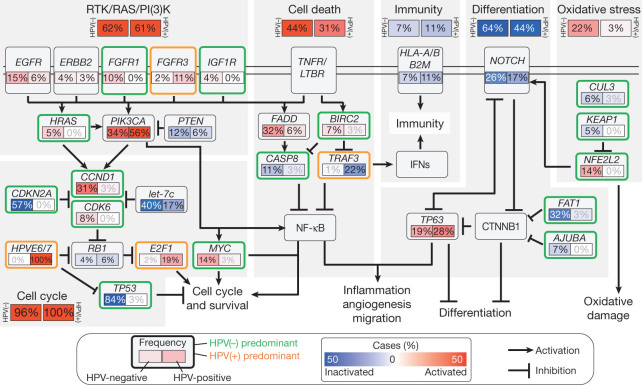
“HPV-mediated cancers are frequently found to have somatic mutations, including those driven by apolipoprotein B mRNA editing catalytic polypeptide-like (APOBEC), copy number variations and large chromosomal rearrangements.”
From The Not-So-Good, the Bad and the Ugly: HPV E5, E6 and E7 Oncoproteins in the Orchestration of Carcinogenesis (2021) · cites this paper as [21]
In the citing paper
“Some of the key genes often mutated in HPV-associated cancers are in PIK3CA (phosphatidylinositol-4,5-bisphosphate 3-kinase catalytic subunit alpha), PTEN (phosphatase and tensin homologue), HLA-A and HLA-B (human leukocyte antigen A and B), TGFβ (transforming growth factor beta), and components of the NOTCH1 and RAS/EGFR/ERK pathways; in contrast mutations are very rarely found in p53 and pRB [13”
From The Not-So-Good, the Bad and the Ugly: HPV E5, E6 and E7 Oncoproteins in the Orchestration of Carcinogenesis (2021) · cites this paper as [21]
In the citing paper
“HPV-positive cancers have fewer somatic alterations and changes in the protein expression profile compared with HPV-negative cancers [21]”
From The Not-So-Good, the Bad and the Ugly: HPV E5, E6 and E7 Oncoproteins in the Orchestration of Carcinogenesis (2021) · cites this paper as [21]
Self-check
Percentages, sums and reported differences re-computed from the paper’s own numbers.
The self-check needs the paper’s full text.
Citations
Cites
Comprehensive genomic characterization of squamous cell lung cancers
The Cancer Genome Atlas Research Network (2012) · Nature · doi:10.1038/nature11404 · Cited by 3,994
Comprehensive molecular characterization of clear cell renal cell carcinoma
The Cancer Genome Atlas Research Network (2013) · Nature · doi:10.1038/nature12222 · Cited by 3,485
Self-citation
Research works printed but not linked: 43
- Ferlay, J. et al. Estimates of worldwide burden of cancer in 2008: GLOBOCAN 2008. Int. J. Cancer 127, 2893–2917 (2010) · DOI
- Ang, K. K. et al. Human papillomavirus and survival of patients with oropharyngeal cancer. N. Engl. J. Med. 363, 24–35 (2010) · DOI
- Stransky, N. et al. The mutational landscape of head and neck squamous cell carcinoma. Science 333, 1157–1160 (2011) · DOI
- Agrawal, N. et al. Exome sequencing of head and neck squamous cell carcinoma reveals inactivating mutations in NOTCH1. Science 333, 1154–1157 (2011) · DOI
- Oganesyan, G. et al. Critical role of TRAF3 in the Toll-like receptor-dependent and -independent antiviral response. Nature 439, 208–211 (2006) · DOI
- Karim, R. et al. Human papillomavirus (HPV) upregulates the cellular deubiquitinase UCHL1 to suppress the keratinocyte’s innate immune response. PLoS Pathog. 9, e1003384 (2013) · DOI
- Eliopoulos, A. G. et al. CD40-induced growth inhibition in epithelial cells is mimicked by Epstein–Barr Virus-encoded LMP1: involvement of TRAF3 as a common mediator. Oncogene 13, 2243–2254 (1996)
- Imbeault, M. et al. Acquisition of host-derived CD40L by HIV-1 in vivo and its functional consequences in the B-cell compartment. J. Virol. 85, 2189–2200 (2011) · DOI
- Ni, C. Z. et al. Molecular basis for CD40 signaling mediated by TRAF3. Proc. Natl Acad. Sci. USA 97, 10395–10399 (2000) · DOI
- Chung, G. T. et al. Constitutive activation of distinct NF-κB signals in EBV-associated nasopharyngeal carcinoma. J. Pathol. 231, 311–322 (2013) · DOI
- Annunziata, C. M. et al. Frequent engagement of the classical and alternative NF-κB pathways by diverse genetic abnormalities in multiple myeloma. Cancer Cell 12, 115–130 (2007) · DOI · Paper page
- Ciriello, G. et al. Emerging landscape of oncogenic signatures across human cancers. Nature Genet. 45, 1127–1133 (2013) · DOI
- Smeets, S. J. et al. Genetic classification of oral and oropharyngeal carcinomas identifies subgroups with a different prognosis. Cellular Oncol. 31, 291–300 (2009)
- Mayo, M. W. et al. Requirement of NF-κB activation to suppress p53-independent apoptosis induced by oncogenic Ras. Science 278, 1812–1815 (1997) · DOI
- Sok, J. C. et al. Mutant epidermal growth factor receptor (EGFRvIII) contributes to head and neck cancer growth and resistance to EGFR targeting. Clin. Cancer Res. 12, 5064–5073 (2006) · DOI
- Kong-Beltran, M. et al. Somatic mutations lead to an oncogenic deletion of met in lung cancer. Cancer Res. 66, 283–289 (2006) · DOI
- Popescu, N. C., DiPaolo, J. A. & Amsbaugh, S. C. Integration sites of human papillomavirus 18 DNA sequences on HeLa cell chromosomes. Cytogenet. Cell Genet. 44, 58–62 (1987) · DOI
- Forbes, S. A. et al. The Catalogue of Somatic Mutations in Cancer (COSMIC). Curr. Protoc. Hum. Genet. Chapter 10, Unit 10.11 (2008)
- Sherry, S. T. et al. dbSNP: the NCBI database of genetic variation. Nucleic Acids Res. 29, 308–311 (2001) · DOI · Paper page
- Morris, L. G. et al. Recurrent somatic mutation of FAT1 in multiple human cancers leads to aberrant Wnt activation. Nature Genet. 45, 253–261 (2013) · DOI
- Haraguchi, K. et al. Ajuba negatively regulates the Wnt signaling pathway by promoting GSK-3β-mediated phosphorylation of β-catenin. Oncogene 27, 274–284 (2008) · DOI
- Nagai, Y. et al. The LIM protein Ajuba is required for ciliogenesis and left-right axis determination in medaka. Biochem. Biophys. Res. Commun. 396, 887–893 (2010) · DOI
- Sun, G. & Irvine, K. D. Ajuba family proteins link JNK to Hippo signaling. Sci. Signal. 6, ra81 (2013)
- Reddy, B. V. & Irvine, K. D. Regulation of Hippo signaling by EGFR-MAPK signaling through Ajuba family proteins. Dev. Cell 24, 459–471 (2013) · DOI
- Kalan, S., Matveyenko, A. & Loayza, D. LIM protein Ajuba participates in the repression of the ATR-mediated DNA damage response. Front. Genet. 4, 95 (2013) · DOI
- Nola, S. et al. Ajuba is required for Rac activation and maintenance of E-cadherin adhesion. J. Cell Biol. 195, 855–871 (2011) · DOI
- Fickie, M. R. et al. Adults with Sotos syndrome: review of 21 adults with molecularly confirmed NSD1 alterations, including a detailed case report of the oldest person. Am. J. Med. Genet. A. 155, 2105–2111 (2011) · DOI
- Wang, G. G., Cai, L., Pasillas, M. P. & Kamps, M. P. NUP98–NSD1 links H3K36 methylation to Hox-A gene activation and leukaemogenesis. Nature Cell Biol. 9, 804–812 (2007) · DOI
- Quintana, R. M. et al. A transposon-based analysis of gene mutations related to skin cancer development. J. Invest. Dermatol. 133, 239–248 (2013) · DOI
- Chung, C. H. et al. Molecular classification of head and neck squamous cell carcinomas using patterns of gene expression. Cancer Cell 5, 489–500 (2004) · DOI · Paper page
- Walter, V. et al. Molecular subtypes in head and neck cancer exhibit distinct patterns of chromosomal gain and loss of canonical cancer genes. PLoS ONE 8, e56823 (2013) · DOI
- Lu, S. L. et al. Loss of transforming growth factor-β type II receptor promotes metastatic head-and-neck squamous cell carcinoma. Genes Dev. 20, 1331–1342 (2006) · DOI
- Oberst, A. & Green, D. R. It cuts both ways: reconciling the dual roles of caspase 8 in cell death and survival. Nature Rev. Mol. Cell Biol. 12, 757–763 (2011) · DOI
- Park, S. J. et al. Opposite role of Ras in tumor necrosis factor-α-induced cell cycle regulation: competition for Raf kinase. Biochem. Biophys. Res. Commun. 287, 1140–1147 (2001) · DOI
- Byers, L. A. et al. An epithelial-mesenchymal transition gene signature predicts resistance to EGFR and PI3K inhibitors and identifies Axl as a therapeutic target for overcoming EGFR inhibitor resistance. Clin. Cancer Res. 19, 279–290 (2013) · DOI · Paper page
- Wong, J. V., Dong, P., Nevins, J. R., Mathey-Prevot, B. & You, L. Network calisthenics: control of E2F dynamics in cell cycle entry. Cell Cycle 10, 3086–3094 (2011) · DOI
- Sanhaji, M. et al. Polo-like kinase 1 inhibitors, mitotic stress and the tumor suppressor p53. Cell Cycle 12, 1340–1351 (2013) · DOI
- Morishita, A. et al. HMGA2 is a driver of tumor metastasis. Cancer Res. 73, 4289–4299 (2013) · DOI
- Bancroft, C. C. et al. Effects of pharmacologic antagonists of epidermal growth factor receptor, PI3K and MEK signal kinases on NF-κB and AP-1 activation and IL-8 and VEGF expression in human head and neck squamous cell carcinoma lines. Int. J. Cancer 99, 538–548 (2002) · DOI
- Yang, X. et al. ΔNp63 versatilely regulates a broad NF-κB gene program and promotes squamous epithelial proliferation, migration, and inflammation. Cancer Res. 71, 3688–3700 (2011) · DOI
- Keating, P. J. et al. Frequency of down-regulation of individual HLA-A and -B alleles in cervical carcinomas in relation to TAP-1 expression. Br. J. Cancer 72, 405–411 (1995) · DOI
- Esteban, F. et al. Lack of MHC class I antigens and tumour aggressiveness of the squamous cell carcinoma of the larynx. Br. J. Cancer 62, 1047–1051 (1990) · DOI
- Dotto, G. P. Crosstalk of Notch with p53 and p63 in cancer growth control. Nature Rev. Cancer 9, 587–595 (2009) · DOI
Cited by
Oncogenic Signaling Pathways in The Cancer Genome Atlas
Sanchez-Vega, F., Mina, M., Armenia, J., Chatila, W. K., Luna, A., La, K. C., Dimitriadoy, S., Liu, D. L., Kantheti, H. S., Saghafinia, S., et al. (2018) · Cell · doi:10.1016/j.cell.2018.03.035 · Cited by 3,201
Puram, S. V., Tirosh, I., Parikh, A. S., Patel, A. P., Yizhak, K., Gillespie, S., Rodman, C., Luo, C. L., Mroz, E. A., Emerick, K. S., et al. (2017) · Cell · doi:10.1016/j.cell.2017.10.044 · Cited by 2,557
Seiwert, T. Y., Burtness, B., Mehra, R., Weiss, J., Berger, R., Eder, J. P., Heath, K., McClanahan, T., Lunceford, J., Gause, C., et al. (2016) · The Lancet Oncology · doi:10.1016/s1470-2045(16)30066-3 · Cited by 1,745
Distinct patterns of somatic genome alterations in lung adenocarcinomas and squamous cell carcinomas
Cancer Genome Atlas Research Network, Campbell, J. D., Alexandrov, A., Kim, J., Wala, J., Berger, A. H., Pedamallu, C. S., Shukla, S. A., Guo, G., Brooks, A. N., et al. (2016) · Nature Genetics · doi:10.1038/ng.3564 · Cited by 1,200
RAS-targeted therapies: is the undruggable drugged?
Moore, A. R., Rosenberg, S. C., McCormick, F., & Malek, S. (2020) · Nature Reviews Drug Discovery · doi:10.1038/s41573-020-0068-6 · Cited by 1,103
Ashley, E. A. (2016) · Nature Reviews Genetics · doi:10.1038/nrg.2016.86 · Cited by 1,116
TGFβ biology in cancer progression and immunotherapy
Derynck, R., Turley, S. J., & Akhurst, R. J. (2021) · Nature Reviews Clinical Oncology · doi:10.1038/s41571-020-0403-1 · Cited by 944
Tebay, L. E., Robertson, H., Durant, S. T., Vitale, S. R., Penning, T. M., Dinkova-Kostova, A. T., & Hayes, J. D. (2015) · Free Radical Biology and Medicine · doi:10.1016/j.freeradbiomed.2015.06.021 · Cited by 837
Metabolic landscape of the tumor microenvironment at single cell resolution
Xiao, Z., Dai, Z., & Locasale, J. W. (2019) · Nature Communications · doi:10.1038/s41467-019-11738-0 · Cited by 524
Rodriguez-Martin, B., Alvarez, E. G., Baez-Ortega, A., Zamora, J., Supek, F., Demeulemeester, J., Santamarina, M., Ju, Y. S., Temes, J., Garcia-Souto, D., et al. (2020) · Nature Genetics · doi:10.1038/s41588-019-0562-0 · Cited by 451
Implications of Genetic and Epigenetic Alterations of CDKN2A (p16 INK4a ) in Cancer
Zhao, R., Choi, B. Y., Lee, M.-H., Bode, A. M., & Dong, Z. (2016) · EBioMedicine · doi:10.1016/j.ebiom.2016.04.017 · Cited by 425
Pan-cancer deconvolution of tumour composition using DNA methylation
Chakravarthy, A., Furness, A., Joshi, K., Ghorani, E., Ford, K., Ward, M. J., King, E. V., Lechner, M., Marafioti, T., Quezada, S. A., et al. (2018) · Nature Communications · doi:10.1038/s41467-018-05570-1 · Cited by 327
The impact of site-specific digital histology signatures on deep learning model accuracy and bias
Howard, F. M., Dolezal, J., Kochanny, S., Schulte, J., Chen, H., Heij, L., Huo, D., Nanda, R., Olopade, O. I., Kather, J. N., et al. (2021) · Nature Communications · doi:10.1038/s41467-021-24698-1 · Cited by 330
Zhou, L., Mudianto, T., Ma, X., Riley, R., & Uppaluri, R. (2020) · Clinical Cancer Research · doi:10.1158/1078-0432.ccr-19-1351 · Cited by 269
Sebestyén, E., Singh, B., Miñana, B., Pagès, A., Mateo, F., Pujana, M. A., Valcárcel, J., & Eyras, E. (2016) · Genome Research · doi:10.1101/gr.199935.115 · Cited by 276
Tipifarnib in Head and Neck Squamous Cell Carcinoma With HRAS Mutations
Ho, A. L., Brana, I., Haddad, R., Bauman, J., Bible, K., Oosting, S., Wong, D. J., Ahn, M.-J., Boni, V., Even, C., et al. (2021) · Journal of Clinical Oncology · doi:10.1200/jco.20.02903 · Cited by 208
PDXNET Consortium, Woo, X. Y., EurOPDX Consortium, Giordano, J., Srivastava, A., Zhao, Z.-M., Lloyd, M. W., de Bruijn, R., Suh, Y.-S., Patidar, R., et al. (2021) · Nature Genetics · doi:10.1038/s41588-020-00750-6 · Cited by 190
Assembly and activation of the Hippo signalome by FAT1 tumor suppressor
Martin, D., Degese, M. S., Vitale-Cross, L., Iglesias-Bartolome, R., Valera, J. L. C., Wang, Z., Feng, X., Yeerna, H., Vadmal, V., Moroishi, T., et al. (2018) · Nature Communications · doi:10.1038/s41467-018-04590-1 · Cited by 174
Basukala, O., & Banks, L. (2021) · Viruses · doi:10.3390/v13101892 · Cited by 137
Conditional Selection of Genomic Alterations Dictates Cancer Evolution and Oncogenic Dependencies
Mina, M., Raynaud, F., Tavernari, D., Battistello, E., Sungalee, S., Saghafinia, S., Laessle, T., Sanchez-Vega, F., Schultz, N., Oricchio, E., et al. (2017) · Cancer Cell · doi:10.1016/j.ccell.2017.06.010 · Cited by 129
Chen, T.-W., Lee, C.-C., Liu, H., Wu, C.-S., Pickering, C. R., Huang, P.-J., Wang, J., Chang, I. Y.-F., Yeh, Y.-M., Chen, C.-D., et al. (2017) · Nature Communications · doi:10.1038/s41467-017-00493-9 · Cited by 115
Nassar, A. H., Adib, E., Abou Alaiwi, S., El Zarif, T., Groha, S., Akl, E. W., Nuzzo, P. V., Mouhieddine, T. H., Perea-Chamblee, T., Taraszka, K., et al. (2022) · Cancer Cell · doi:10.1016/j.ccell.2022.08.022 · Cited by 121
Rosendahl Huber, A., Pleguezuelos-Manzano, C., Puschhof, J., Ubels, J., Boot, C., Saftien, A., Verheul, M., Trabut, L. T., Groenen, N., van Roosmalen, M., et al. (2024) · Cancer Cell · doi:10.1016/j.ccell.2024.02.009 · Cited by 106
Liang, W.-W., Lu, R. J.-H., Jayasinghe, R. G., Foltz, S. M., Porta-Pardo, E., Geffen, Y., Wendl, M. C., Lazcano, R., Kolodziejczak, I., Song, Y., et al. (2023) · Cancer Cell · doi:10.1016/j.ccell.2023.07.013 · Cited by 105
Karamboulas, C., Bruce, J. P., Hope, A. J., Meens, J., Huang, S. H., Erdmann, N., Hyatt, E., Pereira, K., Goldstein, D. P., Weinreb, I., et al. (2018) · Cell Reports · doi:10.1016/j.celrep.2018.10.004 · Cited by 94
Interpretation of cancer mutations using a multiscale map of protein systems
Zheng, F., Kelly, M. R., Ramms, D. J., Heintschel, M. L., Tao, K., Tutuncuoglu, B., Lee, J. J., Ono, K., Foussard, H., Chen, M., et al. (2021) · Science · doi:10.1126/science.abf3067 · Cited by 81
Kang, S.-H., Keam, B., Ahn, Y.-O., Park, H.-R., Kim, M., Kim, T. M., Kim, D.-W., & Heo, D. S. (2019) · OncoImmunology · doi:10.1080/2162402x.2018.1515057 · Cited by 72
In vivo single-cell CRISPR uncovers distinct TNF programmes in tumour evolution
Renz, P. F., Ghoshdastider, U., Baghai Sain, S., Valdivia-Francia, F., Khandekar, A., Ormiston, M., Bernasconi, M., Duré, C., Kretz, J. A., Lee, M., et al. (2024) · Nature · doi:10.1038/s41586-024-07663-y · Cited by 40
Li, J., Feng, X., Sun, C., Zeng, X., Xie, L., Xu, H., Li, T., Wang, R., Xu, X., Zhou, X., et al. (2015) · EBioMedicine · doi:10.1016/j.ebiom.2015.07.004 · Cited by 38
Yap1 Mediates Trametinib Resistance in Head and Neck Squamous Cell Carcinomas
Mudianto, T., Campbell, K. M., Webb, J., Zolkind, P., Skidmore, Z. L., Riley, R., Barnell, E. K., Ozgenc, I., Giri, T., Dunn, G. P., et al. (2021) · Clinical Cancer Research · doi:10.1158/1078-0432.ccr-19-4179 · Cited by 33
Dual PD-L1 and TGF-b blockade in patients with recurrent respiratory papillomatosis
Robbins, Y., Friedman, J., Clavijo, P. E., Sievers, C., Bai, K., Donahue, R. N., Schlom, J., Sinkoe, A., Abdul Sater, H., Gulley, J. L., et al. (2021) · Journal for ImmunoTherapy of Cancer · doi:10.1136/jitc-2021-003113 · Cited by 17
Metadata
- Authors
- The Cancer Genome Atlas Network
- Venue
- Nature
- Published
- 2015-01-28
- Volume
- 517
- Issue
- 7536
- Pages
- 576-582
- ISSN
- 0028-0836
- 1476-4687
- PMC
- PMC4311405
- Cited by
- 3,877
- References
- 2
- Language
- en
About these checks
Registry, retraction, Cites and statistics results are computed, not judged, with one exception: a printed reference that no index lists is read once by a model before it counts as a citation error. Claims are extracted by a model and kept only when their quote appears verbatim in the text; each cited source is then checked, with a model’s help, for whether it says what the claim relies on. Those verdicts can be wrong — read the claim’s quote and the cited source before relying on one.
Statistics and arithmetic are recomputed, not judged.
Confidence bands are shown only for fields calibrated on our annotated test set. None is calibrated yet, so no band is shown.
What counts as a claim ›How we measure confidence ›How we check this ›
Are you an author, or do you see a problem with this page? Request a review
