

The DOI matches its Crossref record; 4 of the 4 references it cites were found in a registry or an index.
Dynamic protein coronas revealed as a modulator of silver nanoparticle sulphidation in vitro
Abstract
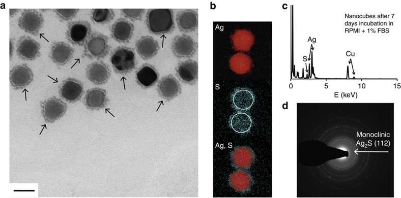
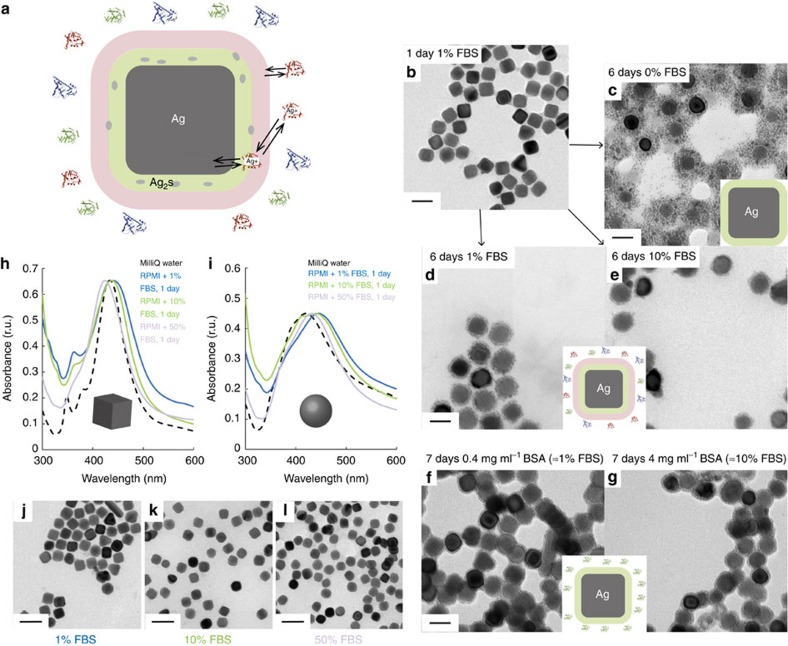
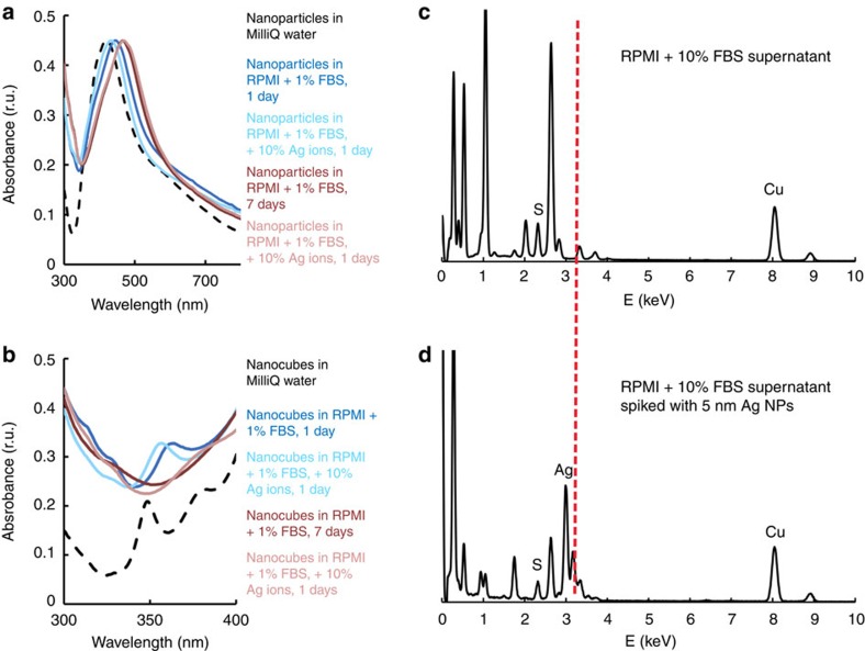
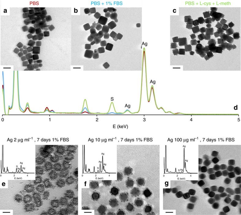
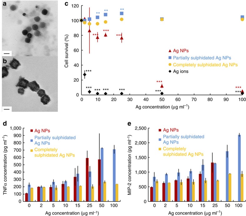
Proteins adsorbing at nanoparticles have been proposed as critical toxicity mediators and are included in ongoing efforts to develop predictive tools for safety assessment. Strongly attached proteins can be isolated, identified and correlated to changes in nanoparticle state, cellular association or toxicity. Weakly attached, rapidly exchanging proteins are also present at nanoparticles, but are difficult to isolate and have hardly been examined. Here we study rapidly exchanging proteins and show for the first time that they have a strong modulatory effect on the biotransformation of silver nanoparticles. Released silver ions, known for their role in particle toxicity, are found to be trapped as silver sulphide nanocrystals within the protein corona at silver nanoparticles in serum-containing cell culture media. The strongly attached corona acts as a site for sulphidation, while the weakly attached proteins reduce nanocrystal formation in a serum-concentration-dependent manner. Sulphidation results in decreased toxicity of Ag NPs.
Overview
The claims and statistics of this paper haven’t been checked yet.
Run the check — free, no account needed. It reads the open-access text and checks the sources it cites.
Integrity signals
Registry
Cites
Statistics
Figures
Verification
Claims the paper makes
The claims and statistics of this paper haven’t been checked yet.
Run the check — free, no account needed. It reads the open-access text and checks the sources it cites.
Claims other papers make about this one
Each is checked against this paper’s own text: does it say what the citing paper says it does?
In the citing paper
“For example, the corona adsorbed to NPs has been shown to slow dissolution by blocking oxidative processes [121] or by promoting sulfidation process and entrapping nanocrystals of Ag2S in the hard corona [122], while in other cases, adsorbed coronas have been found to accelerate dissolution, especially where there is a strong affinity for the metal ions by the proteins and an excess of the prote”
From Particle toxicology and health - where are we? (2019) · cites this paper as [122]
In the citing paper
“The absorbed biomolecule corona can also alter the biopersistence of the NPs, for example, by changing the rate of dissolution of NPs.”
From Particle toxicology and health - where are we? (2019) · cites this paper as [122]
Self-check
Percentages, sums and reported differences re-computed from the paper’s own numbers.
The self-check needs the paper’s full text.
Citations
Cites
Nanoparticle Size and Surface Chemistry Determine Serum Protein Adsorption and Macrophage Uptake
Walkey, C. D., Olsen, J. B., Guo, H., Emili, A., & Chan, W. C. W. (2012) · Journal of the American Chemical Society · doi:10.1021/ja2084338 · Cited by 1,926
Monopoli, M. P., Walczyk, D., Campbell, A., Elia, G., Lynch, I., Baldelli Bombelli, F., & Dawson, K. A. (2011) · Journal of the American Chemical Society · doi:10.1021/ja107583h · Cited by 1,835
Walkey, C. D., & Chan, W. C. W. (2012) · Chem. Soc. Rev. · doi:10.1039/c1cs15233e · Cited by 1,758
Time Evolution of the Nanoparticle Protein Corona
Casals, E., Pfaller, T., Duschl, A., Oostingh, G. J., & Puntes, V. (2010) · ACS Nano · doi:10.1021/nn901372t · Cited by 1,211
Research works printed but not linked: 65
- Monopoli, M. P., Åberg, C., Salvati, A. & Dawson, K. A. Biomolecular coronas provide the biological identity of nanosized materials. Nat. Nanotechnol 7, 779–786 (2012). · DOI · Paper page
- Walczyk, D., Baldelli Bombelli, F., Monopoli, M. P., Lynch, I. & Dawson, K. A. What the cell “sees” in bionanoscience. J. Am. Chem. Soc. 132, 5761–5768 (2010). · DOI
- Tenzer, S. et al. Rapid formation of plasma protein corona critically affects nanoparticle pathophysiology. Nat. Nanotechnol 8, 772–781 (2013). · DOI · Paper page
- Walkey, C. D. et al. Protein corona fingerprinting predicts the cellular interaction of gold and silver nanoparticles. ACS Nano 8, 2439–2455 (2014). · DOI
- Albanese, A. et al. Secreted biomolecules alter the biological identity and cellular interactions of nanoparticles. ACS Nano 8, 5515–5526 (2014). · DOI · Paper page
- Pelaz, B. et al. Interfacing engineered nanoparticles with biological systems: anticipating adverse nano-bio interactions. Small 9, 1573–1584 (2013). · DOI
- Docter, D. et al. The nanoparticles biomolecule corona: lessons learned – challenge accepted? Chem. Soc. Rev. 44, 6094–6121 (2015). · DOI
- Del Pino, P. et al. Protein corona formation around nanoparticles – from the past to the future. Mater. Horiz 1, 301–313 (2014). · DOI
- Röcker, C., Pötzl, M., Zhang, F., Parak, W. J. & Nienhaus, G. U. A quantitative fluorescence study of protein monolayer formation on colloidal nanoparticles. Nat. Nanotechnol 4, 577–580 (2009). · DOI
- Milani, S., Baldelli Bombelli, F., Pitek, A. S., Dawson, K. A. & Rädler, J. Reversible versus irreversible binding of transferrin to polystyrene nanoparticles: soft and hard corona. ACS Nano 6, 2533–2541 (2012). · DOI
- Fedeli, C. et al. The functional dissection of the plasma corona of SiO2-NPs spots histidine rich glycoprotein as a major player able to hamper nanoparticle capture by macrophages. Nanoscale 7, 17710–17728 (2015). · DOI
- Lundqvist, M. et al. Nanoparticle size and surface properties determine the protein corona with possible implications for biological impacts. Proc. Natl Acad. Sci. USA 105, 14265–14270 (2008). · DOI · Paper page
- Eigenheer, R. et al. Silver nanoparticle protein corona composition compared across engineered particle properties and environmentally relevant reaction conditions. Environ. Sci. Nano 1, 238–247 (2014). · DOI
- Nel, A. E. et al. Understanding the biophysicochemical interactions at the nano-bio interface. Nat. Mater. 8, 543–557 (2009). · DOI · Paper page
- Tenzer, S. et al. Nanoparticle size is a critical physic-chemical determinant of the human blood plasma corona: a comprehensive quantitative proteomic analysis. ACS Nano 5, 7155–7167 (2011). · DOI
- Deng, Z. J. et al. Differential plasma protein binding to metal oxide nanoparticles. Nanotechnology 20, 455101–455109 (2009). · DOI
- Lesniak, A. et al. Effects of the presence or absence of a protein corona on silica nanoparticle uptake and impact on cells. ACS Nano 6, 5845–5857 (2012). · DOI · Paper page
- Liu, R., Jiang, W., Walkey, C. D., Chan, W. C. W. & Cohen, Y. Prediction of nanoparticles-cell association based on corona proteins and physicochemical properties. Nanoscale 7, 9664–9675 (2015). · DOI
- Setyawati, M. I., Tay, C. Y., Docter, D., Stauber, R. H. & Leond, D. T. Understanding and exploiting nanoparticles’ intimacy with the blood vessel and blood. Chem. Soc. Rev. 44, 8174–8199 (2015). · DOI
- Miclăuş, T., Bochenkov, V. E., Ogaki, R., Howard, K. A. & Sutherland, D. S. Spatial mapping and quantification of soft and hard protein coronas at silver nanocubes. Nano Lett. 14, 2086–2093 (2014). · DOI
- Beer, C., Foldbjerg, R., Hayashi, Y., Sutherland, D. S. & Autrup, H. Toxicity of silver nanoparticles – nanoparticle or silver ion? Toxicol. Lett. 208, 286–292 (2012). · DOI
- Foldbjerg, R. et al. Silver nanoparticles – wolves in sheep’s clothing? Toxicol. Res 4, 563–575 (2015). · DOI
- Gliga, A. R., Skoglund, S., Wallinder, I. O., Fadeel, B. & Karlsson, H. L. Size-dependent cytotoxicity of silver nanoparticles in human lung cells: the role of cellular uptake, agglomeration and Ag release. Part. Fibre. Toxicol 11, 11–27 (2014). · DOI
- Johnston, H. J. et al. A review of the in vivo and in vitro toxicity of silver and gold particulates: particle attributes and biological mechanism responsible for the observed toxicity. Crit. Rev. Toxicol. 40, 328–346 (2010). · DOI
- Henglein, A. Colloidal silver nanoparticles: photochemical preparation and interaction with O2, CCl4, and some metal ions. Chem. Mater. 10, 444–450 (1998). · DOI
- Lok, C.-N. et al. Silver nanoparticles: partial oxidation and antibacterial activities. J. Biol. Inorg. Chem. 12, 527–534 (2007). · DOI
- Sotiriou, G. A., Meyer, A., Knijnenburg, J. T. N., Panke, S. & Pratsinis, S. E. Quantifying the origin of released Ag+ ions from nanosilver. Langmuir 28, 15929–15936 (2012). · DOI
- Liu, J. & Hurt, R. H. Ion release kinetics and particle persistence in aqueous nano-silver colloids. Environ. Sci. Technol. 44, 2169–2175 (2010). · DOI
- Ma, R. et al. Size-controlled dissolution of organic-coated silver nanoparticles. Environ. Sci. Technol. 46, 752–759 (2012). · DOI
- Liu, J., Pennell, K. G. & Hurt, R. H. Kinetics and mechanism of nanosilver oxysulfidation. Environ. Sci. Technol. 45, 7345–7353 (2011). · DOI
- Reinsch, B. C. et al. Sulfidation of silver nanoparticles decreases Escherichia coli growth inhibition. Environ. Sci. Technol. 46, 6992–7000 (2012). · DOI
- Levard, C. et al. Sulfidation of silver nanoparticles: natural antidote to their toxicity. Environ. Sci. Technol. 47, 13440–13448 (2013). · DOI
- Devi, G. P. et al. Sulfidation of silver nanoparticles reduces its toxicity in zebrafish. Aquat. Toxicol. 158, 149–156 (2015). · DOI
- Starnes, D. L. et al. Impact of sulfidation on the bioavailability and toxicity of silver nanoparticles to Caenorhabditis elegans. Environ. Pollut. 196, 239–246 (2015). · DOI
- Kim, B., Park, C.-S., Murayama, S. & Hochella, M. F. Jr. Discovery and characterization of silver sulfide nanoparticles in final sewage sludge products. Environ. Sci. Technol. 44, 7509–7514 (2010). · DOI
- Kent, R. D., Oser, J. G. & Vikesland, P. J. Controlled evaluation of silver nanoparticle sulfidation in full-scale wastewater treatment plant. Environ. Sci. Technol. 48, 8564–8572 (2014). · DOI
- Thalmann, B., Voegelin, A., Sinnet, B., Morgenroth, E. & Kaegi, R. Sulfidation kinetics of silver nanoparticles reacted with metal sulfides. Environ. Sci. Technol. 48, 4885–4892 (2014). · DOI
- Yu, S.-J., Yin, Y.-G., Chao, J.-B., Shen, M.-H. & Liu, J.-F. Highly dynamic PVP-coated silver nanoparticles in aquatic environments: chemical and morphology change induced by oxidation of Ag0 and reduction of Ag+. Environ. Sci. Technol. 48, 403–411 (2014). · DOI
- Liu, J., Sonshine, D. A., Shervani, S. & Hurt, R. H. Controlled release of biologically active silver from nanosilver surfaces. ACS Nano 4, 6903–6913 (2010). · DOI
- Gondikas, A. P. et al. Cysteine-induced modifications of zero-valent silver nanomaterials: implications for particles surface chemistry, aggregation, dissolution, and silver speciation. Environ. Sci. Technol. 46, 7037–7045 (2012). · DOI
- Jiang, X. et al. Fast intracellular dissolution and persistent cellular uptake of silver nanoparticles in CHO-K1 cells: implication for cytotoxicity. Nanotoxicology 9, 181–189 (2015). · DOI
- Wang, L. et al. Use of synchrotron radiation-analytical techniques to reveal chemical origin of silver-nanoparticle cytotoxicity. ACS Nano 9, 6532–6547 (2015). · DOI
- Chen, S. et al. Sulfidation of silver nanowires inside human alveolar epithelial cells: a potential detoxification mechanism. Nanoscale 5, 9839–9847 (2013). · DOI
- Chen, S. et al. High-resolution analytical electron microscopy reveals cell culture media-induced changes to the chemistry of silver nanowires. Environ. Sci. Technol. 47, 13813–13821 (2013). · DOI
- Choi, O. et al. Role of sulfide and ligand strength in controlling nanosilver toxicity. Water. Res 43, 1879–1886 (2009). · DOI
- Bianchini, A. et al. Evaluation of the effect of reactive sulfide on the acute toxicity of silver (I) to Daphnia magna. Part 2: toxicity results. Environ. Toxicol. Chem. 21, 1294–1300 (2002). · DOI
- Mann, R. M., Ernste, M. J., Bell, R. A., Kramer, J. R. & Wood, C. M. Evaluation of the protective effects of reactive sulfide on the acute toxicity of silver to rainbow trout (Oncorhynchus Mykiss). Enviro. Toxicol. Chem 23, 1204–1210 (2004). · DOI
- Selected Powder Diffraction Data for Metals & Alloys Data Book. 1st edn. vol. 1, JCPDS International Center for Diffraction Data (1982).
- Cia, X., Zeng, J., Zhang, Q., Moran, C. H. & Xia, Y. Recent developments in shape-controlled synthesis of silver nanocrystals. J. Phys. Chem. C 116, 21647–21656 (2012). · DOI
- Wang, Y., Zheng, Y., Huang, C. Z. & Xia, Y. Synthesis of Ag nanocubes 18-32 nm in edge length: the effects of polyol on reduction kinetics, size control and reproducibility. J. Am. Chem. Soc. 135, 1941–1951 (2013). · DOI
- Elechiguerra, J. L. et al. Corrosion at the nanoscale: the case of silver nanowires and nanoparticles. Chem. Mater. 17, 6042–6052 (2005). · DOI
- El-Khairy, L., Ueland, P. M., Refsum, H., Graham, I. M. & Vollset, S. E. Plasma total cysteine as a risk factor for vascular disease. Circulation 103, 2544–2549 (2001). · DOI
- Salemi, G. et al. Blood levels of homocysteine, cysteine, glutathione, folic acid, and vitamin B12 in the acute phase of atherothrombosis stroke. Neural. Sci 30, 361–364 (2009). · DOI
- Levard, C., Hotze, E. M., Lowry, G. V. & Brown, G. E. Jr. Environmental transformations of silver nanoparticles: Impact on stability and toxicity. Environ. Sci. Technol. 46, 6900–6914 (2012). · DOI
- Bopst, M., Haas, C., Car, B. & Engster, H. P. The combined inactivation of tumor necrosis factor and interleukin-6 prevents induction of the major acute phase proteins by endotoxin. Eur. J. Immunol. 28, 4130–4137 (1998). · DOI
- Martinez-Gutierrez, F. et al. Antibacterial activity, inflammatory response, coagulation and cytotoxicity effects of silver nanoparticles. Nanomed. Nanotechnol 8, 328–336 (2012). · DOI
- Park, M.V.D.Z. et al. The effect of particle size on the cytotoxicity, inflammation, developmental toxicity and genotoxicity of silver nanoparticles. Biomaterials 32, 9810–9817 (2011). · DOI
- Driscoll, K. E. TNFα and MIP-2 production: role in particle-induced inflammation and regulation by oxidative stress. Toxicol. Lett. 112-113, 177–184 (2000). · DOI
- Zhang, Q., Li, W., Wen, L.-P., Chen, J. & Xia, Y. Facile synthesis of Ag nanocubes of 30 to 70 nm in edge length with CF3COOAg as a precursor. Chem. Eur. J 16, 10234–10239 (2010). · DOI
- Wiley, B., Sun, Y., Mayers, B. & Xia, Y. Shape-controlled synthesis of metal nanoparticles: the case of silver. Chem. Eur. J 11, 454–463 (2005). · DOI
- Foldbjerg, R., Dang, D. A. & Autrup, H. Cytotoxicity and genotoxicity of silver nanoparticles in the human lung cancer cell line, A549. Arch. Toxicol. 85, 743–750 (2011). · DOI
- Reidy, B., Haase, A., Luch, A., Dawson, K. A. & Lynch, I. Mechanism of silver nanoparticle release, transformation and toxicity: a critical review of current knowledge and recommendations for future studies and applications. Materials 6, 2295–2350 (2013). · DOI
- Helmlinger, J. et al. Silver nanoparticles with different size and shape: equal cytotoxicity, but different antibacterial effects. RSC Adv 6, 18490–18501 (2016). · DOI
- Foldbjerg, R. et al. Global gene expression profiling of human lung epithelial cells after exposure to nanosilver. Toxicol. Sci. 130, 145–157 (2012). · DOI
- Jiang, X. et al. Multi-platform genotoxicity analysis of silver nanoparticles in the model cell line CHO-K1. Toxicol. Lett. 222, 55–63 (2013). · DOI
Cited by
Sorted by citations
Nanoparticle protein corona: from structure and function to therapeutic targeting
Bashiri, G., Padilla, M. S., Swingle, K. L., Shepherd, S. J., Mitchell, M. J., & Wang, K. (2023) · Lab on a Chip · doi:10.1039/d2lc00799a · Cited by 296
Particle toxicology and health - where are we?
Riediker, M., Zink, D., Kreyling, W., Oberdörster, G., Elder, A., Graham, U., Lynch, I., Duschl, A., Ichihara, G., Ichihara, S., et al. (2019) · Particle and Fibre Toxicology · doi:10.1186/s12989-019-0302-8 · Cited by 237
Ho, Y. T., Kamm, R. D., & Kah, J. C. Y. (2018) · Nanoscale · doi:10.1039/c8nr02393j · Cited by 102
Length and diameter-dependent phagocytosis and cytotoxicity of long silver nanowires in macrophages
Wang, F., Wang, Y., Yao, X., Ma, C., Yin, Y., & Song, M. (2019) · Chemosphere · doi:10.1016/j.chemosphere.2019.124565 · Cited by 17
Metadata
- Authors
- Miclăuş, T., Beer, C., Chevallier, J., Scavenius, C., Bochenkov, V. E., Enghild, J. J., & Sutherland, D. S.
- Venue
- Nature Communications
- Published
- 2016-06-09
- Volume
- 7
- Issue
- 1
- ISSN
- 2041-1723
- PMC
- PMC4906166
- Cited by
- 174
- References
- 4
- Language
- en
About these checks
Registry, retraction, Cites and statistics results are computed, not judged, with one exception: a printed reference that no index lists is read once by a model before it counts as a citation error. Claims are extracted by a model and kept only when their quote appears verbatim in the text; each cited source is then checked, with a model’s help, for whether it says what the claim relies on. Those verdicts can be wrong — read the claim’s quote and the cited source before relying on one.
Statistics and arithmetic are recomputed, not judged.
Confidence bands are shown only for fields calibrated on our annotated test set. None is calibrated yet, so no band is shown.
What counts as a claim ›How we measure confidence ›How we check this ›
Are you an author, or do you see a problem with this page? Request a review
How to cite
Cite CoChat for the checks on this page, and the paper for its own findings.
- This page
- CoChat. (2026). Citation check: Dynamic protein coronas revealed as a modulator of silver nanoparticle sulphidation in vitro. https://app.cochat.ai/papers/10.1038/ncomms11770
- The paper
- Miclăuş, T., Beer, C., Chevallier, J., Scavenius, C., Bochenkov, V. E., Enghild, J. J., & Sutherland, D. S. (2016). Dynamic protein coronas revealed as a modulator of silver nanoparticle sulphidation in vitro. Nature Communications. https://doi.org/10.1038/ncomms11770
