

The DOI matches its Crossref record; 26 of the 26 references it cites were found in a registry or an index.
Microglial over-pruning of synapses during development in autism-associated SCN2A-deficient mice and human cerebral organoids
Published version of a preprint
Overview
The claims and statistics of this paper haven’t been checked yet.
Run the check — free, no account needed. It reads the open-access text and checks the sources it cites.
Integrity signals
Registry
Cites
Statistics
Figures
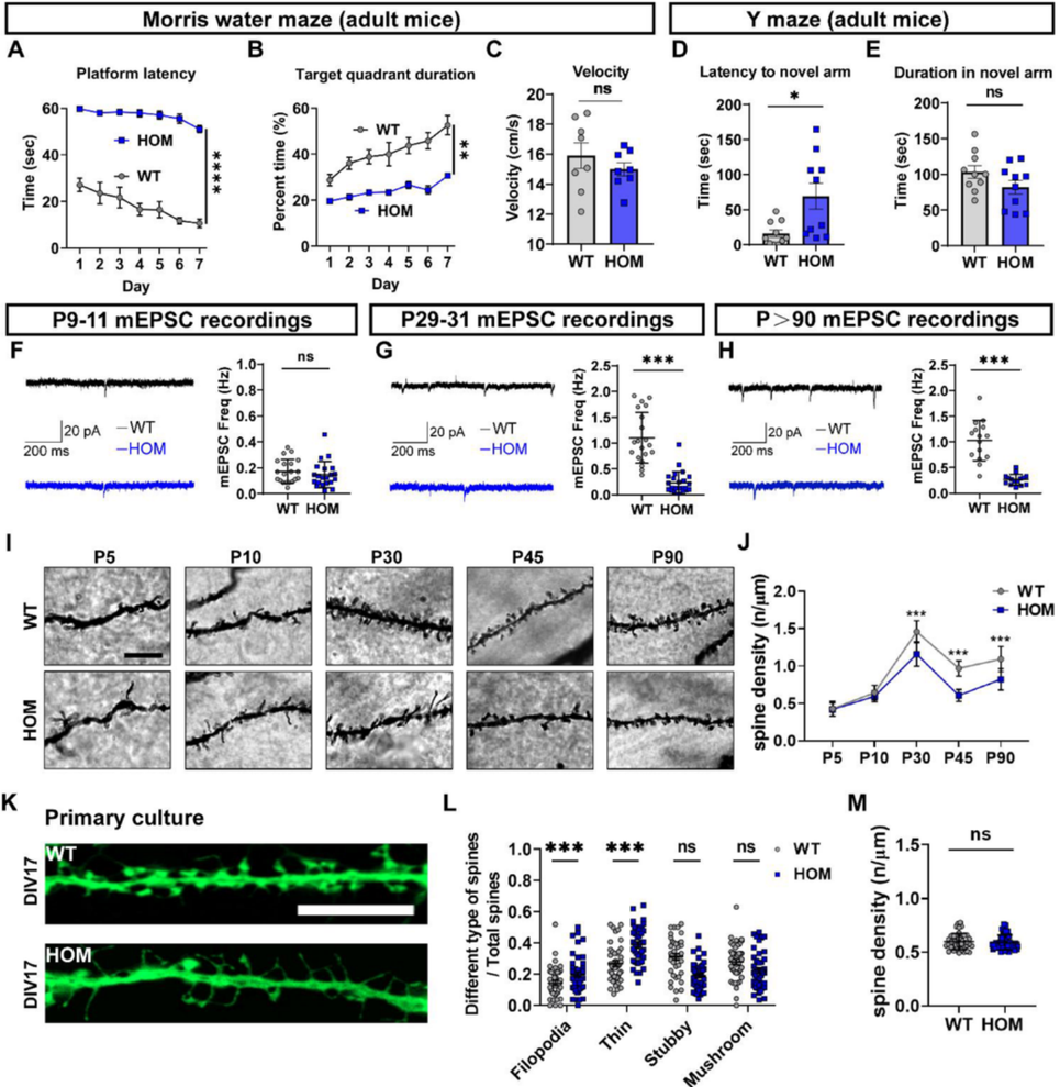
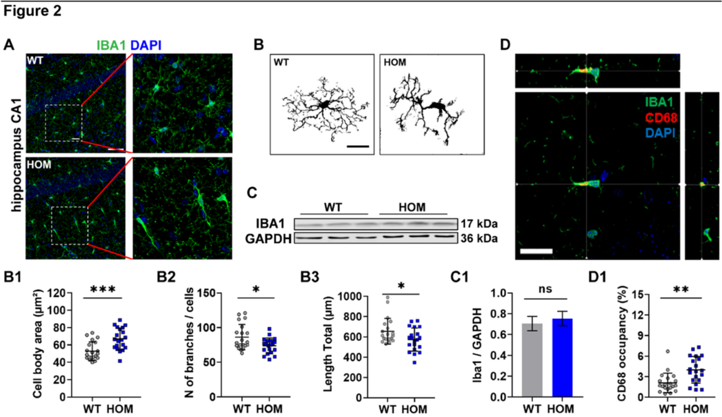
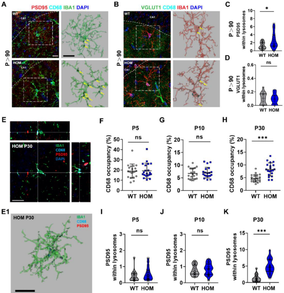
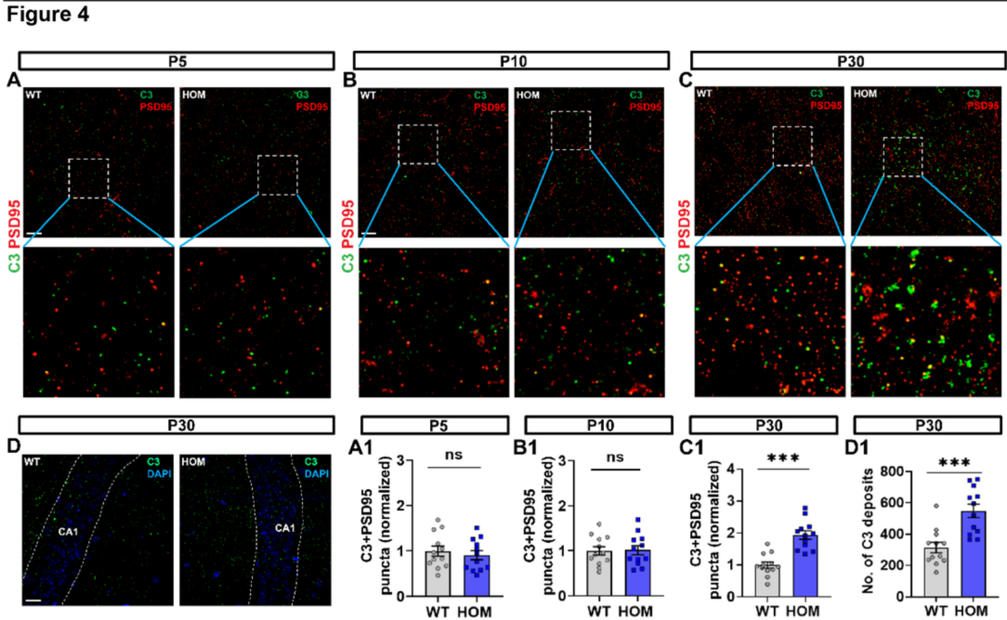
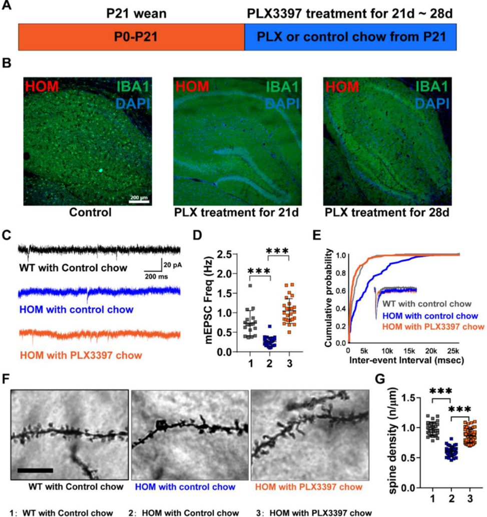
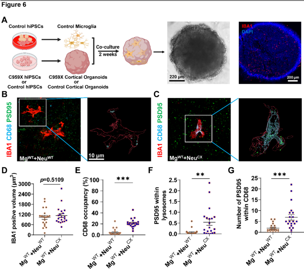
Verification
Claims the paper makes
The claims and statistics of this paper haven’t been checked yet.
Run the check — free, no account needed. It reads the open-access text and checks the sources it cites.
Claims other papers make about this one
Each is checked against this paper’s own text: does it say what the citing paper says it does?
In the citing paper
“Complement proteins, including C1q and C3, are upregulated in AD brain tissues36 and cerebrospinal fluid87,91, where they are pivotal in neuroinflammation and synapse elimination88,92.”
Self-check
Percentages, sums and reported differences re-computed from the paper’s own numbers.
The self-check needs the paper’s full text.
Citations
Cites
Zhang, Y., Chen, K., Sloan, S. A., Bennett, M. L., Scholze, A. R., O'Keeffe, S., Phatnani, H. P., Guarnieri, P., Caneda, C., Ruderisch, N., et al. (2014) · The Journal of Neuroscience · doi:10.1523/jneurosci.1860-14.2014 · Cited by 4,875 · cited 1×
“It is suggested that C3 is mainly released by astrocytes [77] while C1q can be secreted by activated microglia [78, 79].”
Self-citationSynaptic Pruning by Microglia Is Necessary for Normal Brain Development
Paolicelli, R. C., Bolasco, G., Pagani, F., Maggi, L., Scianni, M., Panzanelli, P., Giustetto, M., Ferreira, T. A., Guiducci, E., Dumas, L., et al. (2011) · Science · doi:10.1126/science.1202529 · Cited by 4,244
Microglia Sculpt Postnatal Neural Circuits in an Activity and Complement-Dependent Manner
Schafer, D. P., Lehrman, E. K., Kautzman, A. G., Koyama, R., Mardinly, A. R., Yamasaki, R., Ransohoff, R. M., Greenberg, M. E., Barres, B. A., & Stevens, B. (2012) · Neuron · doi:10.1016/j.neuron.2012.03.026 · Cited by 4,089
Maenner, M. J., Warren, Z., Williams, A. R., Amoakohene, E., Bakian, A. V., Bilder, D. A., Durkin, M. S., Fitzgerald, R. T., Furnier, S. M., Hughes, M. M., et al. (2023) · MMWR. Surveillance Summaries · doi:10.15585/mmwr.ss7202a1 · Cited by 2,706 · cited 1×
“Autism spectrum disorder (ASD) is a major neurodevelopmental disorder that currently affects 1 in 36 children in the United States [1].”
Satterstrom, F. K., Kosmicki, J. A., Wang, J., Breen, M. S., De Rubeis, S., An, J.-Y., Peng, M., Collins, R., Grove, J., Klei, L., et al. (2020) · Cell · doi:10.1016/j.cell.2019.12.036 · Cited by 2,321
Elmore, M. R. P., Najafi, A. R., Koike, M. A., Dagher, N. N., Spangenberg, E. E., Rice, R. A., Kitazawa, M., Matusow, B., Nguyen, H., West, B. L., et al. (2014) · Neuron · doi:10.1016/j.neuron.2014.02.040 · Cited by 1,968
Maenner, M. J., Shaw, K. A., Bakian, A. V., Bilder, D. A., Durkin, M. S., Esler, A., Furnier, S. M., Hallas, L., Hall-Lande, J., Hudson, A., et al. (2021) · MMWR. Surveillance Summaries · doi:10.15585/mmwr.ss7011a1 · Cited by 1,818
A conditional knockout resource for the genome-wide study of mouse gene function
Skarnes, W. C., Rosen, B., West, A. P., Koutsourakis, M., Bushell, W., Iyer, V., Mujica, A. O., Thomas, M., Harrow, J., Cox, T., et al. (2011) · Nature · doi:10.1038/nature10163 · Cited by 1,708
ApoE4 markedly exacerbates tau-mediated neurodegeneration in a mouse model of tauopathy
Initiative, A. D. N., Shi, Y., Yamada, K., Liddelow, S. A., Smith, S. T., Zhao, L., Luo, W., Tsai, R. M., Spina, S., Grinberg, L. T., et al. (2017) · Nature · doi:10.1038/nature24016 · Cited by 1,220
Lui, H., Zhang, J., Makinson, S. R., Cahill, M. K., Kelley, K. W., Huang, H.-Y., Shang, Y., Oldham, M. C., Martens, L. H., Gao, F., et al. (2016) · Cell · doi:10.1016/j.cell.2016.04.001 · Cited by 729
Self-citationLian, H., Yang, L., Cole, A., Sun, L., Chiang, A. C.-A., Fowler, S. W., Shim, D. J., Rodriguez-Rivera, J., Taglialatela, G., Jankowsky, J. L., et al. (2015) · Neuron · doi:10.1016/j.neuron.2014.11.018 · Cited by 697
Filipello, F., Morini, R., Corradini, I., Zerbi, V., Canzi, A., Michalski, B., Erreni, M., Markicevic, M., Starvaggi-Cucuzza, C., Otero, K., et al. (2018) · Immunity · doi:10.1016/j.immuni.2018.04.016 · Cited by 695
Vainchtein, I. D., Chin, G., Cho, F. S., Kelley, K. W., Miller, J. G., Chien, E. C., Liddelow, S. A., Nguyen, P. T., Nakao-Inoue, H., Dorman, L. C., et al. (2018) · Science · doi:10.1126/science.aal3589 · Cited by 657
Microglia contact induces synapse formation in developing somatosensory cortex
Miyamoto, A., Wake, H., Ishikawa, A. W., Eto, K., Shibata, K., Murakoshi, H., Koizumi, S., Moorhouse, A. J., Yoshimura, Y., & Nabekura, J. (2016) · Nature Communications · doi:10.1038/ncomms12540 · Cited by 649
The Role of the Immune System in Autism Spectrum Disorder
Meltzer, A., & Van de Water, J. (2017) · Neuropsychopharmacology · doi:10.1038/npp.2016.158 · Cited by 533
Immune mediators in the brain and peripheral tissues in autism spectrum disorder
Estes, M. L., & McAllister, A. K. (2015) · Nature Reviews Neuroscience · doi:10.1038/nrn3978 · Cited by 514 · cited 2×
“While neurons have been the focus to understand ASD, an altered immune response in the brain has been suggested to be closely associated with ASD, and a neuro-immune interaction could play a role in the disease progression [14, 15].”
McQuade, A., Coburn, M., Tu, C. H., Hasselmann, J., Davtyan, H., & Blurton-Jones, M. (2018) · Molecular Neurodegeneration · doi:10.1186/s13024-018-0297-x · Cited by 449
Progress in Understanding and Treating SCN2A-Mediated Disorders
Sanders, S. J., Campbell, A. J., Cottrell, J. R., Moller, R. S., Wagner, F. F., Auldridge, A. L., Bernier, R. A., Catterall, W. A., Chung, W. K., Empfield, J. R., et al. (2018) · Trends in Neurosciences · doi:10.1016/j.tins.2018.03.011 · Cited by 338 · cited 2×
“2, a major sodium channel expressed in the central nervous system (CNS) mediating the action potential initiation, propagation, and backpropagation [6 – 8].”
Microglia and the brain: Complementary partners in development and disease
Hammond, T. R., Robinton, D., & Stevens, B. (2018) · Annual Review of Cell and Developmental Biology · doi:10.1146/annurev-cellbio-100616-060509 · Cited by 288
Synaptic Signaling in Learning and Memory
Kennedy, M. B. (2016) · Cold Spring Harbor Perspectives in Biology · doi:10.1101/cshperspect.a016824 · Cited by 270
Shaping Neuronal Fate: Functional Heterogeneity of Direct Microglia-Neuron Interactions
Cserép, C., Pósfai, B., & Dénes, Á. (2021) · Neuron · doi:10.1016/j.neuron.2020.11.007 · Cited by 256
How microglia sense and regulate neuronal activity
Umpierre, A. D., & Wu, L. (2021) · Glia · doi:10.1002/glia.23961 · Cited by 237
Self-citationAdult neurogenesis, glia, and the extracellular matrix
Cope, E. C., & Gould, E. (2019) · Cell Stem Cell · doi:10.1016/j.stem.2019.03.023 · Cited by 236
Shaw, K. A., Bilder, D. A., McArthur, D., Williams, A. R., Amoakohene, E., Bakian, A. V., Durkin, M. S., Fitzgerald, R. T., Furnier, S. M., Hughes, M. M., et al. (2023) · MMWR. Surveillance Summaries · doi:10.15585/mmwr.ss7201a1 · Cited by 223
Jin, M., Xu, R., Wang, L., Alam, M. M., Ma, Z., Zhu, S., Martini, A. C., Jadali, A., Bernabucci, M., Xie, P., et al. (2022) · Cell Stem Cell · doi:10.1016/j.stem.2022.06.007 · Cited by 174 · cited 2×
“Additionally, the microglia-organoid model was used to study neurodegenerative disorders [90, 92, 93].”
Self-citationPinto, B., Morelli, G., Rastogi, M., Savardi, A., Fumagalli, A., Petretto, A., Bartolucci, M., Varea, E., Catelani, T., Contestabile, A., et al. (2020) · Neuron · doi:10.1016/j.neuron.2020.09.010 · Cited by 135
Research works printed but not linked: 78
- Rolland T, Cliquet F, Anney RJL, Moreau C, Traut N, Mathieu A, et al. Phenotypic effects of genetic variants associated with autism. Nat Med. 2023;29:1671–80. · DOI
- Qin Y, Kang J, Jiao Z, Wang Y, Wang J, Wang H, et al. Polygenic risk for autism spectrum disorder affects left amygdala activity and negative emotion in schizophrenia. Transl Psychiatry. 2020;10:322. · DOI
- Sanders SJ, Murtha MT, Gupta AR, Murdoch JD, Raubeson MJ, Willsey AJ, et al. De novo mutations revealed by whole-exome sequencing are strongly associated with autism. Nature. 2012;485:237–41. · DOI · Paper page
- Gazina EV, Leaw BT, Richards KL, Wimmer VC, Kim TH, Aumann TD, et al. 'Neonatal’ Nav1.2 reduces neuronal excitability and affects seizure susceptibility and behaviour. Hum Mol Genet. 2015;24:1457–68. · DOI · Paper page
- Sanders SJ, He X, Willsey AJ, Ercan-Sencicek AG, Samocha KE, Cicek AE, et al. Insights into autism spectrum disorder genomic architecture and biology from 71 risk loci. Neuron. 2015;87:1215–33. · DOI · Paper page
- Miyamoto H, Tatsukawa T, Shimohata A, Yamagata T, Suzuki T, Amano K, et al. Impaired cortico-striatal excitatory transmission triggers epilepsy. Nat Commun. 2019;10:1917. · DOI
- Middleton SJ, Kneller EM, Chen S, Ogiwara I, Montal M, Yamakawa K, et al. Altered hippocampal replay is associated with memory impairment in mice heterozygous for the Scn2a gene. Nat Neurosci. 2018;21:996–1003. · DOI
- Tatsukawa T, Raveau M, Ogiwara I, Hattori S, Miyamoto H, Mazaki E, et al. Scn2a haploinsufficient mice display a spectrum of phenotypes affecting anxiety, sociability, memory flexibility and ampakine CX516 rescues their hyperactivity. Mol Autism. 2019;10:15. · DOI
- Eaton M, Zhang J, Ma Z, Park AC, Lietzke E, Romero CM, et al. Generation and basic characterization of a gene-trap knockout mouse model of Scn2a with a substantial reduction of voltage-gated sodium channel Na(v) 1.2 expression. Genes Brain Behav. 2021;20:e12725. · DOI
- Ma Z, Eaton M, Liu Y, Zhang J, Chen X, Tu X, et al. Deficiency of autism-related Scn2a gene in mice disrupts sleep patterns and circadian rhythms. Neurobiol Dis. 2022;168:105690. · DOI
- Suzuki K, Sugihara G, Ouchi Y, Nakamura K, Futatsubashi M, Takebayashi K, et al. Microglial activation in young adults with autism spectrum disorder. JAMA Psychiatry. 2013;70:49–58. · DOI · Paper page
- Zhan Y, Paolicelli RC, Sforazzini F, Weinhard L, Bolasco G, Pagani F, et al. Deficient neuron-microglia signaling results in impaired functional brain connectivity and social behavior. Nat Neurosci. 2014;17:400–6. · DOI · Paper page
- Andoh M, Shibata K, Okamoto K, Onodera J, Morishita K, Miura Y, et al. Exercise reverses behavioral and synaptic abnormalities after maternal inflammation. Cell Rep. 2019;27:2817–25.e2815. · DOI
- Kim HJ, Cho MH, Shim WH, Kim JK, Jeon EY, Kim DH, et al. Deficient autophagy in microglia impairs synaptic pruning and causes social behavioral defects. Mol Psychiatry. 2017;22:1576–84. · DOI · Paper page
- Parkhurst CN, Yang G, Ninan I, Savas JN, Yates JR 3rd, Lafaille JJ, et al. Microglia promote learning-dependent synapse formation through brain-derived neurotrophic factor. Cell. 2013;155:1596–609. · DOI · Paper page
- Jawaid S, Kidd GJ, Wang J, Swetlik C, Dutta R, Trapp BD. Alterations in CA1 hippocampal synapses in a mouse model of fragile X syndrome. Glia. 2018;66:789–800. · DOI
- Schafer DP, Heller CT, Gunner G, Heller M, Gordon C, Hammond T, et al. Microglia contribute to circuit defects in Mecp2 null mice independent of microglia-specific loss of Mecp2 expression. Elife. 2016;5:e15224. · DOI · Paper page
- Arsenault J, Hooper AWM, Gholizadeh S, Kong T, Pacey LK, Koxhioni E, et al. Interregulation between fragile X mental retardation protein and methyl CpG binding protein 2 in the mouse posterior cerebral cortex. Hum Mol Genet. 2021;29:3744–56. · DOI
- Bach S, Shovlin S, Moriarty M, Bardoni B, Tropea D. Rett syndrome and fragile X syndrome: different etiology with common molecular dysfunctions. Front Cell Neurosci. 2021;15:764761. · DOI
- Black JA, Liu S, Waxman SG. Sodium channel activity modulates multiple functions in microglia. Glia. 2009;57:1072–81. · DOI
- Lai MC, Lombardo MV, Baron-Cohen S. Autism. Lancet. 2014;383:896–910. · DOI · Paper page
- Kraeuter AK, Guest PC, Sarnyai Z. The Y-maze for assessment of spatial working and reference memory in mice. Methods Mol Biol. 2019;1916:105–11. · DOI
- Nisar S, Bhat AA, Masoodi T, Hashem S, Akhtar S, Ali TA, et al. Genetics of glutamate and its receptors in autism spectrum disorder. Mol Psychiatry. 2022;27:2380–92. · DOI
- Spratt PWE, Ben-Shalom R, Keeshen CM, Burke KJ Jr, Clarkson RL, Sanders SJ, et al. The autism-associated gene Scn2a contributes to dendritic excitability and synaptic function in the prefrontal cortex. Neuron. 2019;103:673–85.e675. · DOI · Paper page
- Shin W, Kweon H, Kang R, Kim D, Kim K, Kang M, et al. Scn2a haploinsufficiency in mice suppresses hippocampal neuronal excitability, excitatory synaptic drive, and long-term potentiation, and spatial learning and memory. Front Mol Neurosci. 2019;12:145. · DOI
- Zhang J, Chen X, Eaton M, Wu J, Ma Z, Lai S, et al. Severe deficiency of the voltage-gated sodium channel Na(V)1.2 elevates neuronal excitability in adult mice. Cell Rep. 2021;36:109495. · DOI
- Posfai B, Cserep C, Orsolits B, Denes A. New insights into microglia-neuron interactions: a neuron’s perspective. Neuroscience. 2019;405:103–17. · DOI · Paper page
- Choudhury ME, Miyanishi K, Takeda H, Islam A, Matsuoka N, Kubo M, et al. Phagocytic elimination of synapses by microglia during sleep. Glia. 2020;68:44–59. · DOI
- Matta SM, Hill-Yardin EL, Crack PJ. The influence of neuroinflammation in autism spectrum disorder. Brain Behav Immun. 2019;79:75–90. · DOI
- Andoh M, Koyama R. Microglia regulate synaptic development and plasticity. Dev Neurobiol. 2021;81:568–90. · DOI
- Stevens B, Allen NJ, Vazquez LE, Howell GR, Christopherson KS, Nouri N, et al. The classical complement cascade mediates CNS synapse elimination. Cell. 2007;131:1164–78. · DOI · Paper page
- Wallace J, Lord J, Dissing-Olesen L, Stevens B, Murthy VN. Microglial depletion disrupts normal functional development of adult-born neurons in the olfactory bulb. Elife. 2020;9:e50531. · DOI
- Nguyen PT, Dorman LC, Pan S, Vainchtein ID, Han RT, Nakao-Inoue H, et al. Microglial remodeling of the extracellular matrix promotes synapse plasticity. Cell. 2020;182:388–403 e315. · DOI · Paper page
- Ben-Shalom R, Keeshen CM, Berrios KN, An JY, Sanders SJ, Bender KJ. Opposing effects on Na(V)1.2 function underlie differences between SCN2A variants observed in individuals with autism spectrum disorder or infantile seizures. Biol Psychiatry. 2017;82:224–32. · DOI · Paper page
- Ramos DM, Skarnes WC, Singleton AB, Cookson MR, Ward ME. Tackling neurodegenerative diseases with genomic engineering: a new stem cell initiative from the NIH. Neuron. 2021;109:1080–3. · DOI · Paper page
- Pantazis CB, Yang A, Lara E, McDonough JA, Blauwendraat C, Peng L, et al. A reference human induced pluripotent stem cell line for large-scale collaborative studies. Cell Stem Cell. 2022;29:1685–702 e1622. · DOI · Paper page
- Que Z, Olivero-Acosta MI, Zhang J, Eaton M, Tukker AM, Chen X, et al. Hyperexcitability and pharmacological responsiveness of cortical neurons derived from human iPSCs carrying epilepsy-associated sodium channel Nav1.2-L1342P genetic variant. J Neurosci. 2021;41:10194–208. · DOI
- Skarnes WC, Pellegrino E, McDonough JA. Improving homology-directed repair efficiency in human stem cells. Methods. 2019;164-165:18–28. · DOI
- Thion MS, Ginhoux F, Garel S. Microglia and early brain development: an intimate journey. Science. 2018;362:185–9. · DOI
- Tseng CJ, McDougle CJ, Hooker JM, Zurcher NR. Epigenetics of autism spectrum disorder: histone deacetylases. Biol Psychiatry. 2022;91:922–33. · DOI
- Hegarty JP 2nd, Pegoraro LFL, Lazzeroni LC, Raman MM, Hallmayer JF, Monterrey JC, et al. Genetic and environmental influences on structural brain measures in twins with autism spectrum disorder. Mol Psychiatry. 2020;25:2556–66. · DOI
- Spratt PWE, Alexander RPD, Ben-Shalom R, Sahagun A, Kyoung H, Keeshen CM, et al. Paradoxical hyperexcitability from Na(V)1.2 sodium channel loss in neocortical pyramidal cells. Cell Rep. 2021;36:109483. · DOI
- Ogiwara I, Miyamoto H, Tatsukawa T, Yamagata T, Nakayama T, Atapour N, et al. Nav1.2 haplodeficiency in excitatory neurons causes absence-like seizures in mice. Commun Biol. 2018;1:96. · DOI
- Witcher KG, Bray CE, Chunchai T, Zhao F, O’Neil SM, Gordillo AJ, et al. Traumatic brain injury causes chronic cortical inflammation and neuronal dysfunction mediated by microglia. J Neurosci. 2021;41:1597–616. · DOI · Paper page
- Hui CW, St-Pierre A, El Hajj H, Remy Y, Hebert SS, Luheshi GN, et al. Prenatal immune challenge in mice leads to partly sex-dependent behavioral, microglial, and molecular abnormalities associated with schizophrenia. Front Mol Neurosci. 2018;11:13. · DOI
- Sarn N, Jaini R, Thacker S, Lee H, Dutta R, Eng C. Cytoplasmic-predominant Pten increases microglial activation and synaptic pruning in a murine model with autism-like phenotype. Mol Psychiatry. 2021;26:1458–71. · DOI
- Bennett FC, Molofsky AV. The immune system and psychiatric disease: a basic science perspective. Clin Exp Immunol. 2019;197:294–307. · DOI
- Saitoh BY, Tanaka E, Yamamoto N, Kruining DV, Iinuma K, Nakamuta Y, et al. Early postnatal allergic airway inflammation induces dystrophic microglia leading to excitatory postsynaptic surplus and autism-like behavior. Brain Behav Immun. 2021;95:362–80. · DOI · Paper page
- Williams JA, Burgess S, Suckling J, Lalousis PA, Batool F, Griffiths SL, et al. Inflammation and brain structure in schizophrenia and other neuropsychiatric disorders: a Mendelian randomization study. JAMA Psychiatry. 2022;79:498–507. · DOI
- Onore CE, Schwartzer JJ, Careaga M, Berman RF, Ashwood P. Maternal immune activation leads to activated inflammatory macrophages in offspring. Brain Behav Immun. 2014;38:220–6. · DOI · Paper page
- Cieslik M, Gassowska-Dobrowolska M, Jesko H, Czapski GA, Wilkaniec A, Zawadzka A, et al. Maternal immune activation induces neuroinflammation and cortical synaptic deficits in the adolescent rat offspring. Int J Mol Sci. 2020;21:4097. · DOI
- Werneburg S, Jung J, Kunjamma RB, Ha SK, Luciano NJ, Willis CM, et al. Targeted complement inhibition at synapses prevents microglial synaptic engulfment and synapse loss in demyelinating disease. Immunity. 2020;52:167–82 e167. · DOI · Paper page
- Weinhard L, di Bartolomei G, Bolasco G, Machado P, Schieber NL, Neniskyte U, et al. Microglia remodel synapses by presynaptic trogocytosis and spine head filopodia induction. Nat Commun. 2018;9:1228. · DOI · Paper page
- Cong Q, Soteros BM, Wollet M, Kim JH, Sia GM. The endogenous neuronal complement inhibitor SRPX2 protects against complement-mediated synapse elimination during development. Nat Neurosci. 2020;23:1067–78. · DOI
- Hammond JW, Bellizzi MJ, Ware C, Qiu WQ, Saminathan P, Li H, et al. Complement-dependent synapse loss and microgliosis in a mouse model of multiple sclerosis. Brain Behav Immun. 2020;87:739–50. · DOI
- Cao P, Chen C, Liu A, Shan Q, Zhu X, Jia C, et al. Early-life inflammation promotes depressive symptoms in adolescence via microglial engulfment of dendritic spines. Neuron. 2021;109:2573–89.e2579. · DOI
- Xu ZX, Kim GH, Tan JW, Riso AE, Sun Y, Xu EY, et al. Elevated protein synthesis in microglia causes autism-like synaptic and behavioral aberrations. Nat Commun. 2020;11:1797. · DOI
- Ding X, Wang J, Huang M, Chen Z, Liu J, Zhang Q, et al. Loss of microglial SIRPalpha promotes synaptic pruning in preclinical models of neurodegeneration. Nat Commun. 2021;12:2030. · DOI · Paper page
- Paolicelli RC, Jawaid A, Henstridge CM, Valeri A, Merlini M, Robinson JL, et al. TDP-43 depletion in microglia promotes amyloid clearance but also induces synapse loss. Neuron. 2017;95:297–308.e296. · DOI · Paper page
- Druart M, Le Magueresse C. Emerging roles of complement in psychiatric disorders. Front Psychiatry. 2019;10:573. · DOI
- Wei Y, Chen T, Bosco DB, Xie M, Zheng J, Dheer A, et al. The complement C3-C3aR pathway mediates microglia–astrocyte interaction following status epilepticus. Glia. 2021;69:1155–69. · DOI
- Wu J, Bie B, Foss JF, Naguib M. Amyloid fibril-induced astrocytic glutamate transporter disruption contributes to complement C1q-mediated microglial pruning of glutamatergic synapses. Mol Neurobiol. 2020;57:2290–2300. · DOI
- De Schepper S, Crowley G, Hong S. Understanding microglial diversity and implications for neuronal function in health and disease. Dev Neurobiol. 2021;81:507–23. · DOI
- Lehrman EK, Wilton DK, Litvina EY, Welsh CA, Chang ST, Frouin A, et al. CD47 protects synapses from excess microglia-mediated pruning during development. Neuron. 2018;100:120–34.e126. · DOI
- Li T, Chiou B, Gilman CK, Luo R, Koshi T, Yu D, et al. A splicing isoform of GPR56 mediates microglial synaptic refinement via phosphatidylserine binding. EMBO J. 2020;39:e104136. · DOI
- Scott-Hewitt N, Perrucci F, Morini R, Erreni M, Mahoney M, Witkowska A, et al. Local externalization of phosphatidylserine mediates developmental synaptic pruning by microglia. EMBO J. 2020;39:e105380. · DOI
- Eyo U, Molofsky AV. Defining microglial-synapse interactions. Science. 2023;381:1155–6. · DOI
- Kasahara Y, Koyama R, Ikegaya Y. Depth and time-dependent heterogeneity of microglia in mouse hippocampal slice cultures. Neurosci Res. 2016;111:64–69. · DOI
- Zhang W, Jiang J, Xu Z, Yan H, Tang B, Liu C, et al. Microglia-containing human brain organoids for the study of brain development and pathology. Mol Psychiatry. 2023;28:96–107. · DOI
- Ginhoux F, Garel S. The mysterious origins of microglia. Nat Neurosci. 2018;21:897–9. · DOI
- Abud EM, Ramirez RN, Martinez ES, Healy LM, Nguyen CHH, Newman SA, et al. iPSC-derived human microglia-like cells to study neurological diseases. Neuron. 2017;94:278–93.e279. · DOI · Paper page
- Sabate-Soler S, Nickels SL, Saraiva C, Berger E, Dubonyte U, Barmpa K, et al. Microglia integration into human midbrain organoids leads to increased neuronal maturation and functionality. Glia. 2022;70:1267–88. · DOI
- Xu R, Boreland AJ, Li X, Erickson C, Jin M, Atkins C, et al. Developing human pluripotent stem cell-based cerebral organoids with a controllable microglia ratio for modeling brain development and pathology. Stem Cell Rep. 2021;16:1923–37. · DOI
- Cakir B, Xiang Y, Tanaka Y, Kural MH, Parent M, Kang YJ, et al. Engineering of human brain organoids with a functional vascular-like system. Nat Methods. 2019;16:1169–75. · DOI
- Skarnes WC, von Melchner H, Wurst W, Hicks G, Nord AS, Cox T, et al. A public gene trap resource for mouse functional genomics. Nat Genet. 2004;36:543–4. · DOI
- Zhang J, Zhang C, Chen X, Wang B, Ma W, Yang Y, et al. PKA-RIIbeta autophosphorylation modulates PKA activity and seizure phenotypes in mice. Commun Biol. 2021;4:263. · DOI
- Miura Y, Li MY, Revah O, Yoon SJ, Narazaki G, Pasca SP. Engineering brain assembloids to interrogate human neural circuits. Nat Protoc. 2022;17:15–35. · DOI
- Madhu V, Dighe AS, Cui Q, Deal DN. Dual inhibition of activin/nodal/TGF-beta and BMP signaling pathways by SB431542 and dorsomorphin induces neuronal differentiation of human adipose derived stem cells. Stem Cells Int. 2016;2016:1035374. · DOI
Cited by
Sorted by citations
Niu, T., Wang, S., Zhang, H., Zhu, W., Liu, K., Zhou, X., Sun, R., Niu, D., & Yuan, Y. (2025) · Experimental & Molecular Medicine · doi:10.1038/s12276-025-01534-w · Cited by 7
Metadata
- Authors
- Wu, J., Zhang, J., Chen, X., Wettschurack, K., Que, Z., Deming, B. A., Olivero-Acosta, M. I., Cui, N., Eaton, M., Zhao, Y., Li, S. M., Suzuki, M., Chen, I., Xiao, T., Halurkar, M. S., Mandal, P., Yuan, C., Xu, R., Koss, W. A., Du, D., Chen, F., Wu, L.-J., & Yang, Y.
- Venue
- Molecular Psychiatry
- Published
- 2024-03-18
- Volume
- 29
- Issue
- 8
- Pages
- 2424-2437
- ISSN
- 1359-4184
- 1476-5578
- Cited by
- 75
- References
- 26
- Language
- en
- Licence
- Funders
- U.S. Department of Health & Human Services | NIH | National Institute of Neurological Disorders and Stroke (R01NS123154, R01NS117585)
- the FamilieSCN2A Foundation for the Hodgkin-Huxley Award
About these checks
Registry, retraction, Cites and statistics results are computed, not judged, with one exception: a printed reference that no index lists is read once by a model before it counts as a citation error. Claims are extracted by a model and kept only when their quote appears verbatim in the text; each cited source is then checked, with a model’s help, for whether it says what the claim relies on. Those verdicts can be wrong — read the claim’s quote and the cited source before relying on one.
Statistics and arithmetic are recomputed, not judged.
Confidence bands are shown only for fields calibrated on our annotated test set. None is calibrated yet, so no band is shown.
What counts as a claim ›How we measure confidence ›How we check this ›
Are you an author, or do you see a problem with this page? Request a review
