

The DOI matches its Crossref record; 12 of the 12 references it cites were found in a registry or an index.
Vascular normalisation as the stepping stone into tumour microenvironment transformation
Abstract
A functional vascular system is indispensable for drug delivery and fundamental for responsiveness of the tumour microenvironment to such medication. At the same time, the progression of a tumour is defined by the interactions of the cancer cells with their surrounding environment, including neovessels, and the vascular network continues to be the major route for the dissemination of tumour cells in cancer, facilitating metastasis. So how can this apparent conflict be reconciled? Vessel normalisation—in which redundant structures are pruned and the abnormal vasculature is stabilised and remodelled—is generally considered to be beneficial in the course of anti-cancer treatments. A causality between normalised vasculature and improved response to medication and treatment is observed. For this reason, it is important to discern the consequence of vessel normalisation on the tumour microenvironment and to modulate the vasculature advantageously. This article will highlight the challenges of controlled neovascular remodelling and outline how vascular normalisation can shape disease management.
Overview
The claims and statistics of this paper haven’t been checked yet.
Run the check — free, no account needed. It reads the open-access text and checks the sources it cites.
Integrity signals
Registry
Cites
Statistics
Figures
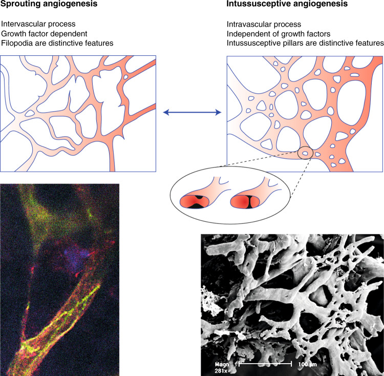
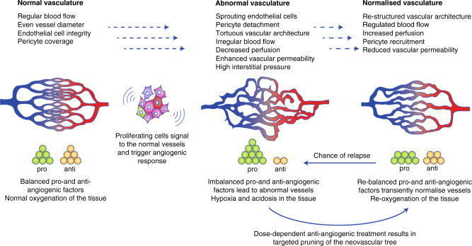
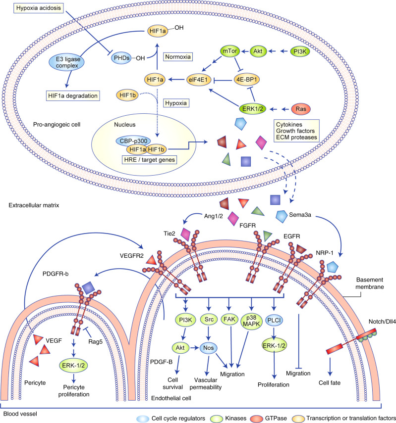
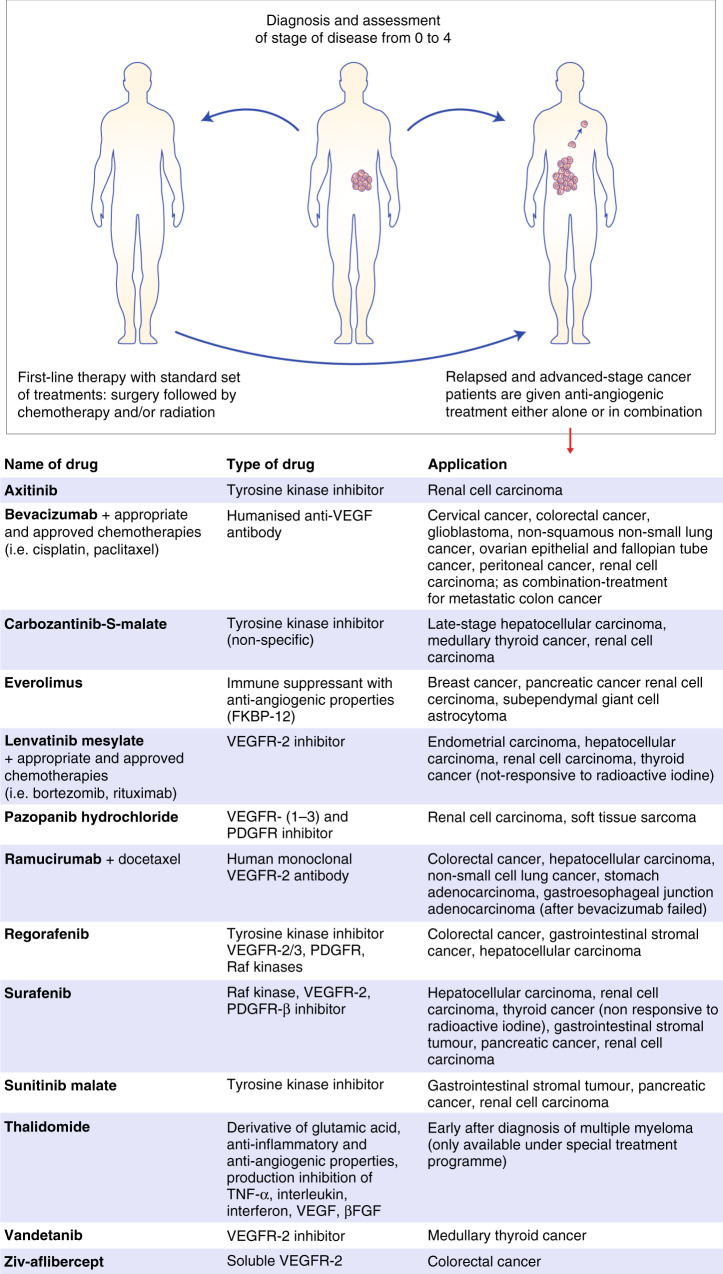
Verification
Claims the paper makes
The claims and statistics of this paper haven’t been checked yet.
Run the check — free, no account needed. It reads the open-access text and checks the sources it cites.
Claims other papers make about this one
Each is checked against this paper’s own text: does it say what the citing paper says it does?
In the citing paper
“The concept of “vascular normalization” proposes that judicious, often low-dose anti-angiogenic modulation can transiently restore vessel integrity and perfusion, thereby enlarging the delivery window for drugs and nanocarriers [43].”
From Overcoming tumor microenvironment barriers: transformable and bioinspired nanomedicine strategies for deep tumor penetration (2026) · cites this paper as [43]
Self-check
Percentages, sums and reported differences re-computed from the paper’s own numbers.
The self-check needs the paper’s full text.
Citations
Cites
PD-1 blockade induces responses by inhibiting adaptive immune resistance
Tumeh, P. C., Harview, C. L., Yearley, J. H., Shintaku, I. P., Taylor, E. J. M., Robert, L., Chmielowski, B., Spasic, M., Henry, G., Ciobanu, V., et al. (2014) · Nature · doi:10.1038/nature13954 · Cited by 6,609 · cited 2×
“Integrin adhesion via the β1 chain seems essential for cancer cells to adhere to endothelial cells to both invade and escape from the vascular system through intercalation.(133,104) In the brain, glioblastoma cells with cytoskeletal actin extensions, called flectopodia, adhere to the blood vessels by connecting in an…”
BackgroundNormalization of Tumor Vasculature: An Emerging Concept in Antiangiogenic Therapy
Jain, R. K. (2005) · Science · doi:10.1126/science.1104819 · Cited by 5,527
Predictive correlates of response to the anti-PD-L1 antibody MPDL3280A in cancer patients
Herbst, R. S., Soria, J.-C., Kowanetz, M., Fine, G. D., Hamid, O., Gordon, M. S., Sosman, J. A., McDermott, D. F., Powderly, J. D., Gettinger, S. N., et al. (2014) · Nature · doi:10.1038/nature14011 · Cited by 5,231
Tumors: Wounds That Do Not Heal
Flier, J. S., Underhill, L. H., & Dvorak, H. F. (1986) · New England Journal of Medicine · doi:10.1056/nejm198612253152606 · Cited by 4,391 · cited 1×
“Consequently, the inflammatory response in a tumour no longer has the positive effect of an acute reaction to harmful stimuli but becomes a chronic inflammatory site; to quote Harold Dvorak, “tumours are wounds that never heal”.(95,96) That may explain why a majority of patients do not benefit from anti-cancer immunot…”
BackgroundCouzin-Frankel, J. (2013) · Science · doi:10.1126/science.342.6165.1432 · Cited by 2,051
Corzo, C. A., Condamine, T., Lu, L., Cotter, M. J., Youn, J.-I., Cheng, P., Cho, H.-I., Celis, E., Quiceno, D. G., Padhya, T., et al. (2010) · Journal of Experimental Medicine · doi:10.1084/jem.20100587 · Cited by 1,173
Mutual regulation of tumour vessel normalization and immunostimulatory reprogramming
Tian, L., Goldstein, A., Wang, H., Ching Lo, H., Sun Kim, I., Welte, T., Sheng, K., Dobrolecki, L. E., Zhang, X., Putluri, N., et al. (2017) · Nature · doi:10.1038/nature21724 · Cited by 841
Why are tumour blood vessels abnormal and why is it important to know?
Nagy, J. A., Chang, S.-H., Dvorak, A. M., & Dvorak, H. F. (2009) · British Journal of Cancer · doi:10.1038/sj.bjc.6604929 · Cited by 611
Park, J.-S., Kim, I.-K., Han, S., Park, I., Kim, C., Bae, J., Oh, S. J., Lee, S., Kim, J. H., Woo, D.-C., et al. (2016) · Cancer Cell · doi:10.1016/j.ccell.2016.10.018 · Cited by 327
Modulation of murine breast tumor vascularity, hypoxia and chemotherapeutic response by exercise
Betof, A. S., Lascola, C. D., Weitzel, D., Landon, C., Scarbrough, P. M., Devi, G. R., Palmer, G., Jones, L. W., & Dewhirst, M. W. (2015) · JNCI: Journal of the National Cancer Institute · doi:10.1093/jnci/djv040 · Cited by 309
Tolaney, S. M., Boucher, Y., Duda, D. G., Martin, J. D., Seano, G., Ancukiewicz, M., Barry, W. T., Goel, S., Lahdenrata, J., Isakoff, S. J., et al. (2015) · Proceedings of the National Academy of Sciences · doi:10.1073/pnas.1518808112 · Cited by 258
Jumping the barrier: VE‐cadherin, VEGF and other angiogenic modifiers in cancer
Le Guelte, A., Dwyer, J., & Gavard, J. (2011) · Biology of the Cell · doi:10.1042/bc20110069 · Cited by 91
Research works printed but not linked: 136
- Franses, J. W. & Edelman, E. R. The evolution of endothelial regulatory paradigms in cancer biology and vascular repair. Cancer Res. 71, 7339 (2011). · DOI
- Harvey, W. An anatomical disquisition on the motion of the heart and blood in animals*. Ann. Noninvasive Electrocardiol. 5, 196–203 (2020). · DOI
- Meyer, J. Über die Neubildung von Blutgefäßen in plastischen Exudaten serösen Membranen und in Hautwunden. Ann Charité 4, 41–140 (1852).
- Clark, E. R. & Clark, E. L. Observations on changes in blood vascular endothelium in the living animal. Am. J. Anat. 57, 385–438 (1935). · DOI
- Clark, E. R. & Clark, E. L. Microscopic observations on the growth of blood capillaries in the living mammal. Am. J. Anat. 64, 251–301 (1939). · DOI
- Ribatti, D., Nico, B. & Crivellato, E. The development of the vascular system: a historical overview. in Vascular Morphogenesis: Methods and Protocols (ed. Ribatti, D.) 1–14 (Springer New York, 2015). · DOI
- Patan, S. Vasculogenesis and angiogenesis. Cancer Treat. Res. 117, 3–32 (2004). · DOI
- Patan, S. Vasculogenesis and angiogenesis as mechanisms of vascular network formation, growth and remodeling. J. Neurooncol. 50, 1–15 (2000). · DOI
- Chen, Q., Jiang, L., Li, C., Hu, D., Bu, J.-W., Cai, D. et al. Haemodynamics-driven developmental pruning of brain vasculature in zebrafish. PLoS Biol. 10, e1001374 (2012). · DOI
- Asahara, T., Murohara, T., Sullivan, A., Silver, M., van der Zee, R., Li, T. et al. Isolation of putative progenitor endothelial cells for angiogenesis. Science 275, 964–967 (1997). · DOI · Paper page
- Rafii, S. & Lyden, D. Therapeutic stem and progenitor cell transplantation for organ vascularization and regeneration. Nat. Med. 9, 702–712 (2003). · DOI
- Larrivée, B., Niessen, K., Pollet, I., Corbel, S. Y., Long, M., Rossi, F. M. et al. Minimal contribution of marrow-derived endothelial precursors to tumor vasculature. J. Immunol. 175, 2890 (2005). · DOI
- Marçola, M. & Rodrigues, C. E. Endothelial progenitor cells in tumor angiogenesis: another brick in the wall. Stem Cells Int. 2015, 832649 (2015). · DOI
- Moschetta, M., Mishima, Y., Sahin, I., Manier, S., Glavey, S., Vacca, A. et al. Role of endothelial progenitor cells in cancer progression. Biochimica et Biophysica Acta 1846, 26–39 (2014).
- Tanaka, S., Ueno, T., Ishiguro, H., Morita, S. & Toi, M. The lack of increases in circulating endothelial progenitor cell as a negative predictor for pathological response to neoadjuvant chemotherapy in breast cancer patients. npj Precis. Oncol. 1, 6 (2017). · DOI
- Mentzer, S. J. & Konerding, M. A. Intussusceptive angiogenesis: expansion and remodeling of microvascular networks. Angiogenesis 17, 499–509 (2014). · DOI
- Burri, P. H. & Djonov, V. Intussusceptive angiogenesis–the alternative to capillary sprouting. Mol. Aspects Med. 23, S1–S27 (2002). · DOI
- Burri, P. H., Hlushchuk, R. & Djonov, V. Intussusceptive angiogenesis: its emergence, its characteristics, and its significance. Dev. Dyn. 231, 474–488 (2004). · DOI
- Algire, G. H. C. & Vascular, H. W. reactions of normal and malignant tissues in vivo. J. Natl Cancer Inst. 6, 73 (1945). · DOI
- Tannock, I. F. The relation between cell proliferation and the vascular system in a transplanted mouse mammary tumour. Br. J. Cancer 22, 258–273 (1968). · DOI · Paper page
- Folkman, J., Merler, E., Abernathy, C. & Williams, G. Isolation of a tumor factor responsible for angiogenesis. J. Exp. Med. 133, 275–288 (1971). · DOI · Paper page
- Leung, D. W., Cachianes, G., Kuang, W. J., Goeddel, D. V. & Ferrara, N. Vascular endothelial growth factor is a secreted angiogenic mitogen. Science 246, 1306–1309 (1989). · DOI
- Plouët, J., Schilling, J. & Gospodarowicz, D. Isolation and characterization of a newly identified endothelial cell mitogen produced by AtT-20 cells. EMBO J. 8, 3801–3806 (1989). · DOI
- Senger, D. R., Galli, S. J., Dvorak, A. M., Perruzzi, C. A., Harvey, V. S. & Dvorak, H. F. Tumor cells secrete a vascular permeability factor that promotes accumulation of ascites fluid. Science 219, 983–985 (1983). · DOI
- Kerbel, R. S. Tumor angiogenesis. New Engl. J. Med. 358, 2039–2049 (2008). · DOI
- Sennino, B. & McDonald, D. M. Controlling escape from angiogenesis inhibitors. Nat. Rev. Cancer 12, 699–709 (2012). · DOI
- Overgaard, J. & Horsman, M. R. Modification of hypoxia-induced radioresistance in tumors by the use of oxygen and sensitizers. Semin. Radiat. Oncol. 6, 10–21 (1996). · DOI
- Krzywinska, E. & Stockmann, C. Hypoxia, metabolism and immune cell function. Biomedicines 6, 56 (2018). · DOI
- Ohga, N., Ishikawa, S., Maishi, N., Akiyama, K., Hida, Y., Kawamoto, T. et al. Heterogeneity of tumor endothelial cells: comparison between tumor endothelial cells isolated from high- and low-metastatic tumors. Am. J. Pathol. 180, 1294–1307 (2012). · DOI
- Matsumoto, S., Batra, S., Saito, K., Yasui, H., Choudhuri, R., Gadisetti, C. et al. Antiangiogenic agent sunitinib transiently increases tumor oxygenation and suppresses cycling hypoxia. Cancer Res. 71, 6350–6359 (2011). · DOI
- Morikawa, S., Baluk, P., Kaidoh, T., Haskell, A., Jain, R. K. & McDonald, D. M. Abnormalities in pericytes on blood vessels and endothelial sprouts in tumors. Am. J. Pathol. 160, 985–1000 (2002). · DOI
- Claffey, K. P., Brown, L. F., del Aguila, L. F., Tognazzi, K., Yeo, K. T., Manseau, E. J. et al. Expression of vascular permeability factor/vascular endothelial growth factor by melanoma cells increases tumor growth, angiogenesis, and experimental metastasis. Cancer Res. 56, 172–181 (1996).
- Baluk, P., Morikawa, S., Haskell, A., Mancuso, M. & McDonald, D. M. Abnormalities of basement membrane on blood vessels and endothelial sprouts in tumors. Am. J. Pathol. 163, 1801–1815 (2003). · DOI · Paper page
- Nina Kristine, R., Aphirak, J., Tore, L. & Catharina de Lange, D. Macromolecular diffusion in the extracellular matrix measured by fluorescence correlation spectroscopy. J. Biomed. Optics 13, 1–9 (2008).
- Ariffin, A. B., Forde, P. F., Jahangeer, S., Soden, D. M. & Hinchion, J. Releasing pressure in tumors: what do we know so far and where do we go from here? A review. Cancer Res. 74, 2655–2662 (2014). · DOI
- Le Serve, A. W. & Hellmann, K. Metastases and the normalization of tumour blood vessels by ICRF 159: a new type of drug action. Br. Med. J. 1, 597–601 (1972). · DOI
- Rybak, S. M., Sanovich, E., Hollingshead, M. G., Borgel, S. D., Newton, D. L., Melillo, G. et al. “Vasocrine” formation of tumor cell-lined vascular spaces: implications for rational design of antiangiogenic therapies. Cancer Res. 63, 2812–2819 (2003).
- Jain, R. K. Molecular regulation of vessel maturation. Nat. Med. 9, 685–693 (2003). · DOI
- Carmeliet, P. & Jain, R. K. Principles and mechanisms of vessel normalization for cancer and other angiogenic diseases. Nat. Rev. Drug Discov. 10, 417–427 (2011). · DOI
- Jain, R. K. Normalizing tumor vasculature with anti-angiogenic therapy: a new paradigm for combination therapy. Nat. Med. 7, 987–989 (2001). · DOI · Paper page
- Schwartz, M. A., Vestweber, D. & Simons, M. A unifying concept in vascular health and disease. Science 360, 270–271 (2018). · DOI
- Schreiter, J., Meyer, S., Schmidt, C., Schulz, R. M. & Langer, S. Dorsal skinfold chamber models in mice. GMS Interdiscip Plast. Reconstr. Surg. DGPW 6, Doc10, https://doi.org/10.3205/iprs000112 (2017). · DOI
- Tong, R. T., Boucher, Y., Kozin, S. V., Winkler, F., Hicklin, D. J. & Jain, R. K. Vascular normalization by vascular endothelial growth factor receptor 2 blockade induces a pressure gradient across the vasculature and improves drug penetration in tumors. Cancer Res. 64, 3731 (2004). · DOI · Paper page
- Yang, J., Liao, C., Liu, Y., Yang, G., Ke, T., Ding, Y. et al. MR imaging biomarkers evaluating vascular normalization window after anti-vessel treatment. Oncotarget 9, 11964–11976 (2017). · DOI
- Bohndiek, S. E., Sasportas, L. S., Machtaler, S., Jokerst, J. V., Hori, S. & Gambhir, S. S. Photoacoustic tomography detects early vessel regression and normalization during ovarian tumor response to the antiangiogenic therapy trebananib. J. Nucl. Med. 56, 1942–1947 (2015). · DOI
- Goel, S., Duda, D. G., Xu, L., Munn, L. L., Boucher, Y., Fukumura, D. et al. Normalization of the vasculature for treatment of cancer and other diseases. Physiol. Rev. 91, 1071–1121 (2011). · DOI · Paper page
- Winkler, F., Kozin, S. V., Tong, R. T., Chae, S.-S., Booth, M. F., Garkavtsev, I. et al. Kinetics of vascular normalization by VEGFR2 blockade governs brain tumor response to radiation: Role of oxygenation, angiopoietin-1, and matrix metalloproteinases. Cancer Cell 6, 553–563 (2004).
- Willett, C. G., Kozin, S. V., Duda, D. G., di Tomaso, E., Kozak, K. R., Boucher, Y. et al. Combined vascular endothelial growth factor-targeted therapy and radiotherapy for rectal cancer: theory and clinical practice. Semin. Oncol. 33, S35–S40 (2006). · DOI
- Collet, G., Lamerant-Fayel, N., Tertil, M., El Hafny-Rahbi, B., Stepniewski, J., Guichard, A. et al. Hypoxia-regulated overexpression of soluble VEGFR2 controls angiogenesis and inhibits tumor growth. Mol. Cancer Ther. 13, 165–178 (2014). · DOI
- Boudria, A., Abou Faycal, C., Jia, T., Gout, S., Keramidas, M., Didier, C. et al. VEGF165b, a splice variant of VEGF-A, promotes lung tumor progression and escape from anti-angiogenic therapies through a β1 integrin/VEGFR autocrine loop. Oncogene 38, 1050–1066 (2019). · DOI · Paper page
- Woolard, J., Wang, W. Y., Bevan, H. S., Qiu, Y., Morbidelli, L., Pritchard-Jones, R. O. et al. VEGF165b, an inhibitory vascular endothelial growth factor splice variant: mechanism of action, in vivo effect on angiogenesis and endogenous protein expression. Cancer Res. 64, 7822–7835 (2004). · DOI · Paper page
- Nowak, D. G., Woolard, J., Amin, E. M., Konopatskaya, O., Saleem, M. A., Churchill, A. J. et al. Expression of pro- and anti-angiogenic isoforms of VEGF is differentially regulated by splicing and growth factors. J. Cell Sci. 121, 3487 (2008). · DOI
- Mavrou, A. & Oltean, S. SRPK1 inhibition in prostate cancer: A novel anti-angiogenic treatment through modulation of VEGF alternative splicing. Pharmacol Res. 107, 276–281 (2016). · DOI
- Magnussen, A. L., Rennel, E. S., Hua, J., Bevan, H. S., Beazley Long, N., Lehrling, C. et al. VEGF-A165b is cytoprotective and antiangiogenic in the retina. Investig. Ophthalmol. Vis. Sci. 51, 4273–4281 (2010). · DOI
- Liu, J., Liao, S., Diop-Frimpong, B., Chen, W., Goel, S., Naxerova, K. et al. TGF-β blockade improves the distribution and efficacy of therapeutics in breast carcinoma by normalizing the tumor stroma. Proc. Natl Acad. Sci. USA 109, 16618–16623 (2012). · DOI
- Dieterich, L. C., Mellberg, S., Langenkamp, E., Zhang, L., Zieba, A., Salomäki, H. et al. Transcriptional profiling of human glioblastoma vessels indicates a key role of VEGF-A and TGFβ2 in vascular abnormalization. J. Pathol. 228, 378–390 (2012). · DOI
- Qayum, N., Muschel, R. J., Im, J. H., Balathasan, L., Koch, C. J., Patel, S. et al. Tumor vascular changes mediated by inhibition of oncogenic signaling. Cancer Res. 69, 6347–6354 (2009). · DOI
- Sawada, J., Urakami, T., Li, F., Urakami, A., Zhu, W., Fukuda, M. et al. Small GTPase R-Ras regulates integrity and functionality of tumor blood vessels. Cancer Cell 22, 235–249 (2012). · DOI
- Hida, K., Maishi, N., Annan, D. A. & Hida, Y. Contribution of tumor endothelial cells in cancer progression. Int. J. Mol. Sci. 19, 1272 (2018). · DOI
- Hamzah, J., Jugold, M., Kiessling, F., Rigby, P., Manzur, M., Marti, H. H. et al. Vascular normalization in Rgs5-deficient tumours promotes immune destruction. Nature 453, 410–414 (2008). · DOI
- Cox, D. B., Platt, R. J. & Zhang, F. Therapeutic genome editing: prospects and challenges. Nat. Med. 21, 121–131 (2015). · DOI · Paper page
- Falcón, B. L., Hashizume, H., Koumoutsakos, P., Chou, J., Bready, J. V., Coxon, A. et al. Contrasting actions of selective inhibitors of angiopoietin-1 and angiopoietin-2 on the normalization of tumor blood vessels. Am. J. Pathol. 175, 2159–2170 (2009). · DOI
- Alto, L. T. & Terman, J. R. Semaphorins and their signaling mechanisms. Methods Mol. Biol. 1493, 1–25 (2017). · DOI
- Casazza, A., Fu, X., Johansson, I., Capparuccia, L., Andersson, F., Giustacchini, A. et al. Systemic and targeted delivery of semaphorin 3A inhibits tumor angiogenesis and progression in mouse tumor models. Arterioscler. Thromb. Vasc. Biol. 31, 741–749 (2011). · DOI
- Serini, G., Bussolino, F., Maione, F. & Giraudo, E. Class 3 semaphorins: physiological vascular normalizing agents for anti-cancer therapy. J. Intern. Med. 273, 138–155 (2013). · DOI
- Fogal, V., Sugahara, K. N., Ruoslahti, E. & Christian, S. Cell surface nucleolin antagonist causes endothelial cell apoptosis and normalization of tumor vasculature. Angiogenesis 12, 91–100 (2009). · DOI
- Baron, M. An overview of the Notch signalling pathway. Semin. Cell Dev. Biol. 14, 113–119 (2003). · DOI
- Hellstrom, M., Phng, L. K., Hofmann, J. J., Wallgard, E., Coultas, L., Lindblom, P. et al. Dll4 signalling through Notch1 regulates formation of tip cells during angiogenesis. Nature 445, 776–780 (2007). · DOI · Paper page
- Phng, L. K. & Gerhardt, H. Angiogenesis: a team effort coordinated by notch. Dev. Cell 16, 196–208 (2009). · DOI
- Maes, H., Kuchnio, A., Peric, A., Moens, S., Nys, K., De Bock, K. et al. Tumor vessel normalization by chloroquine independent of autophagy. Cancer Cell 26, 190–206 (2014). · DOI
- Trindade, A., Djokovic, D., Gigante, J., Mendonça, L. & Duarte, A. Endothelial Dll4 overexpression reduces vascular response and inhibits tumor growth and metastasization in vivo. BMC Cancer 17, 189- (2017). · DOI
- Polet, F. & Feron, O. Endothelial cell metabolism and tumour angiogenesis: glucose and glutamine as essential fuels and lactate as the driving force. J. Intern. Med. 273, 156–165 (2013). · DOI
- Zhang, L., Li, S., Li, L., Chen, Z. & Yang, Y. COX‑2 inhibition in the endothelium induces glucose metabolism normalization and impairs tumor progression. Mol. Med. Rep. 17, 2937–2944 (2018).
- Cantelmo, A. R., Conradi, L. C., Brajic, A., Goveia, J., Kalucka, J., Pircher, A. et al. Inhibition of the glycolytic activator PFKFB3 in endothelium induces tumor vessel normalization, impairs metastasis, and improves chemotherapy. Cancer Cell 30, 968–985 (2016). · DOI · Paper page
- Kocher, M., Treuer, H., Voges, J., Hoevels, M., Sturm, V. & Muller, R. P. Computer simulation of cytotoxic and vascular effects of radiosurgery in solid and necrotic brain metastases. Radiother. Oncol. 54, 149–156 (2000). · DOI
- Potiron, V. A., Abderrahmani, R., Clément-Colmou, K., Marionneau-Lambot, S., Oullier, T., Paris, F. et al. Improved functionality of the vasculature during conventionally fractionated radiation therapy of prostate cancer. PLoS ONE 8, e84076 (2013). · DOI
- Koelwyn, G. J., Quail, D. F., Zhang, X., White, R. M. & Jones, L. W. Exercise-dependent regulation of the tumour microenvironment. Nat. Rev. Cancer 17, 620 (2017). · DOI
- Di Francescomarino, S., Sciartilli, A., Di Valerio, V., Di Baldassarre, A. & Gallina, S. The effect of physical exercise on endothelial function. Sports Med. 39, 797–812 (2009). · DOI
- Resnick, N., Yahav, H., Shay-Salit, A., Shushy, M., Schubert, S., Zilberman, L. C. et al. Fluid shear stress and the vascular endothelium: for better and for worse. Prog. Biophys. Mol. Biol. 81, 177–199 (2003). · DOI
- Schadler, K. L., Thomas, N. J., Galie, P. A., Bhang, D. H., Roby, K. C., Addai, P. et al. Tumor vessel normalization after aerobic exercise enhances chemotherapeutic efficacy. Oncotarget 7, 65429–65440 (2016). · DOI · Paper page
- McCarthy, M. Antiangiogenesis drug promising for metastatic colorectal cancer. Lancet 361, 1959 (2003). · DOI
- Gonzalez, H., Hagerling, C. & Werb, Z. Roles of the immune system in cancer: from tumor initiation to metastatic progression. Genes Dev. 32, 1267–1284 (2018). · DOI · Paper page
- Al-Soudi, A., Kaaij, M. H. & Tas, S. W. Endothelial cells: from innocent bystanders to active participants in immune responses. Autoimmun. Rev. 16, 951–962 (2017). · DOI
- Funaro, A., Ferrero, E., Mehta, K. & Malavasi, F. Schematic portrait of human CD38 and related molecules. Chem. Immunol. 75, 256–273 (2000).
- Rothermel, A. L., Wang, Y., Schechner, J., Mook-Kanamori, B., Aird, W. C., Pober, J. S. et al. Endothelial cells present antigens in vivo. BMC Immunol. 5, 5 (2004). · DOI
- Mai, J., Virtue, A., Shen, J., Wang, H. & Yang, X.-F. An evolving new paradigm: endothelial cells—conditional innate immune cells. J. Hematol. Oncol. 6, 61 (2013). · DOI
- Masopust, D. & Schenkel, J. M. The integration of T cell migration, differentiation and function. Nat. Rev. Immunol. 13, 309–320 (2013). · DOI
- Huang, Y., Kim, B. Y. S., Chan, C. K., Hahn, S. M., Weissman, I. L. & Jiang, W. Improving immune–vascular crosstalk for cancer immunotherapy. Nat. Rev. Immunol. 18, 195 (2018). · DOI · Paper page
- Barzilai, S., Yadav, S. K., Morrell, S., Roncato, F., Klein, E., Stoler-Barak, L. et al. Leukocytes breach endothelial barriers by insertion of nuclear lobes and disassembly of endothelial actin filaments. Cell Rep. 18, 685–699 (2017). · DOI
- Dvorak, H. F. Tumors: wounds that do not heal—a historical perspective with a focus on the fundamental roles of increased vascular permeability and clotting. Semin. Thromb. Hemost. 45, 576–592 (2019). · DOI
- Mpekris, F., Voutouri, C., Baish, J. W., Duda, D. G., Munn, L. L., Stylianopoulos, T. et al. Combining microenvironment normalization strategies to improve cancer immunotherapy. Proc. Natl Acad. Sci. USA 117, 3728 (2020). · DOI
- Noman, M. Z., Hasmim, M., Messai, Y., Terry, S., Kieda, C., Janji, B. et al. Hypoxia: a key player in antitumor immune response. A Review in the theme: cellular responses to hypoxia. Am. J. Physiol. Cell Physiol. 309, C569–C579 (2015). · DOI
- Doedens, A. L., Stockmann, C., Rubinstein, M. P., Liao, D., Zhang, N., DeNardo, D. G. et al. Macrophage expression of hypoxia-inducible factor-1 alpha suppresses T-cell function and promotes tumor progression. Cancer Res. 70, 7465–7475 (2010). · DOI · Paper page
- McEver, R. P. & Zhu, C. Rolling cell adhesion. Annu. Rev. Cell Dev. Biol. 26, 363–396 (2010). · DOI
- Huang, H., Langenkamp, E., Georganaki, M., Loskog, A., Fuchs, P. F., Dieterich, L. C. et al. VEGF suppresses T-lymphocyte infiltration in the tumor microenvironment through inhibition of NF-kappaB-induced endothelial activation. Faseb J. 29, 227–238 (2015). · DOI
- Pardoll, D. M. The blockade of immune checkpoints in cancer immunotherapy. Nat. Rev. Cancer 12, 252–264 (2012). · DOI · Paper page
- Li, Y., Li, F., Jiang, F., Lv, X., Zhang, R., Lu, A. et al. A mini-review for cancer immunotherapy: molecular understanding of PD-1/PD-L1 pathway: translational blockade of immune checkpoints. Int. J. Mol. Sci. 17, 1151 (2016). · DOI
- Carter, T., Shaw, H., Cohn-Brown, D., Chester, K. & Mulholland, P. Ipilimumab and bevacizumab in glioblastoma. Clin. Oncol. 28, 622–626 (2016). · DOI
- Wu, X., Li, J., Connolly, E. M., Liao, X., Ouyang, J., Giobbie-Hurder, A. et al. Combined anti-VEGF and anti-CTLA-4 therapy elicits humoral immunity to galectin-1 which is associated with favorable clinical outcomes. Cancer Immunol. Res. 5, 446–454 (2017). · DOI
- Di Tacchio, M., Macas, J., Weissenberger, J., Sommer, K., Bähr, O., Steinbach, J. P. et al. Tumor vessel normalization, immuno-stimulatory reprogramming, and improved survival in glioblastoma with combined inhibition of PD-1, angiopoietin-2, and VEGF. Cancer Immunol. Res. 7, 1910–1927 (2019). · DOI
- Davis, B. P. & Rothenberg, M. E. Eosinophils and cancer. Cancer Immunol. Res. 2, 1–8 (2014). · DOI
- Carretero, R., Sektioglu, I. M., Garbi, N., Salgado, O. C., Beckhove, P. & Hämmerling, G. J. Eosinophils orchestrate cancer rejection by normalizing tumor vessels and enhancing infiltration of CD8+ T cells. Nat. Immunol. 16, 609 (2015). · DOI · Paper page
- Sanchez, L. R., Borriello, L., Entenberg, D., Condeelis, J. S., Oktay, M. H. & Karagiannis, G. S. The emerging roles of macrophages in cancer metastasis and response to chemotherapy. J. Leukoc. Biol. 106, 259–274 (2019). · DOI
- Arwert, E. N., Harney, A. S., Entenberg, D., Wang, Y., Sahai, E., Pollard, J. W. et al. A unidirectional transition from migratory to perivascular macrophage is required for tumor cell intravasation. Cell Rep. 23, 1239–1248 (2018). · DOI
- Harney, A. S., Karagiannis, G. S., Pignatelli, J., Smith, B. D., Kadioglu, E., Wise, S. C. et al. The selective Tie2 inhibitor rebastinib blocks recruitment and function of Tie2(Hi) macrophages in breast cancer and pancreatic neuroendocrine tumors. Mol. Cancer Ther. 16, 2486–2501 (2017). · DOI
- Nishide, S., Matsunaga, S., Shiota, M., Yamaguchi, T., Kitajima, S., Maekawa, Y. et al. Controlling the phenotype of tumor-infiltrating macrophages via the PHD-HIF axis inhibits tumor growth in a mouse model. iScience 19, 940–954 (2019). · DOI
- Sato, N., Nariuchi, H., Tsuruoka, N., Nishihara, T., Beitz, J. G., Calabresi, P. et al. Actions of TNF and IFN-gamma on angiogenesis in vitro. J. Investig. Dermatol. 95, 85s–89ss (1990). · DOI
- Sun, T., Yang, Y., Luo, X., Cheng, Y., Zhang, M., Wang, K. et al. Inhibition of tumor angiogenesis by interferon-gamma by suppression of tumor-associated macrophage differentiation. Oncol. Res. 21, 227–235 (2014). · DOI
- Kwee, B. J., Seo, B. R., Najibi, A. J., Li, A. W., Shih, T.-Y., White, D. et al. Treating ischemia via recruitment of antigen-specific T cells. Sci. Adv. 5, eaav6313 (2019). · DOI
- Johansson-Percival, A., He, B. & Ganss, R. Immunomodulation of tumor vessels: it takes two to tango. Trends Immunol. 39, 801–814 (2018). · DOI
- Batchelor, T. T., Sorensen, A. G., di Tomaso, E., Zhang, W.-T., Duda, D. G., Cohen, K. S. et al. AZD2171, a pan-VEGF receptor tyrosine kinase inhibitor, normalizes tumor vasculature and alleviates edema in glioblastoma patients. Cancer Cell 11, 83–95 (2007). · DOI · Paper page
- Sorensen, A. G., Batchelor, T. T., Zhang, W.-T., Chen, P.-J., Yeo, P., Wang, M. et al. A “vascular normalization index” as potential mechanistic biomarker to predict survival after a single dose of cediranib in recurrent glioblastoma patients. Cancer Res. 69, 5296–5300 (2009). · DOI
- Batchelor, T. T., Gerstner, E. R., Emblem, K. E., Duda, D. G., Kalpathy-Cramer, J., Snuderl, M. et al. Improved tumor oxygenation and survival in glioblastoma patients who show increased blood perfusion after cediranib and chemoradiation. Proc. Natl Acad. Sci. USA 110, 19059–19064 (2013). · DOI
- von Baumgarten, L., Brucker, D., Tirniceru, A., Kienast, Y., Grau, S., Burgold, S. et al. Bevacizumab has differential and dose-dependent effects on glioma blood vessels and tumor cells. Clin. Cancer Res. 17, 6192–6205 (2011). · DOI
- Keunen, O., Johansson, M., Oudin, A., Sanzey, M., Rahim, S. A., Fack, F. et al. Anti-VEGF treatment reduces blood supply and increases tumor cell invasion in glioblastoma. Proc. Natl Acad. Sci. USA 108, 3749–3754 (2011). · DOI
- Li, Y., Ali, S., Clarke, J. & Cha, S. Bevacizumab in recurrent glioma: patterns of treatment failure and implications. Brain Tumor Res. Treat. 5, 1–9 (2017). · DOI
- Claes, A., Wesseling, P., Jeuken, J., Maass, C., Heerschap, A. & Leenders, W. P. Antiangiogenic compounds interfere with chemotherapy of brain tumors due to vessel normalization. Mol. Cancer Ther. 7, 71–78 (2008). · DOI
- Boujelben, A., Watson, M., McDougall, S., Yen, Y.-F., Gerstner, E. R., Catana, C. et al. Multimodality imaging and mathematical modelling of drug delivery to glioblastomas. Interface Focus 6, 20160039 (2016). · DOI
- Donnem, T., Reynolds, A. R., Kuczynski, E. A., Gatter, K., Vermeulen, P. B., Kerbel, R. S. et al. Non-angiogenic tumours and their influence on cancer biology. Nat. Rev. Cancer 18, 323–336 (2018). · DOI
- García-Gómez, P. & Valiente, M. Vascular co-option in brain metastasis. Angiogenesis 23, 3–8 (2020). · DOI
- Holash, J., Maisonpierre, P. C., Compton, D., Boland, P., Alexander, C. R., Zagzag, D. et al. Vessel cooption, regression, and growth in tumors mediated by angiopoietins and VEGF. Science 284, 1994 (1999). · DOI
- Reymond, N., Im, J. H., Garg, R., Vega, F. M., Borda d’Agua, B., Riou, P. et al. Cdc42 promotes transendothelial migration of cancer cells through β1 integrin. J. Cell Biol. 199, 653–668 (2012). · DOI
- Watkins, S., Robel, S., Kimbrough, I. F., Robert, S. M., Ellis-Davies, G. & Sontheimer, H. Disruption of astrocyte-vascular coupling and the blood-brain barrier by invading glioma cells. Nat. Commun. 5, 4196 (2014). · DOI
- Jezzard, P., Matthews, P. M. & Smith, S. M. Functional Magnetic Resonance Imaging: An Introduction to Methods (Oxford University Press, 2001). · DOI
- Yang, J., Zhang, G., Li, Q., Liao, C., Huang, L., Ke, T. et al. Photoacoustic imaging for the evaluation of early tumor response to antivascular treatment. Quant. Imaging Med. Surg. 9, 160–170 (2019). · DOI
- Brown, E., Brunker, J. & Bohndiek, S. E. Photoacoustic imaging as a tool to probe the tumour microenvironment. Dis. Model. Mech. 12, dmm039636 (2019). · DOI
- Matsumoto, S., Saito, K., Takakusagi, Y., Matsuo, M., Munasinghe, J. P., Morris, H. D. et al. In vivo imaging of tumor physiological, metabolic, and redox changes in response to the anti-angiogenic agent sunitinib: longitudinal assessment to identify transient vascular renormalization. Antioxid. Redox Signal 21, 1145–1155 (2014). · DOI
- Xu, S., Han, P., Huang, M., Wu, J. C., Chang, C., Tsao, P. S. et al. In vivo, ex vivo, and in vitro studies on apelin’s effect on myocardial glucose uptake. Peptides 37, 320–326 (2012). · DOI
- Cox, C. M., D’Agostino, S. L., Miller, M. K., Heimark, R. L. & Krieg, P. A. Apelin, the ligand for the endothelial G-protein-coupled receptor, APJ, is a potent angiogenic factor required for normal vascular development of the frog embryo. Dev. Biol. 296, 177–189 (2006). · DOI
- Eyries, M., Siegfried, G., Ciumas, M., Montagne, K., Agrapart, M., Lebrin, F. et al. Hypoxia-induced apelin expression regulates endothelial cell proliferation and regenerative angiogenesis. Circ Res. 103, 432–440 (2008). · DOI
- Zhang, L., Takara, K., Yamakawa, D., Kidoya, H. & Takakura, N. Apelin as a marker for monitoring the tumor vessel normalization window during antiangiogenic therapy. Cancer Sci. 107, 36–44 (2016). · DOI
- Yamakawa, D., Jia, W., Kidoya, H., Hosojima, S., Torigata, M., Zhang, L. et al. Visualization of proliferative vascular endothelial cells in tumors in vivo by imaging their partner of Sld5-1 promoter activity. Am. J. Pathol. 188, 1300–1314 (2018). · DOI
- Boumahdi, S. & de Sauvage, F. J. The great escape: tumour cell plasticity in resistance to targeted therapy. Nat Rev. Drug Discov. 19, 39–56 (2020). · DOI
- Standring, S., Standring, S. & Gray, H. Gray’s Anatomy E-Book: The Anatomical Basis of Clinical Practice (Elsevier, 2015).
- Aird, W. C. Endothelial cell heterogeneity. Cold Spring Harb. Perspect. Med. 2, a006429 (2012). · DOI
- Hida, K., Hida, Y., Amin, D. N., Flint, A. F., Panigrahy, D., Morton, C. C. et al. Tumor-associated endothelial cells with cytogenetic abnormalities. Cancer Res. 64, 8249 (2004). · DOI
Cited by
Sorted by citations
Vascularised organoids: Recent advances and applications in cancer research
Zhou, R., Brislinger, D., Fuchs, J., Lyons, A., Langthaler, S., Hauser, C. A. E., & Baumgartner, C. (2025) · Clinical and Translational Medicine · doi:10.1002/ctm2.70258 · Cited by 41
Sheng, J., Yuan, W., Zhang, M., Chen, Z., Qi, Y., Zhao, Y., & Zhang, S. (2026) · Journal of Nanobiotechnology · doi:10.1186/s12951-026-04028-7 · Cited by 35
Multifaceted Interplay between Hormones, Growth Factors and Hypoxia in the Tumor Microenvironment
Lappano, R., Todd, L. A., Stanic, M., Cai, Q., Maggiolini, M., Marincola, F., & Pietrobon, V. (2022) · Cancers · doi:10.3390/cancers14030539 · Cited by 32
Hypoxic microenvironment in cancer: role in metabolic reprogramming
Sureka, N., Maheshwari, R., Agravat, A., Singhal, S., Zaman, S., Rishi, B., Siraj, F., Zaheer, S., & Misra, A. (2026) · Frontiers in Oncology · doi:10.3389/fonc.2026.1771365 · Cited by 11
Metadata
- Authors
- Magnussen, A. L., & Mills, I. G.
- Venue
- British Journal of Cancer
- Published
- 2021-04-07
- Volume
- 125
- Issue
- 3
- Pages
- 324-336
- ISSN
- 0007-0920
- 1532-1827
- PMC
- PMC8329166
- Cited by
- 146
- References
- 12
- Language
- en
About these checks
Registry, retraction, Cites and statistics results are computed, not judged, with one exception: a printed reference that no index lists is read once by a model before it counts as a citation error. Claims are extracted by a model and kept only when their quote appears verbatim in the text; each cited source is then checked, with a model’s help, for whether it says what the claim relies on. Those verdicts can be wrong — read the claim’s quote and the cited source before relying on one.
Statistics and arithmetic are recomputed, not judged.
Confidence bands are shown only for fields calibrated on our annotated test set. None is calibrated yet, so no band is shown.
What counts as a claim ›How we measure confidence ›How we check this ›
Are you an author, or do you see a problem with this page? Request a review
