

The DOI matches its Crossref record; 51 of the 51 references it cites were found in a registry or an index.
Galectin-3 promotes Aβ oligomerization and Aβ toxicity in a mouse model of Alzheimer’s disease
Open accessAbstract
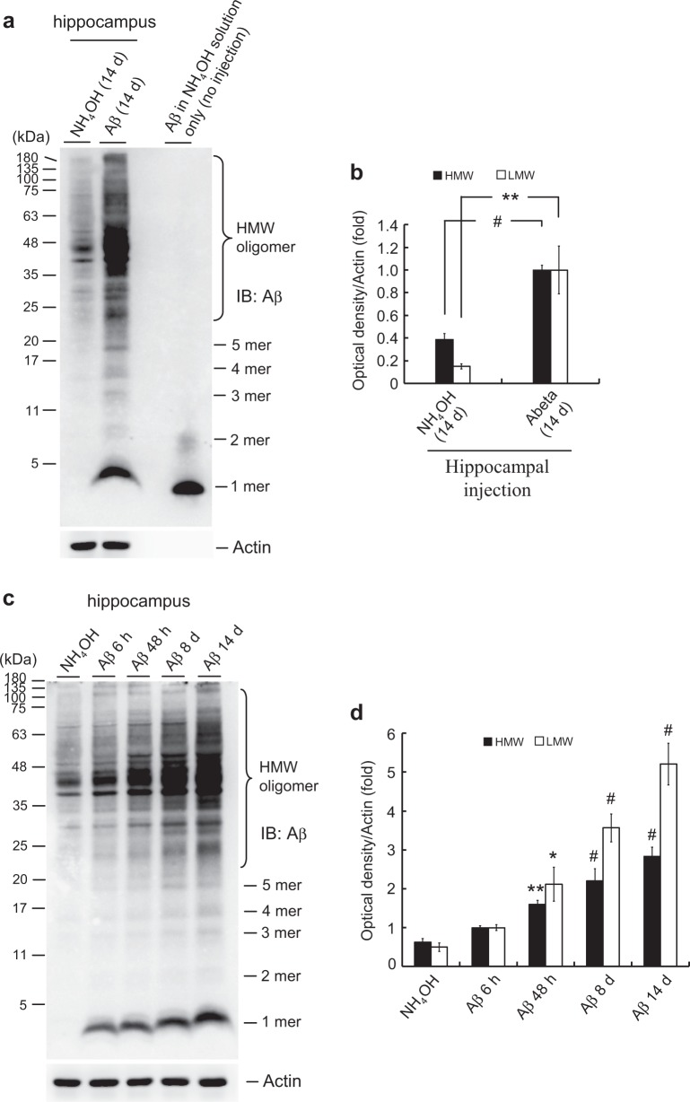
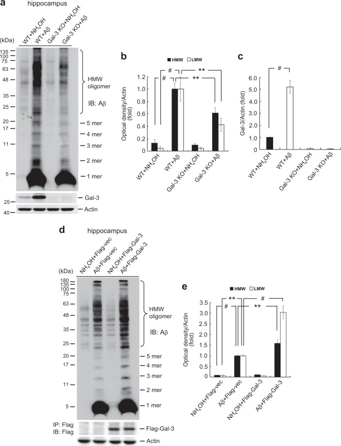
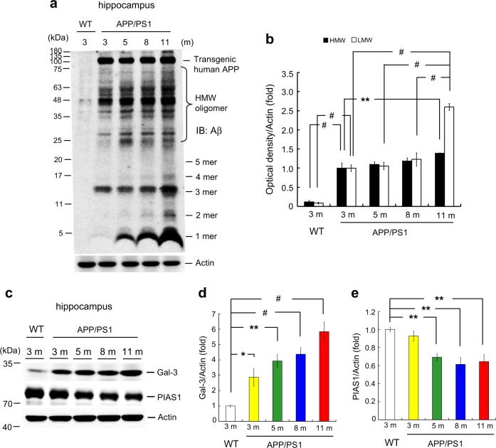
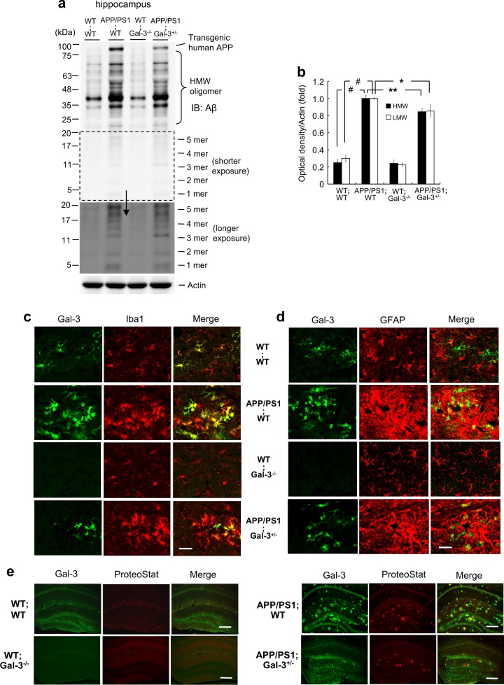
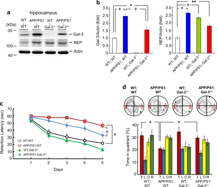
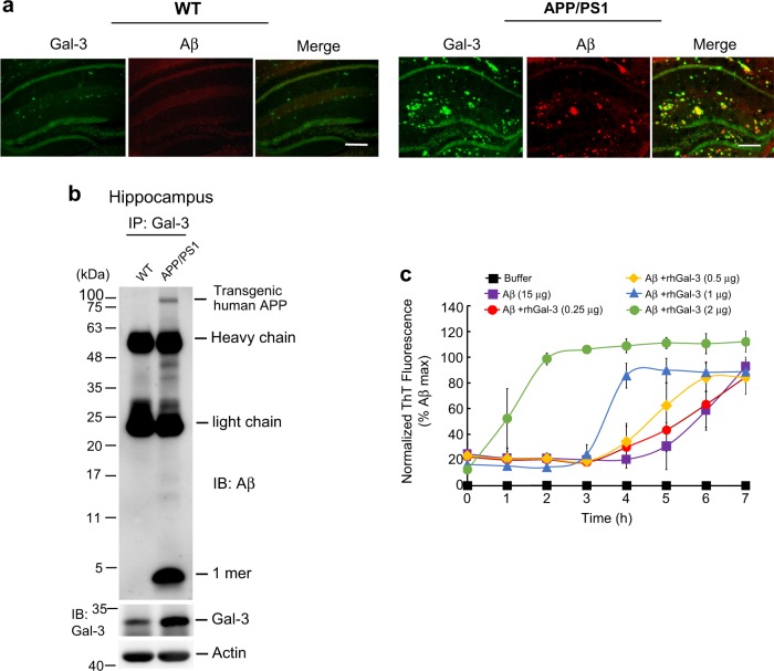
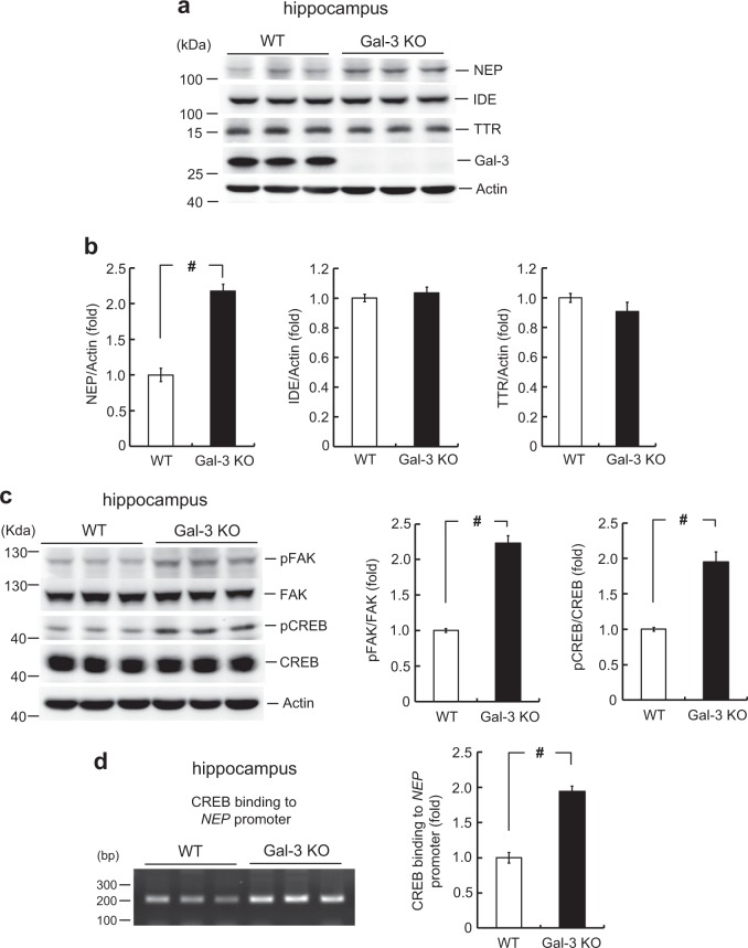
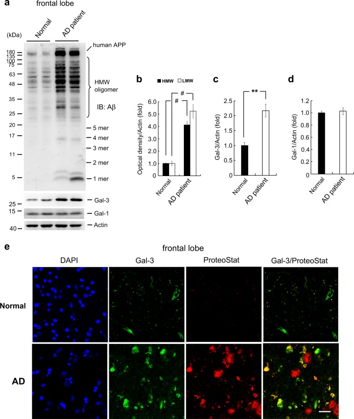
Amyloid-β (Aβ) oligomers largely initiate the cascade underlying the pathology of Alzheimer’s disease (AD). Galectin-3 (Gal-3), which is a member of the galectin protein family, promotes inflammatory responses and enhances the homotypic aggregation of cancer cells. Here, we examined the role and action mechanism of Gal-3 in Aβ oligomerization and Aβ toxicities. Wild-type (WT) and Gal-3-knockout (KO) mice, APP/PS1;WT mice, APP/PS1;Gal-3 +/− mice and brain tissues from normal subjects and AD patients were used. We found that Aβ oligomerization is reduced in Gal-3 KO mice injected with Aβ, whereas overexpression of Gal-3 enhances Aβ oligomerization in the hippocampi of Aβ-injected mice. Gal-3 expression shows an age-dependent increase that parallels endogenous Aβ oligomerization in APP/PS1 mice. Moreover, Aβ oligomerization, Iba1 expression, GFAP expression and amyloid plaque accumulation are reduced in APP/PS1;Gal-3 +/− mice compared with APP/PS1;WT mice. APP/PS1;Gal-3 +/− mice also show better acquisition and retention performance compared to APP/PS1;WT mice. In studying the mechanism underlying Gal-3-promoted Aβ oligomerization, we found that Gal-3 primarily co-localizes with Iba1, and that microglia-secreted Gal-3 directly interacts with Aβ. Gal-3 also interacts with triggering receptor expressed on myeloid cells-2, which then mediates the ability of Gal-3 to activate microglia for further Gal-3 expression. Immunohistochemical analyses show that the distribution of Gal-3 overlaps with that of endogenous Aβ in APP/PS1 mice and partially overlaps with that of amyloid plaque. Moreover, the expression of the Aβ-degrading enzyme, neprilysin, is increased in Gal-3 KO mice and this is associated with enhanced integrin-mediated signaling. Consistently, Gal-3 expression is also increased in the frontal lobe of AD patients, in parallel with Aβ oligomerization. Because Gal-3 expression is dramatically increased as early as 3 months of age in APP/PS1 mice and anti-Aβ oligomerization is believed to protect against Aβ toxicity, Gal-3 could be considered a novel therapeutic target in efforts to combat AD.
Overview
The claims and statistics of this paper haven’t been checked yet.
Run the check — free, no account needed. It reads the open-access text and checks the sources it cites.
Integrity signals
Registry
Cites
Statistics
Figures
Verification
Claims the paper makes
The claims and statistics of this paper haven’t been checked yet.
Run the check — free, no account needed. It reads the open-access text and checks the sources it cites.
Claims other papers make about this one
Each is checked against this paper’s own text: does it say what the citing paper says it does?
The cited source says what the claim attributes to it.
In the citing paper
“Although LGALS3 was reported to promote Aβ oligomerization and toxicity, these studies were performed using either global Lgals3 -KO mice or Aβ monomers precultured with LGALS3 before injection into the brain 52,53 .”
From APOE4 impairs the microglial response in Alzheimer’s disease by inducing TGFβ-mediated checkpoints (2023) · cites this paper as [53]
Self-check
Percentages, sums and reported differences re-computed from the paper’s own numbers.
The self-check needs the paper’s full text.
Citations
Cites
McKhann, G. M., Knopman, D. S., Chertkow, H., Hyman, B. T., Jack, C. R., Kawas, C. H., Klunk, W. E., Koroshetz, W. J., Manly, J. J., Mayeux, R., et al. (2011) · Alzheimer's & Dementia · doi:10.1016/j.jalz.2011.03.005 · Cited by 18,657
Mild cognitive impairment as a diagnostic entity
Petersen, R. C. (2004) · Journal of Internal Medicine · doi:10.1111/j.1365-2796.2004.01388.x · Cited by 7,864
Farris, W., Mansourian, S., Chang, Y., Lindsley, L., Eckman, E. A., Frosch, M. P., Eckman, C. B., Tanzi, R. E., Selkoe, D. J., & Guénette, S. Y. (2003) · Proceedings of the National Academy of Sciences · doi:10.1073/pnas.0230450100 · Cited by 1,526
Galectins. Structure and function of a large family of animal lectins.
Barondes, S. H., Cooper, D. N., Gitt, M. A., & Leffler, H. (1994) · Journal of Biological Chemistry · doi:10.1016/s0021-9258(17)31891-4 · Cited by 1,498
Galectins as modulators of tumour progression
Liu, F., & Rabinovich, G. A. (2005) · Nature reviews. Cancer · doi:10.1038/nrc1527 · Cited by 1,435
Galectins: A family of animal β-galactoside-binding lectins
Barondes, S. H., Castronovo, V., Cooper, D. N. W., Cummings, R. D., Drickamer, K., Felzi, T., Gitt, M. A., Hirabayashi, J., Hughes, C., Kasai, K.-I., et al. (1994) · Cell · doi:10.1016/0092-8674(94)90498-7 · Cited by 1,223
Metabolic Regulation of Brain Aβ by Neprilysin
Iwata, N., Tsubuki, S., Takaki, Y., Shirotani, K., Lu, B., Gerard, N. P., Gerard, C. J., Hama, E., Lee, H.-J., & Saido, T. C. (2001) · Science · doi:10.1126/science.1059946 · Cited by 982
Amyloidogenic processing of the Alzheimer β-amyloid precursor protein depends on lipid rafts
Ehehalt, R., Keller, P., Haass, C., Thiele, C., & Simons, K. (2003) · The Journal of Cell Biology · doi:10.1083/jcb.200207113 · Cited by 932
The Amyloid-β Oligomer Hypothesis: Beginning of the Third Decade
Cline, E. N., Bicca, M. A., Viola, K. L., & Klein, W. L. (2018) · Journal of Alzheimer's Disease · doi:10.3233/jad-179941 · Cited by 870
Iwata, N., Tsubuki, S., Takaki, Y., Watanabe, K., Sekiguchi, M., Hosoki, E., Kawashima-Morishima, M., Lee, H.-J., Hama, E., Sekine-Aizawa, Y., et al. (2000) · Nature Medicine · doi:10.1038/72237 · Cited by 867
Cited by
Sorted by citations
Synaptic degeneration in Alzheimer disease
Tzioras, M., McGeachan, R. I., Durrant, C. S., & Spires-Jones, T. L. (2023) · Nature Reviews Neurology · doi:10.1038/s41582-022-00749-z · Cited by 504
APOE4 impairs the microglial response in Alzheimer’s disease by inducing TGFβ-mediated checkpoints
Yin, Z., Rosenzweig, N., Kleemann, K. L., Zhang, X., Brandão, W., Margeta, M. A., Schroeder, C., Sivanathan, K. N., Silveira, S., Gauthier, C., et al. (2023) · Nature Immunology · doi:10.1038/s41590-023-01627-6 · Cited by 202
“Although LGALS3 was reported to promote Aβ oligomerization and toxicity, these studies were performed using either global Lgals3 -KO mice or Aβ monomers precultured with LGALS3 before injection into the brain 52,53 .”
ResultSialylation and Galectin-3 in Microglia-Mediated Neuroinflammation and Neurodegeneration
Puigdellívol, M., Allendorf, D. H., & Brown, G. C. (2020) · Frontiers in Cellular Neuroscience · doi:10.3389/fncel.2020.00162 · Cited by 145
Neurobiochemical Cross-talk Between COVID-19 and Alzheimer’s Disease
Rahman, M. A., Islam, K., Rahman, S., & Alamin, M. (2020) · Molecular Neurobiology · doi:10.1007/s12035-020-02177-w · Cited by 121
COVID-19 and Alzheimer’s Disease
Ciaccio, M., Sasso, B. L., Scazzone, C., Gambino, C. M., Ciaccio, A. M., Bivona, G., Piccoli, T., Giglio, R. V., & Agnello, L. (2021) · Brain Sciences · doi:10.3390/brainsci11030305 · Cited by 116
Galectin-3: a key player in microglia-mediated neuroinflammation and Alzheimer's disease
Tan, Y., Zheng, Y., Xu, D., Sun, Z., Yang, H., & Yin, Q. (2021) · Cell & Bioscience · doi:10.1186/s13578-021-00592-7 · Cited by 100
Microglia dynamics in aging-related neurobehavioral and neuroinflammatory diseases
Javanmehr, N., Saleki, K., Alijanizadeh, P., & Rezaei, N. (2022) · Journal of Neuroinflammation · doi:10.1186/s12974-022-02637-1 · Cited by 106
Novel Galectin-3 Roles in Neurogenesis, Inflammation and Neurological Diseases
Soares, L. C., Al‐Dalahmah, O., Hillis, J. M., Young, C. C., Asbed, I., Sakaguchi, M., O’Neill, E. E., & Szele, F. G. (2021) · Cells · doi:10.3390/cells10113047 · Cited by 96
Gao, J.-M., Zhang, X., Shu, G., Chen, N., Zhang, J., Xu, F., Li, F., Liu, Y., Wei, Y., He, Y., et al. (2022) · Acta Pharmacologica Sinica · doi:10.1038/s41401-022-00888-5 · Cited by 95
Dementia and COVID-19, a Bidirectional Liaison: Risk Factors, Biomarkers, and Optimal Health Care
Toniolo, S., the Management Group of the EAN Dementia and Cognitive Disorders Scientific Panel, Scarioni, M., Lorenzo, F. D., Hort, J., Georges, J., Tomić, S., Nobili, F. M., & Frederiksen, K. S. (2021) · Journal of Alzheimer s Disease · doi:10.3233/jad-210335 · Cited by 86
Cai, W., Li, L., Sang, S., Pan, X., & Zhong, C. (2023) · Neuroscience Bulletin · doi:10.1007/s12264-022-00985-9 · Cited by 83
Galectin-3: Roles in Neurodevelopment, Neuroinflammation, and Behavior
Srejovic, I. M., Selaković, D., Jovicic, N. U., Jakovljevic, V. L., Lukić, M. L., & Rosic, G. L. (2020) · Biomolecules · doi:10.3390/biom10050798 · Cited by 77
Smit, T., Deshayes, N. A. C., Borchelt, D. R., Kamphuis, W., Middeldorp, J., & Hol, E. M. (2021) · Glia · doi:10.1002/glia.23981 · Cited by 71
Melchiorri, D., Merlo, S., Micallef, B., Borg, J.-J., & Dráfi, F. (2023) · Frontiers in Pharmacology · doi:10.3389/fphar.2023.1196413 · Cited by 73
Emre, C., Do, K. V., Jun, B., Hjorth, E., Alcalde, S. G., Kautzmann, M.-A. I., Gordon, W. C., Nilsson, P., Bazan, N. G., & Schultzberg, M. (2021) · Acta Neuropathologica Communications · doi:10.1186/s40478-021-01216-4 · Cited by 75
Chen, Y., Lad, E. M., Mathew, R., Shiraki, N., Littleton, S., Chen, Y., Hou, J., Schlepckow, K., Degan, S., Chew, L. A., et al. (2024) · The Journal of Experimental Medicine · doi:10.1084/jem.20231011 · Cited by 72
Self-citationJayaswamy, P. K., Vijaykrishnaraj, M., Patil, P., Alexander, L. M., Kellarai, A., & Shetty, P. K. (2022) · Ageing Research Reviews · doi:10.1016/j.arr.2022.101791 · Cited by 71
Boza-Serrano, A., Vrillon, A., Minta, K., Paulus, A., Camprubí-Ferrer, L., Garcia, M., Andreasson, U., Antonell, A., Wennström, M., Gouras, G., et al. (2022) · Acta Neuropathologica · doi:10.1007/s00401-022-02469-6 · Cited by 66
Kim, D. W., Tu, K. J., Wei, A., Lau, A., Gonzalez-Gil, A., Cao, T., Braunstein, K. E., Ling, J. P., Troncoso, J. C., Wong, P. C., et al. (2022) · Molecular Neurodegeneration · doi:10.1186/s13024-022-00589-x · Cited by 71
Alzheimer's disease: The role of proteins in formation, mechanisms, and new therapeutic approaches
Gholami, A. (2023) · Neuroscience Letters · doi:10.1016/j.neulet.2023.137532 · Cited by 52
Yin, Q., Chen, J., Ma, S., Dong, C., Zhang, Y., Hou, X., Li, S., & Liu, B. (2020) · Journal of Inflammation Research · doi:10.2147/jir.s273858 · Cited by 46
Microglia‐derived galectin‐3 in neuroinflammation; a bittersweet ligand?
Rahimian, R., Béland, L., Sato, S., & Križ, J. (2021) · Medicinal Research Reviews · doi:10.1002/med.21784 · Cited by 44
Shi, H., Yin, Z., Koronyo, Y., Fuchs, D.-T. S., Sheyn, J., Davis, M. R., Wilson, J. W., Margeta, M. A., Pitts, K. M., Herron, S., et al. (2022) · Acta Neuropathologica Communications · doi:10.1186/s40478-022-01439-z · Cited by 45
Annadurai, N., Sanctis, J. B. D., Hajdúch, M., & Das, V. (2021) · Experimental Neurology · doi:10.1016/j.expneurol.2021.113756 · Cited by 41
SARS-CoV-2 infection increases the gene expression profile for Alzheimer’s disease risk
Green, R. J., Mayilsamy, K., McGill, A. R., Martinez, T. E., Chandran, B., Blair, L. J., Bickford, P. C., Mohapatra, S. S., & Mohapatra, S. (2022) · Molecular Therapy — Methods & Clinical Development · doi:10.1016/j.omtm.2022.09.007 · Cited by 42
Galectin-3 aggravates microglial activation and tau transmission in tauopathy
Siew, J. J., Chen, H.-M., Chiu, F., Lee, C., Chang, Y., Chen, H.-L., Nguyen, T. N. A., Liao, H.-T., Liu, M., Hagar, H. C., et al. (2023) · Journal of Clinical Investigation · doi:10.1172/jci165523 · Cited by 47
COVID-19 as a Risk Factor for Alzheimer’s Disease
Golzari-Sorkheh, M., Weaver, D. F., & Reed, M. A. (2023) · Journal of Alzheimer's Disease · doi:10.3233/jad-220800 · Cited by 39
Lim, H., Lee, D., Choi, W.-K., Choi, S. J., Oh, W., & Kim, D. H. (2020) · Stem Cells International · doi:10.1155/2020/8878412 · Cited by 36
Microglial TREM2 at the Intersection of Brain Aging and Alzheimer’s Disease
Qu, W., & Li, L. (2021) · The Neuroscientist · doi:10.1177/10738584211040786 · Cited by 38
Glycan Mimetics from Natural Products: New Therapeutic Opportunities for Neurodegenerative Disease
Wang, W., Gopal, S., Pocock, R., & Xiao, Z. (2019) · Molecules · doi:10.3390/molecules24244604 · Cited by 36
Nio‐Kobayashi, J., & Itabashi, T. (2021) · Frontiers in Neuroanatomy · doi:10.3389/fnana.2021.767330 · Cited by 36
Piperidine: A Versatile Heterocyclic Ring for Developing Monoamine Oxidase Inhibitors
Jayan, J., Chandran, N., Thekkantavida, A. C., Abdelgawad, M. A., Ghoneim, M. M., Shaker, M. E., Uniyal, P., Benny, F., Zachariah, S. M., Kumar, S., et al. (2023) · ACS Omega · doi:10.1021/acsomega.3c05883 · Cited by 34
Galectins in the brain: advances in neuroinflammation, neuroprotection and therapeutic opportunities
Barake, F., Soza, A., & González, A. (2020) · Current Opinion in Neurology · doi:10.1097/wco.0000000000000812 · Cited by 33
Galectin-3 in Microglia-Mediated Neuroinflammation: Implications for Central Nervous System Diseases
Tian, Y.-K., Ye, D.-W., Ge, M.-M., Chen, N., Zhou, Y.-Q., & Yang, H. (2022) · Current Neuropharmacology · doi:10.2174/1570159x20666220201094547 · Cited by 31
King, D. R., Salako, D. C., Arthur-Bentil, S. K., Rubin, A. E., Italiya, J. B., Tan, J. S., Macris, D. G., Neely, H. K., Palka, J. M., Grodin, J. L., et al. (2021) · Journal of Affective Disorders · doi:10.1016/j.jad.2020.12.050 · Cited by 29
Galectin-3 Involvement in Cognitive Processes for New Therapeutic Considerations
Mijailović, N. R., Vesić, K., Arsenijević, D., Milojević‐Rakić, M., & Borovčanin, M. (2022) · Frontiers in Cellular Neuroscience · doi:10.3389/fncel.2022.923811 · Cited by 29
Therapeutic potential of targeting galectins – A biomaterials-focused perspective
Martín‐Saldaña, S., Chevalier, M. T., & Pandit, A. S. (2022) · Biomaterials · doi:10.1016/j.biomaterials.2022.121585 · Cited by 32
Progress in the Pathogenesis of Diabetic Encephalopathy: The Key Role of Neuroinflammation
Luo, Y., Zhu, J., Hu, Z., Luo, W., Du, X., Hu, H., & Peng, S. (2024) · Diabetes/Metabolism Research and Reviews · doi:10.1002/dmrr.3841 · Cited by 29
Torshizi, A. D., Truong, D. T., Hou, L., Smets, B., Whelan, C. D., & Li, S. (2024) · Nature Communications · doi:10.1038/s41467-024-50718-x · Cited by 27
Dhandapani, L., Salzer, M. C., Duran, J. M., Zaffagnini, G., de Guirior, C., Martínez‐Zamora, M. Á., & Böke, E. (2021) · Journal of Cell Science · doi:10.1242/jcs.259394 · Cited by 26
Quinoline-Pyrazole Scaffold as a Novel Ligand of Galectin-3 and Suppressor of TREM2 Signaling
Gabr, M. T., Rehman, A. U., & Chen, H. (2020) · ACS Medicinal Chemistry Letters · doi:10.1021/acsmedchemlett.0c00330 · Cited by 23
Novel Selective Galectin-3 Antagonists Are Cytotoxic to Acute Lymphoblastic Leukemia
Bum‐Erdene, K., Collins, P. M., Hugo, M. W., Tarighat, S. S., Fei, F., Kishor, C., Leffler, H., Nilsson, U. J., Groffen, J., Grice, I. D., et al. (2022) · Journal of Medicinal Chemistry · doi:10.1021/acs.jmedchem.1c01296 · Cited by 25
Yan, Q., Lv, J., Shen, X., Yang, J., Yang, J.-Z., Nie, R.-F., Lu, J., Huang, Y.-J., Wang, J.-Y., & Shen, X. (2022) · Acta Pharmacologica Sinica · doi:10.1038/s41401-021-00857-4 · Cited by 24
Wang, Z., Tang, X., Swaminathan, S. K., Kandimalla, K. K., & Kalari, K. R. (2022) · npj Systems Biology and Applications · doi:10.1038/s41540-022-00235-8 · Cited by 24
Ramos-Martínez, E. G., Ramos-Martínez, I. E., Sánchez‐Betancourt, J. I., Ramos-Martínez, J. C., Peña‐Corona, S. I., Valencia‐Ortega, J., Saucedo, R., Almeida-Aguirre, E. K. P., & Cerbón, M. A. (2022) · Biomolecules · doi:10.3390/biom12081062 · Cited by 23
Lee, M. J., Wang, C., Carroll, M. J., Brubaker, D. K., Hyman, B. T., & Lauffenburger, D. A. (2021) · Frontiers in Neuroscience · doi:10.3389/fnins.2021.727784 · Cited by 19
Neuroinflammation and galectins: a key relationship in neurodegenerative diseases
Hernández, E. R., Olvera, B. A., González, D. C., Marín, O. G., Mercado, D. P., Gil, L. V., Hernández-Zimbrón, L. F., Sánchez‐Salgado, J. L., Limón, I. D., & Zenteno, E. (2022) · Glycoconjugate Journal · doi:10.1007/s10719-022-10064-w · Cited by 19
Ong, J., Sasaki, K., Ferdousi, F., Suresh, M., Isoda, H., & Szele, F. G. (2025) · Stem Cells · doi:10.1093/stmcls/sxae091 · Cited by 18
Alzheimer's disease: Is there a role for galectins?
da Rosa, M. M., Ferreira, M. D. A., Lima, C. A. D. O., Mendonça, A. C. S., Silva, Y. M., Sharjeel, M., Rêgo, M. J. B. D. M., Pereira, M. C., & Pitta, M. G. D. R. (2021) · European Journal of Pharmacology · doi:10.1016/j.ejphar.2021.174437 · Cited by 17
Galectin-3 Binding to α5β1 Integrin in Pore Suspended Biomembranes
Sarangi, N. K., Shafaq‐Zadah, M., Berselli, G. B., Robinson, J. B. D., Dransart, E., Cicco, A. D., Lévy, D., Johannes, L., & Keyes, T. E. (2022) · The Journal of Physical Chemistry B · doi:10.1021/acs.jpcb.2c05717 · Cited by 15
Li, H., Fan, Y., Shen, Y., Xu, H., Zhang, H., Chen, F., & Feng, S. (2024) · ACS Applied Materials & Interfaces · doi:10.1021/acsami.4c01622 · Cited by 17
Experimental approaches for altering the expression of Abeta‐degrading enzymes
Loeffler, D. A. (2023) · Journal of Neurochemistry · doi:10.1111/jnc.15762 · Cited by 18
Galectin-3 in T cell-mediated immunopathology and autoimmunity
Srejovic, I. M., & Lukić, M. L. (2021) · Immunology Letters · doi:10.1016/j.imlet.2021.03.009 · Cited by 15
Chen, F., Wu, X., Yang, J., Yu, X., Liu, B., & Yan, Z. (2022) · European Journal of Pharmacology · doi:10.1016/j.ejphar.2022.175360 · Cited by 14
Alzheimer’s disease and immunotherapy
Qin, W., Pang, Y., Nie, S., Quan, M., & Jia, J. (2024) · Current Medicine · doi:10.1007/s44194-024-00035-8 · Cited by 15
Xiao, C., Chen, T., Yuan, M., Li, Y., & Wang, F. (2023) · Plant Foods for Human Nutrition · doi:10.1007/s11130-023-01059-5 · Cited by 14
An insight into the TAM system in Alzheimer’s disease
Zhou, S., Li, Y., Zhang, Z., & Yuan, Y. (2023) · International Immunopharmacology · doi:10.1016/j.intimp.2023.109791 · Cited by 14
Liu, X., Wu, W., Li, X., Wang, C., Chai, K., Yuan, F., Zheng, H., Yao, Y., Li, C., Ye, Z., et al. (2024) · Neural Regeneration Research · doi:10.4103/nrr.nrr-d-23-01890 · Cited by 13
de Lima, B. R. F., Patriota, L. L. D. S., Marinho, A. D. O., da Costa, J. A., Napoleão, T. H., da Rosa, M. M., & Paiva, P. M. G. (2022) · Pharmaceuticals · doi:10.3390/ph15111364 · Cited by 16
Cai, C., Wang, L., Li, S., Lou, S.-C., Luo, J., Fu, D., & Chen, T. (2022) · Journal of Neuroscience · doi:10.1523/jneurosci.1989-21.2022 · Cited by 13
Peng, Q., Zhang, G., Guo, X., Dai, L., Xiong, M., Zhang, Z., Chen, L., & Zhang, Z. (2022) · Frontiers in Molecular Neuroscience · doi:10.3389/fnmol.2022.1046992 · Cited by 13
Kell, D., & Pretorius, E. (2024) · bioRxiv · doi:10.3390/molecules30030668 · Cited by 11
Ratiometric Imaging of Mitochondrial Hydrogen Peroxide in Aβ42-Mediated Neurotoxicity
Li, X., Zhang, Y., & Ai, H. (2022) · ACS Sensors · doi:10.1021/acssensors.1c01381 · Cited by 12
Guo, X., Zhang, G., Peng, Q., Liu, H., Ye, K., Xiao, T., Liu, C., Zhang, X., Li, S., Wang, H., et al. (2025) · Brain Behavior and Immunity · doi:10.1016/j.bbi.2025.106158 · Cited by 12
Simões, J. L. B., Sobierai, L. D., Leal, I. F., dos Santos, M. V. R., Coiado, J. V., & Bagatini, M. D. (2022) · Neuroscience · doi:10.1016/j.neuroscience.2022.12.007 · Cited by 11
AI-driven discovery of brain-penetrant Galectin-3 inhibitors for Alzheimer's disease therapy
Liu, X., Xu, J., Zheng, S., Yang, Y., Xie, Y., Liu, J., Zhong, J., Zhang, H., Chen, J., Dai, C., et al. (2025) · Pharmacological Research · doi:10.1016/j.phrs.2025.107834 · Cited by 10
Liu, X., Chen, P., Wu, W., Zhong, M., Dong, S., Lin, H., Dai, C., Zhang, Z., Lin, S., Che, C., et al. (2024) · Life Sciences · doi:10.1016/j.lfs.2024.123085 · Cited by 9
Górski, M. K., Buczkowska, M., Grajek, M. K., Garbicz, J., Całyniuk, B., Szynal, K., Głuszek, A., & Polaniak, R. (2022) · Frontiers in Psychology · doi:10.3389/fpsyg.2021.766675 · Cited by 9
Microglia‐derived Galectin‐9 drives amyloid‐β pathology in Alzheimer's disease
Zhang, G., Peng, Q., Guo, X., Pan, L., Xiong, M., Zhang, X., Dai, L., Zhang, Z., Xiao, T., He, J., et al. (2024) · Aging Cell · doi:10.1111/acel.14396 · Cited by 10
Wojtas, A., Rathore, S., Dammer, E. B., Yu, L., Seifar, F., Gadhavi, J. D., Shantaraman, A., Duong, D. M., Wu, F., Liu, Y., et al. (2025) · SSRN Electronic Journal · doi:10.1101/2025.10.08.25337413 · Cited by 9
Self-citationYuan, J., Dong, X., Gao, Y. L., & Nao, J. (2025) · Neurotherapeutics · doi:10.1016/j.neurot.2025.e00744 · Cited by 7
Glucose Metabolism, Lactate, Lactylation and Alzheimer’s Disease
Hai, S., Hou, Y., Zhang, M., Gao, X., Yang, T., Shang, X., & Sun, X. (2025) · Aging and Disease · doi:10.14336/ad.2025.0338 · Cited by 9
Borovčanin, M., Vesić, K., Jovanović, M., & Mijailović, N. R. (2021) · World Journal of Diabetes · doi:10.4239/wjd.v12.i10.1731 · Cited by 6
Zhao, Y., Yang, W.-Q., Yu, L., Yang, J., Zhu, H.-R., & Zhang, L. (2022) · Experimental and Therapeutic Medicine · doi:10.3892/etm.2022.11248 · Cited by 6
Microglial galectin-3 increases with aging in the mouse hippocampus
Shin, H. J., Lee, S. J., An, H. S., Choi, H. N., Jeong, E. A., Lee, J. W., Kim, K. E., Choi, B.-H., Yun, S. P., Kang, D., et al. (2025) · Korean Journal of Physiology and Pharmacology · doi:10.4196/kjpp.24.196 · Cited by 7
Association between sleep patterns and galectin-3 in a Chinese community population
Liu, L., Zhen, J., Liu, S., Ren, L., Zhao, G., Liang, J., Xu, A., Li, C., Wu, J., & Cheung, B. M. Y. (2024) · BMC Public Health · doi:10.1186/s12889-024-18811-4 · Cited by 5
Implications of Mucin-Type O-Glycosylation in Alzheimer’s Disease
Navarro, N. V., Mundim, G. D. N., & Čudić, M. (2025) · Molecules · doi:10.3390/molecules30091895 · Cited by 5
Sweet Aging: Glycocalyx and Galectins in CNS Aging and Neurodegenerative Disorders
Légrádi, Á., & Mir, M. Y. (2025) · International Journal of Molecular Sciences · doi:10.3390/ijms26104699 · Cited by 5
Etxeberría, A., Lee, S.-H., Kuhn, J., Callow, M. G., Novikova, G., Fortin, J., Choy, M. K., Xie, L., Imperio, J., Vito, S., et al. (2026) · Neuron · doi:10.1016/j.neuron.2026.02.023 · Cited by 4
Jayaswamy, P. K., Ambrish, T., Sangamesh, V. C., Kabekkodu, S. P., Kellarai, A., Shetty, J., & Shetty, P. (2026) · Ecotoxicology and Environmental Safety · doi:10.1016/j.ecoenv.2026.119775 · Cited by 4
Shin, H. J., Kim, K. E., Jeong, E. A., An, H. S., Lee, S. J., Lee, J., & Roh, G. S. (2023) · Biochemical and Biophysical Research Communications · doi:10.1016/j.bbrc.2023.05.026 · Cited by 7
Zhou, X., Dai, N., Yu, D., Niu, T., & Wang, S. (2024) · Frontiers in Medicine · doi:10.3389/fmed.2024.1443133 · Cited by 4
Zhong, F., Yue, R., Chen, J.-X., Wang, D., Ma, S., & Chen, S. (2025) · Journal of Medicinal Chemistry · doi:10.1021/acs.jmedchem.5c00271 · Cited by 4
Li, W., Zhang, Y., Li, C., Jiang, M., Wang, D., Chao, L., & Yang, Y. (2025) · Frontiers in Immunology · doi:10.3389/fimmu.2025.1615540 · Cited by 3
Could Galectin-3 be a key player in the etiology of neuromyelitis optica spectrum disorder?
Ramakrishnan, P. (2020) · Medical Hypotheses · doi:10.1016/j.mehy.2020.110450 · Cited by 3
Zhao, Y., Xing, W., Chen, W., & Wang, Y. (2025) · Frontiers in Immunology · doi:10.3389/fimmu.2025.1560438 · Cited by 2
Lee, N., Youn, K., Kwon, H., Lee, J., Lee, E., Jung, J. W., Moon, M., Lee, D., Kim, D. H., Ho, C., et al. (2025) · Food & Function · doi:10.1039/d5fo04217h · Cited by 2
Self-citationSiew, J. J., & Chern, Y. (2026) · Neurobiology of Disease · doi:10.1016/j.nbd.2026.107410 · Cited by 2
The Link Between COVID-19 and Alzheimer Disease Through Neuroinflammation
Nouraeinejad, A. (2023) · Clinical Medicine & Research · doi:10.3121/cmr.2023.1841 · Cited by 3
Huang, C.-J., Ho, S.-Y., Wu, S., Wang, Y., & Dong, G. (2024) · Journal of the Taiwan Institute of Chemical Engineers · doi:10.1016/j.jtice.2024.105570 · Cited by 2
Mouse oocytes do not contain a Balbiani bodyPreprint
Dhandapani, L., Salzer, M. C., Duran, J. M., Zaffagnini, G., Guirior, C. D., Martínez-Zamora, M. A., & Böke, E. (2021) · bioRxiv (Cold Spring Harbor Laboratory) · doi:10.1101/2021.02.01.429160 · Cited by 2
The bidirectional links between coronavirus disease 2019 and Alzheimer’s disease
Nouraeinejad, A. (2024) · International Journal of Neuroscience · doi:10.1080/00207454.2024.2327403 · Cited by 1
Cheng, K.-M., Hsu, W.-L., Ma, Y., Liu, Y., & Lee, E. H. Y. (2024) · Cellular and Molecular Life Sciences · doi:10.1007/s00018-024-05321-0 · Cited by 1
Self-citationWang, S., Yuan, Y., Guo, X., Qin, M., Chen, J., Jiang, D., Feng, Y., & Mao, L. (2025) · Brain Behavior and Immunity · doi:10.1016/j.bbi.2025.106101 · Cited by 1
Akash, A., Ahamed, M., Porel, P., & Aran, K. R. (2025) · Brain Research · doi:10.1016/j.brainres.2025.149949 · Cited by 2
Galectin-3 shapes microglial phenotype through endogenous and exogenous mechanisms
Camprubí-Ferrer, L., Yang, Y., Fernández‐Calle, R., Boza‐Serrano, A., García‐Revilla, J., Frontiñán-Rubio, J., & Deierborg, T. (2025) · Frontiers in Cellular Neuroscience · doi:10.3389/fncel.2025.1729776 · Cited by 1
Berbamine hydrochloride as a brain-penetrant galectin-3 inhibitor in a model of Huntington’s disease
Siew, J. J., Lee, C., Chen, H.-M., Lin, H., Liu, Y.-L., Lee, Y.-H., Wu, Y. X., Chen, H.-L., Liu, F., Lin, C.-H., et al. (2026) · Brain · doi:10.1093/brain/awag069 · Cited by 3
Alzheimer's disease and COVID-19
Koberskaya, N. N., & Roshchin, P. (2022) · Neurology neuropsychiatry Psychosomatics · doi:10.14412/2074-2711-2022-6-89-97 · Cited by 1
Ai-Driven Discovery of Brain-Penetrant Galectin-3 Inhibitors for Alzheimer's Disease TherapyPreprint
Ye, Z., Liu, X., Xu, J., Zheng, S., Zhong, J., Yang, Y., Xie, Y., Zhang, H., Chen, J., Dai, C., et al. (2025) · SSRN Electronic Journal · doi:10.2139/ssrn.5144196 · Cited by 1
Xiao, C., Chen, T., Yuan, M., Li, Y., & Wang, F. (2022) · Research Square · doi:10.21203/rs.3.rs-2224001/v1
Metadata
- Authors
- Tao, C.-C., Cheng, K.-M., Ma, Y.-L., Hsu, W.-L., Chen, Y.-C., Fuh, J.-L., Lee, W.-J., Chao, C.-C., & Lee, E. H. Y.
- Affiliations
- Institute of Biomedical Sciences, Academia Sinica (TW)
- Taipei Veterans General Hospital (TW)
- Taichung Veterans General Hospital (TW)
- National Chengchi University (TW)
- Corresponding author
- Lee, Eminy H. Y.
- ORCID
- Venue
- Cell Death & Differentiation
- Published
- 2019-05-24
- Volume
- 27
- Issue
- 1
- Pages
- 192-209
- ISSN
- 1350-9047 (ISSN-L)
- 1476-5403
- PMC
- PMC7206130
- OpenAlex
- W2944988109
- Cited by
- 138
- References
- 51
- Citations by year
- 2026: 12 · 2025: 21 · 2024: 32 · 2023: 13 · 2022: 30 · 2021: 20 · 2020: 9 · 2019: 1
- FWCI
- 10.46
- Type
- journal article
- Language
- en
- Open access
- hybrid
- doi.org
- Locations
- Cell Death and Differentiation (publishedVersion, cc-by)
- PubMed Central (publishedVersion, cc-by)
- PubMed (publishedVersion)
- Funders
- Ministry of Science and Technology, Taiwan (MOST 106-2320-B-001-005-MY3)
- Institute of Biomedical Sciences, Academia Sinica, Taiwan
- Topics
- Life Sciences › Neuroscience › Cellular and Molecular Neuroscience › Alzheimer's disease research and treatments
- Life Sciences › Neuroscience › Neurology › Neuroinflammation and Neurodegeneration Mechanisms
- Life Sciences › Immunology and Microbiology › Immunology › Galectins and Cancer Biology
- Keywords
- amyloid-beta oligomers, Alzheimer's disease, amyloid-beta toxicity, microglia, TREM2, neprilysin, APP/PS1 mice, amyloid plaques, hippocampus, integrin signaling, galectin-3 knockout mice, cognitive impairment, therapeutic target
- MeSH terms
- Age Factors, Alzheimer Disease, Animals, Blood Proteins, Calcium-Binding Proteins, Disease Models, Animal, Female, Glial Fibrillary Acidic Protein, Hippocampus, Male, Memory, Mice, Transgenic, Microfilament Proteins, Peptide Fragments, Neprilysin, Signal Transduction, Integrins, Amyloid beta-Peptides, Rats, Sprague-Dawley, Mice, Knockout, Galectins, Galectin 3, Mice, Plaque, Amyloid, Amyloidogenic Proteins
About these checks
Registry, retraction, Cites and statistics results are computed, not judged, with one exception: a printed reference that no index lists is read once by a model before it counts as a citation error. Claims are extracted by a model and kept only when their quote appears verbatim in the text; each cited source is then checked, with a model’s help, for whether it says what the claim relies on. Those verdicts can be wrong — read the claim’s quote and the cited source before relying on one.
Statistics and arithmetic are recomputed, not judged.
Confidence bands are shown only for fields calibrated on our annotated test set. None is calibrated yet, so no band is shown.
What counts as a claim ›How we measure confidence ›How we check this ›
Are you an author, or do you see a problem with this page? Request a review
