

The DOI matches its Crossref record; 35 of the 35 references it cites were found in a registry or an index.
A tool for functional brain imaging with lifespan compliance
Open accessAbstract
The human brain undergoes significant functional and structural changes in the first decades of life, as the foundations for human cognition are laid down. However, non-invasive imaging techniques to investigate brain function throughout neurodevelopment are limited due to growth in head-size with age and substantial head movement in young participants. Experimental designs to probe brain function are also limited by the unnatural environment typical brain imaging systems impose. However, developments in quantum technology allowed fabrication of a new generation of wearable magnetoencephalography (MEG) technology with the potential to revolutionise electrophysiological measures of brain activity. Here we demonstrate a lifespan-compliant MEG system, showing recordings of high fidelity data in toddlers, young children, teenagers and adults. We show how this system can support new types of experimental paradigm involving naturalistic learning. This work reveals a new approach to functional imaging, providing a robust platform for investigation of neurodevelopment in health and disease.
Overview
The claims and statistics of this paper haven’t been checked yet.
Run the check — free, no account needed. It reads the open-access text and checks the sources it cites.
Integrity signals
Registry
Cites
Statistics
Figures
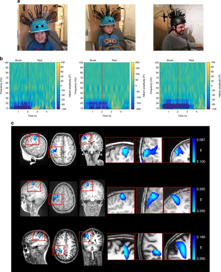
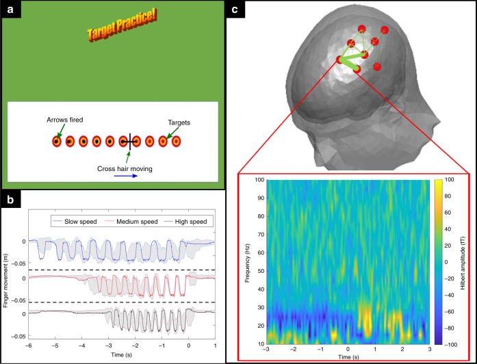
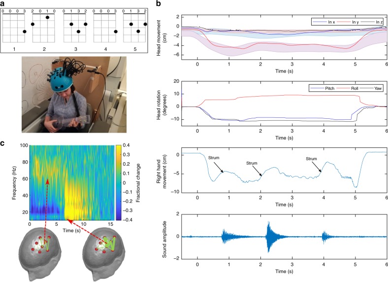
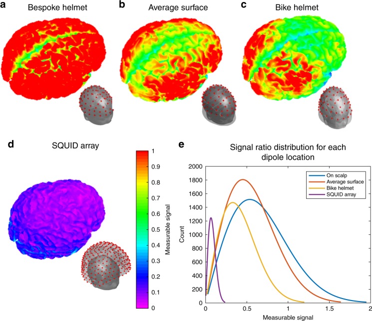
Verification
Claims the paper makes
The claims and statistics of this paper haven’t been checked yet.
Run the check — free, no account needed. It reads the open-access text and checks the sources it cites.
Claims other papers make about this one
Each is checked against this paper’s own text: does it say what the citing paper says it does?
The cited source says what the claim attributes to it.
In the citing paper
“and enable scanning across the lifespan [31,32].”
From Naturalistic Hyperscanning with Wearable Magnetoencephalography (2023) · cites this paper as [31]
Self-check
Percentages, sums and reported differences re-computed from the paper’s own numbers.
The self-check needs the paper’s full text.
Citations
Cites
Event-related EEG/MEG synchronization and desynchronization: basic principles
Pfurtscheller, G., & Lopes da Silva, F. H. (1999) · Clinical Neurophysiology · doi:10.1016/s1388-2457(99)00141-8 · Cited by 7,328
Hämäläinen, M., Hari, R., Ilmoniemi, R. J., Knuutila, J., & Lounasmaa, O. V. (1993) · Reviews of Modern Physics · doi:10.1103/revmodphys.65.413 · Cited by 4,684
Van Veen, B. D., Van Drongelen, W., Yuchtman, M., & Suzuki, A. (1997) · IEEE Transactions on Biomedical Engineering · doi:10.1109/10.623056 · Cited by 2,561
Basic mathematical and electromagnetic concepts of the biomagnetic inverse problem.
Sarvas, J. (1987) · Physics in Medicine and Biology · doi:10.1088/0031-9155/32/1/004 · Cited by 1,945
A subfemtotesla multichannel atomic magnetometer
Kominis, I. K., Kornack, T. W., Allred, J. C., & Romalis, M. V. (2003) · Nature · doi:10.1038/nature01484 · Cited by 1,631
Moving magnetoencephalography towards real-world applications with a wearable system
Boto, E., Holmes, N., Leggett, J., Roberts, G., Shah, V., Meyer, S. S., Muñoz, L. D., Mullinger, K. J., Tierney, T. M., Bestmann, S., et al. (2018) · Nature · doi:10.1038/nature26147 · Cited by 1,377 · cited 5×
“Having derived this inner surface, it was fitted, visually, to the MRI of the same 2-year-old used in (1).”
BackgroundMethodInfluentialSelf-citationHigh-Sensitivity Atomic Magnetometer Unaffected by Spin-Exchange Relaxation
Allred, J. C., Lyman, R. N., Kornack, T. W., & Romalis, M. V. (2002) · Physical Review Letters · doi:10.1103/physrevlett.89.130801 · Cited by 1,049
Magnetoencephalography for Brain Electrophysiology and Imaging
Baillet, S. (2017) · Nature Neuroscience · doi:10.1038/nn.4504 · Cited by 992
Ultrahigh sensitivity magnetic field and magnetization measurements with an atomic magnetometer
Dang, H. B., Maloof, A. C., & Romalis, M. V. (2010) · Applied Physics Letters · doi:10.1063/1.3491215 · Cited by 891
Cohen, D. (1972) · Science · doi:10.1126/science.175.4022.664 · Cited by 785
High-Frequency Brain Activity and Muscle Artifacts in MEG/EEG: A Review and Recommendations
Muthukumaraswamy, S. D. (2013) · Frontiers in Human Neuroscience · doi:10.3389/fnhum.2013.00138 · Cited by 697
Multidimensional divide-and-conquer
Bentley, J. L. (1980) · Communications of the ACM · doi:10.1145/358841.358850 · Cited by 672
(1999) · Medical Entomology and Zoology · Cited by 587
Post-movement beta rebound is generated in motor cortex: Evidence from neuromagnetic recordings
Jurkiewicz, M. T., Gaetz, W. C., Bostan, A. C., & Cheyne, D. O. (2006) · NeuroImage · doi:10.1016/j.neuroimage.2006.06.005 · Cited by 485
Boto, E., Meyer, S. S., Shah, V., Alem, O., Knappe, S., Kruger, P., Fromhold, T. M., Lim, M., Glover, P. M., Morris, P. G., et al. (2017) · NeuroImage · doi:10.1016/j.neuroimage.2017.01.034 · Cited by 471
Self-citationMagnetoencephalography with an atomic magnetometer
Xia, H., Baranga, A. B.-A., Hoffman, D., & Romalis, M. V. (2006) · Applied Physics Letters · doi:10.1063/1.2392722 · Cited by 454
Optically pumped magnetometers: from quantum origins to multi-channel magnetoencephalography
Tierney, T. M., Holmes, N., Mellor, S., López, J. D., Roberts, G., Hill, R. M., Boto, E., Leggett, J., Shah, V., Brookes, M. J., et al. (2019) · NeuroImage · doi:10.1016/j.neuroimage.2019.05.063 · Cited by 400
Self-citationMP RAGE: a three-dimensional, T1-weighted, gradient-echo sequence--initial experience in the brain.
Brant-Zawadzki, M. N., Gillan, G. D., & Nitz, W. R. (1992) · Radiology · doi:10.1148/radiology.182.3.1535892 · Cited by 345
A compact, high performance atomic magnetometer for biomedical applications
Shah, V. K., & Wakai, R. T. (2013) · Physics in Medicine and Biology · doi:10.1088/0031-9155/58/22/8153 · Cited by 322
Self-citationMagnetoencephalography with a chip-scale atomic magnetometer
Sander, T. H., Preusser, J., Mhaskar, R. R., Kitching, J. E., Trahms, L., & Knappe, S. A. (2012) · Biomedical Optics Express · doi:10.1364/boe.3.000981 · Cited by 293
Holmes, N., Leggett, J., Boto, E., Roberts, G., Hill, R. M., Tierney, T. M., Shah, V., Barnes, G. R., Brookes, M. J., & Bowtell, R. (2018) · NeuroImage · doi:10.1016/j.neuroimage.2018.07.028 · Cited by 250
Self-citationMagnetoencephalography with a two-color pump-probe, fiber-coupled atomic magnetometer
Johnson, C., Schwindt, P. D. D., & Weisend, M. P. (2010) · Applied Physics Letters · doi:10.1063/1.3522648 · Cited by 210
On the Potential of a New Generation of Magnetometers for MEG: A Beamformer Simulation Study
Boto, E., Bowtell, R., Krüger, P., Fromhold, T. M., Morris, P. G., Meyer, S. S., Barnes, G. R., & Brookes, M. J. (2016) · PLOS ONE · doi:10.1371/journal.pone.0157655 · Cited by 194
Self-citationHeinrichs‐Graham, E., & Wilson, T. W. (2016) · NeuroImage · doi:10.1016/j.neuroimage.2016.04.032 · Cited by 183
Knuckey, L. M. (2006) · Australasian Physical & Engineering Sciences in Medicine · Cited by 154
Wearable neuroimaging: Combining and contrasting magnetoencephalography and electroencephalography
Boto, E., Seedat, Z. A., Holmes, N., Leggett, J., Hill, R. M., Roberts, G., Shah, V., Fromhold, T. M., Mullinger, K. J., Tierney, T. M., et al. (2019) · NeuroImage · doi:10.1016/j.neuroimage.2019.116099 · Cited by 136
Self-citationMulti-sensor magnetoencephalography with atomic magnetometers
Johnson, C., Schwindt, P. D. D., & Weisend, M. P. (2013) · Physics in Medicine and Biology · doi:10.1088/0031-9155/58/17/6065 · Cited by 131
Evidence for a motor gamma-band network governing response interference
Gaetz, W. C., Liu, C., Zhu, H., Bloy, L., & Roberts, T. P. L. (2013) · NeuroImage · doi:10.1016/j.neuroimage.2013.02.013 · Cited by 76
Kamada, K., Sato, D., Ito, Y., Natsukawa, H., Okano, K., Mizutani, N., & Kobayashi, T. (2015) · Japanese Journal of Applied Physics · doi:10.7567/jjap.54.026601 · Cited by 74
Tierney, T. M., Holmes, N., Meyer, S. S., Boto, E., Roberts, G., Leggett, J., Buck, S., Duque‐Muñoz, L., Vladimir Litvak, A., Bestmann, S., et al. (2018) · NeuroImage · doi:10.1016/j.neuroimage.2018.07.035 · Cited by 66
Self-citationLiao, Y., Acar, Z. A., Makeig, S., & Deák, G. O. (2015) · NeuroImage · doi:10.1016/j.neuroimage.2015.02.055 · Cited by 60
Magnetoencephalography of Epilepsy with a Microfabricated Atomic Magnetrode
Alem, O., Benison, A. M., Barth, D. S., Kitching, J. E., & Knappe, S. A. (2014) · Journal of Neuroscience · doi:10.1523/jneurosci.3495-14.2014 · Cited by 52
Holliday, I. E., Barnes, G. R., Hillebrand, A., & Singh, K. D. (2003) · Human Brain Mapping · doi:10.1002/hbm.10133 · Cited by 41
Self-citationClaus, S. P., Velis, D. N., Lopes, F. H. D. S., Viergever, M. A., & Kalitzin, S. (2012) · Epilepsy Research · doi:10.1016/j.eplepsyres.2012.02.002 · Cited by 35
American Chemical Journal, April
Gilpin, J. E. (1897) · Science · doi:10.1126/science.5.121.664 · Cited by 6
Cited by
Sorted by citations
Quantum sensors for biomedical applications
Aslam, N., Zhou, H., Urbach, E., Turner, M. J., Walsworth, R. L., Lukin, M. D., & Park, H. (2023) · Nature Reviews Physics · doi:10.1038/s42254-023-00558-3 · Cited by 553
Brookes, M. J., Leggett, J., Rea, M., Hill, R. M., Holmes, N., Boto, E., & Bowtell, R. W. (2022) · Trends in Neurosciences · doi:10.1016/j.tins.2022.05.008 · Cited by 438
Self-citationDelivering transformative action in paediatric pain: a Lancet Child & Adolescent Health Commission
Eccleston, C., Fisher, E., Howard, R. F., Slater, R., Forgeron, P. A., Palermo, T. M., Birnie, K. A., Anderson, B. J., Chambers, C. T., Crombez, G., et al. (2020) · The Lancet Child & Adolescent Health · doi:10.1016/s2352-4642(20)30277-7 · Cited by 361
Self-citationMulti-Channel Whole-Head OPM-MEG: Helmet Design and a Comparison with a Conventional System
Hill, R. M., Boto, E., Rea, M., Holmes, N., Leggett, J., Coles, L. A., Papastavrou, M., Everton, S. K., Hunt, B. A. E., Sims, D., et al. (2020) · NeuroImage · doi:10.1016/j.neuroimage.2020.116995 · Cited by 285
Self-citationPortable Magnetometry for Detection of Biomagnetism in Ambient Environments
Limes, M. E., Foley, E. L., Kornack, T. W., Caliga, S., McBride, S., Braun, A., Lee, W., Lucivero, V. G., & Romalis, M. V. (2020) · Physical Review Applied · doi:10.1103/physrevapplied.14.011002 · Cited by 257
Metadata
- Authors
- Hill, R. M., Boto, E., Holmes, N., Hartley, C., Seedat, Z. A., Leggett, J., Roberts, G., Shah, V., Tierney, T. M., Woolrich, M. W., Stagg, C. J., Barnes, G. R., Bowtell, R., Slater, R., & Brookes, M. J.
- Affiliations
- University of Nottingham (GB)
- John Radcliffe Hospital (GB)
- QuSpin (United States) (US)
- Wellcome Centre for Human Neuroimaging (GB)
- Warneford Hospital (GB)
- Corresponding author
- Brookes, Matthew J.
- ORCID
- Venue
- Nature Communications
- Published
- 2019-11-05
- Volume
- 10
- Issue
- 1
- Pages
- 4785-4785
- ISSN
- 2041-1723 (ISSN-L)
- PMC
- PMC6831615
- OpenAlex
- W2969234156
- Cited by
- 180
- References
- 35
- Citations by year
- 2026: 12 · 2025: 35 · 2024: 31 · 2023: 31 · 2022: 31 · 2021: 25 · 2020: 15
- FWCI
- 7.80
- Type
- journal article
- Language
- en
- Update notices
- correction (2019-12-04)
- Open access
- gold
- doi.org
- Locations
- Nature Communications (publishedVersion, cc-by)
- PubMed Central (publishedVersion, cc-by)
- PubMed (publishedVersion)
- Repository@Nottingham (University of Nottingham) (publishedVersion, other-oa)
- Repository@Nottingham (University of Nottingham) (submittedVersion, cc-by)
- RePEc: Research Papers in Economics (submittedVersion)
- DOAJ (DOAJ: Directory of Open Access Journals) (submittedVersion, cc-by-sa)
- DOAJ (DOAJ: Directory of Open Access Journals) (submittedVersion)
- UCL Discovery (University College London) (submittedVersion)
- Funders
- Wellcome Trust (203257/Z/16/Z, 203257/B/16/Z)
- Topics
- Physical Sciences › Physics and Astronomy › Atomic and Molecular Physics, and Optics › Atomic and Subatomic Physics Research
- Health Sciences › Medicine › Radiology, Nuclear Medicine and Imaging › Advanced MRI Techniques and Applications
- Life Sciences › Neuroscience › Cognitive Neuroscience › EEG and Brain-Computer Interfaces
- Keywords
- quantum technologies, functional neuroimaging, toddlers, children, adolescents, head movement, head growth, naturalistic learning, non-invasive brain imaging
- MeSH terms
- Adolescent, Adult, Brain, Brain Mapping, Child, Child, Preschool, Cognition, Electrophysiology, Female, Humans, Infant, Male, Magnetoencephalography, Young Adult
About these checks
Registry, retraction, Cites and statistics results are computed, not judged, with one exception: a printed reference that no index lists is read once by a model before it counts as a citation error. Claims are extracted by a model and kept only when their quote appears verbatim in the text; each cited source is then checked, with a model’s help, for whether it says what the claim relies on. Those verdicts can be wrong — read the claim’s quote and the cited source before relying on one.
Statistics and arithmetic are recomputed, not judged.
Confidence bands are shown only for fields calibrated on our annotated test set. None is calibrated yet, so no band is shown.
What counts as a claim ›How we measure confidence ›How we check this ›
Are you an author, or do you see a problem with this page? Request a review
