

The DOI matches its Crossref record; 70 of the 70 references it cites were found in a registry or an index.
Brain-to-cervical lymph node signaling after stroke.
Open accessAbstract
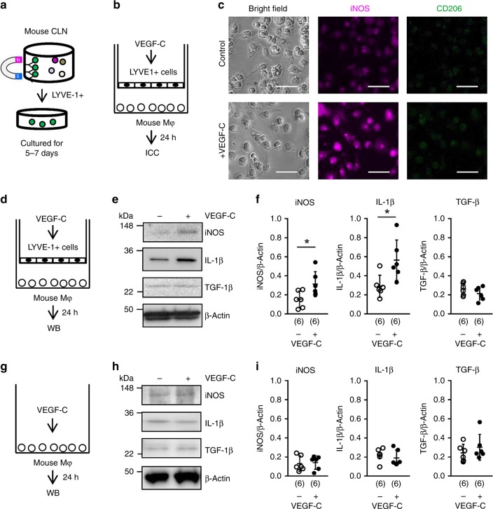
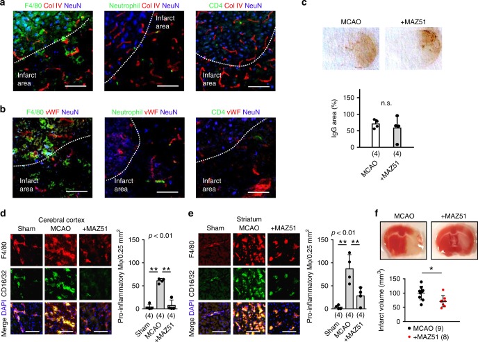
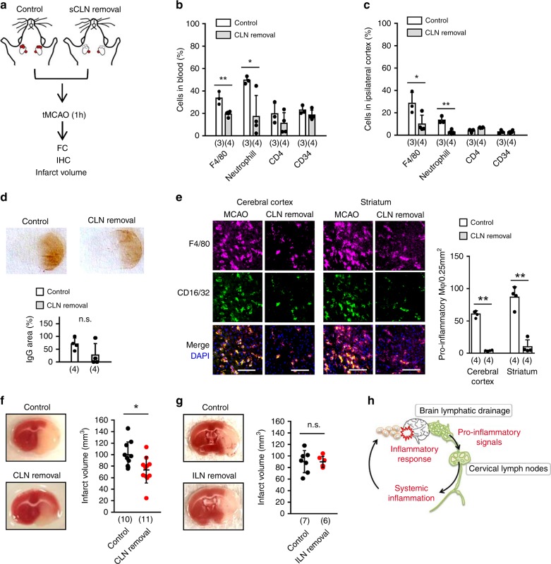
After stroke, peripheral immune cells are activated and these systemic responses may amplify brain damage, but how the injured brain sends out signals to trigger systemic inflammation remains unclear. Here we show that a brain-to-cervical lymph node (CLN) pathway is involved. In rats subjected to focal cerebral ischemia, lymphatic endothelial cells proliferate and macrophages are rapidly activated in CLNs within 24 h, in part via VEGF-C/VEGFR3 signalling. Microarray analyses of isolated lymphatic endothelium from CLNs of ischemic mice confirm the activation of transmembrane tyrosine kinase pathways. Blockade of VEGFR3 reduces lymphatic endothelial activation, decreases pro-inflammatory macrophages, and reduces brain infarction. In vitro, VEGF-C/VEGFR3 signalling in lymphatic endothelial cells enhances inflammatory responses in co-cultured macrophages. Lastly, surgical removal of CLNs in mice significantly reduces infarction after focal cerebral ischemia. These findings suggest that modulating the brain-to-CLN pathway may offer therapeutic opportunities to ameliorate systemic inflammation and brain injury after stroke.
Overview
The claims and statistics of this paper haven’t been checked yet.
Run the check — free, no account needed. It reads the open-access text and checks the sources it cites.
Integrity signals
Registry
Cites
Statistics
Figures
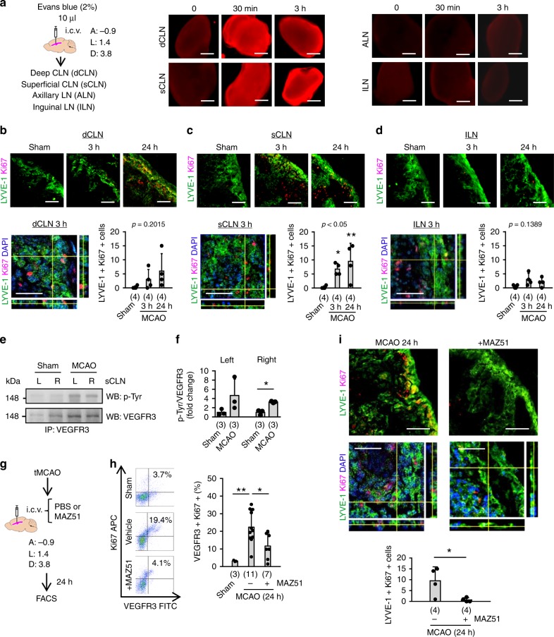
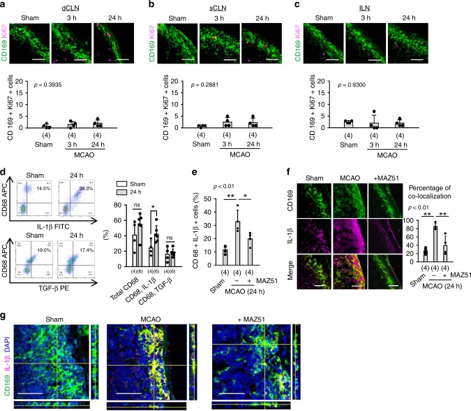
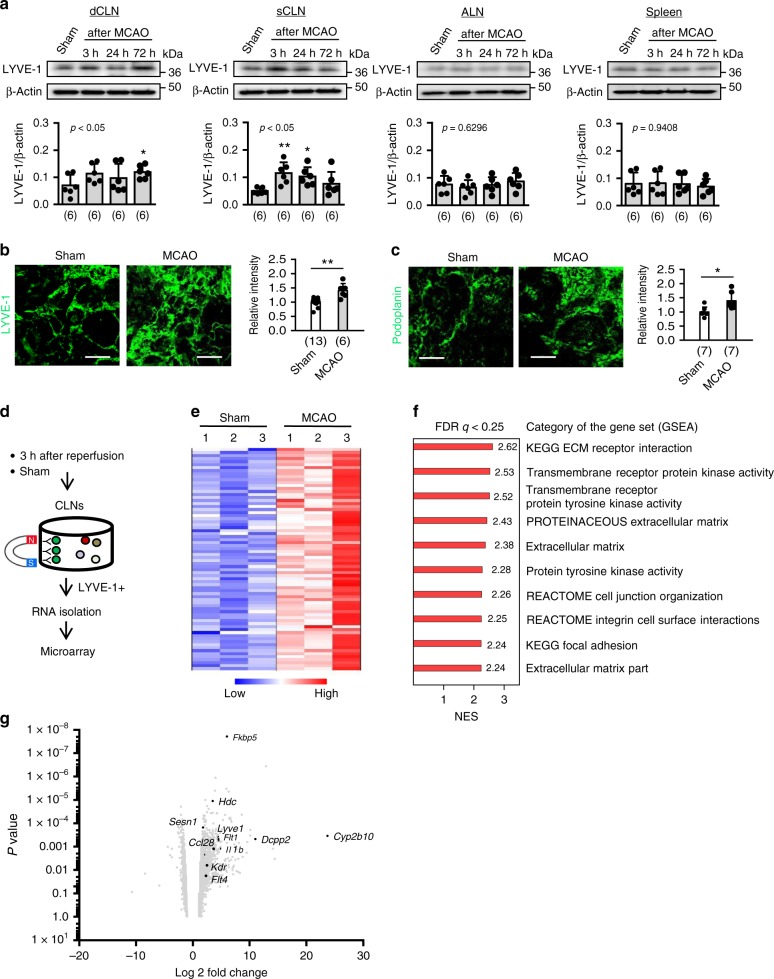
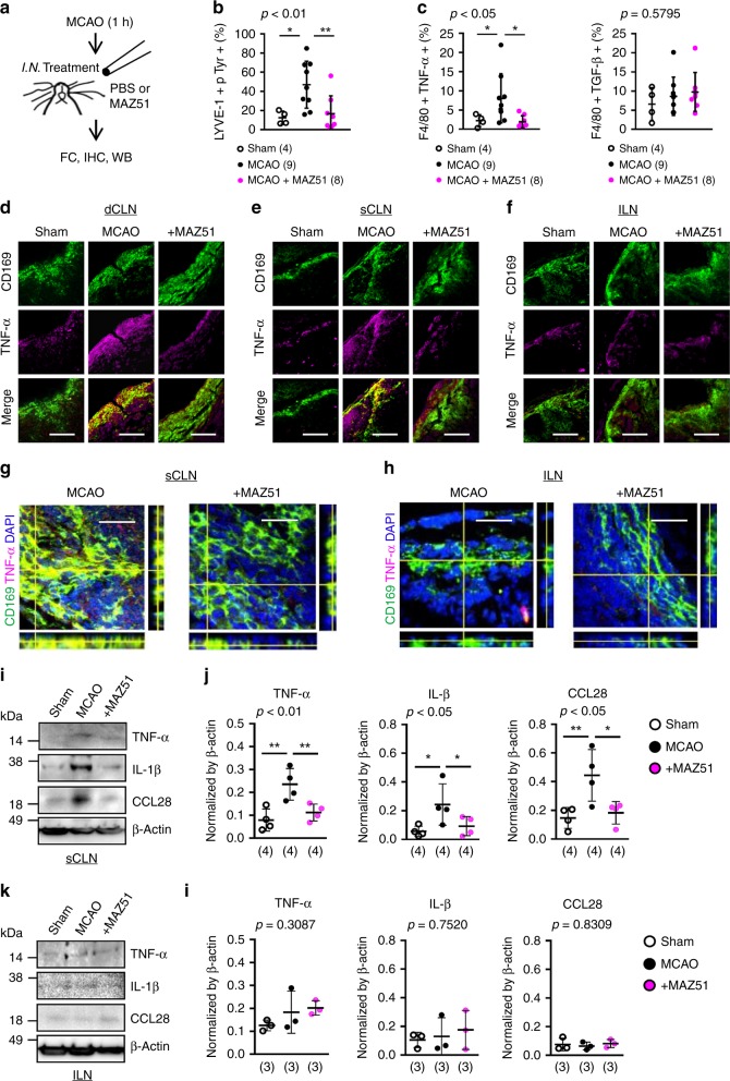
Verification
Claims the paper makes
The claims and statistics of this paper haven’t been checked yet.
Run the check — free, no account needed. It reads the open-access text and checks the sources it cites.
Claims other papers make about this one
Each is checked against this paper’s own text: does it say what the citing paper says it does?
In the citing paper
“Blockage of VEGFR3 (expressed by lymphatic endothelial cells) in mice following focal cerebral ischemia resulted in reduced inflammatory response and infarct”
From Living on the Edge of the CNS: Meninges Cell Diversity in Health and Disease (2021) · cites this paper as Esposito et al., 2019
Self-check
Percentages, sums and reported differences re-computed from the paper’s own numbers.
The self-check needs the paper’s full text.
Citations
Cites
Structural and functional features of central nervous system lymphatic vessels
Louveau, A., Smirnov, I., Keyes, T. J., Eccles, J. D., Rouhani, S. J., Peske, J. D., Derecki, N. C., Castle, D., Mandell, J. W., Lee, K. S., et al. (2015) · Nature · doi:10.1038/nature14432 · Cited by 4,413
Blood–brain barrier breakdown in Alzheimer disease and other neurodegenerative disorders
Sweeney, M. D., Sagare, A. P., & Zlokovic, B. V. (2018) · Nature Reviews Neurology · doi:10.1038/nrneurol.2017.188 · Cited by 3,076
The immunology of stroke: from mechanisms to translation
Iadecola, C., & Anrather, J. (2011) · Nature Medicine · doi:10.1038/nm.2399 · Cited by 2,596
A dural lymphatic vascular system that drains brain interstitial fluid and macromolecules
Aspelund, A., Antila, S., Proulx, S. T., Karlsen, T. V., Karaman, S., Detmar, M., Wiig, H., & Alitalo, K. (2015) · Journal of Experimental Medicine · doi:10.1084/jem.20142290 · Cited by 2,198
Transfer of mitochondria from astrocytes to neurons after stroke
Hayakawa, K., Esposito, E., Wang, X., Terasaki, Y., Liu, Y., Xing, C., Ji, X., & Lo, E. H. (2016) · Nature · doi:10.1038/nature18928 · Cited by 1,423
Self-citationLymphangiogenesis in development and human disease
Alitalo, K. K., Tammela, T., & Petrova, T. V. (2005) · Nature · doi:10.1038/nature04480 · Cited by 1,287
A new look at cerebrospinal fluid circulation
Brinker, T., Stopa, E., Morrison, J., & Klinge, P. (2014) · Fluids and Barriers of the CNS · doi:10.1186/2045-8118-11-10 · Cited by 819
Meningeal lymphatic vessels at the skull base drain cerebrospinal fluid
Ahn, J. H., Cho, H., Kim, J.-H., Kim, S. H., Ham, J.-S., Park, I., Suh, S. H., Hong, S. P., Song, J.-H., Hong, Y.-K., et al. (2019) · Nature · doi:10.1038/s41586-019-1419-5 · Cited by 811
Ma, Q., Ineichen, B. V., Detmar, M., & Proulx, S. T. (2017) · Nature Communications · doi:10.1038/s41467-017-01484-6 · Cited by 690
Kukk, E., Lymboussaki, A., Taira, S., Kaipainen, A., Jeltsch, M., Joukov, V., & Alitalo, K. K. (1996) · Development · doi:10.1242/dev.122.12.3829 · Cited by 687
Cervical lymphatics, the blood-brain barrier and the immunoreactivity of the brain: a new view
Cserr, H. F., & Knopf, P. M. (1992) · Immunology Today · doi:10.1016/0167-5699(92)90027-5 · Cited by 648
Johnston, M., Zakharov, A., Papaiconomou, C., Salmasi, G., & Armstrong, D. (2004) · Cerebrospinal Fluid Research · doi:10.1186/1743-8454-1-2 · Cited by 572
Lymphatic drainage of the brain and the pathophysiology of neurological disease
Weller, R. O., Djuanda, E., Yow, H.-Y., & Carare, R. O. (2009) · Acta Neuropathologica · doi:10.1007/s00401-008-0457-0 · Cited by 488
Cai, R., Pan, C., Ghasemigharagoz, A., Todorov, M. I., Förstera, B., Zhao, S., Bhatia, H. S., Parra-Damas, A., Mrowka, L., Theodorou, D., et al. (2019) · Nature Neuroscience · doi:10.1038/s41593-018-0301-3 · Cited by 470
Cerebral Vascular Disease and Neurovascular Injury in Ischemic Stroke
Hu, X., Silva, T. M. D., Chen, J., & Faraci, F. M. (2017) · Circulation Research · doi:10.1161/circresaha.116.308427 · Cited by 456
Effector T-cell trafficking between the leptomeninges and the cerebrospinal fluid
Schläger, C., Körner, H., Krueger, M., Vidoli, S., Haberl, M., Mielke, D., Brylla, E., Issekutz, T., Cabañas, C., Nelson, P. J., et al. (2016) · Nature · doi:10.1038/nature16939 · Cited by 372
The function and structure of the cerebrospinal fluid outflow system
Pollay, M. (2010) · Cerebrospinal Fluid Research · doi:10.1186/1743-8454-7-9 · Cited by 371
New developments in clinical aspects of lymphatic disease
Mortimer, P. S., & Rockson, S. G. (2014) · Journal of Clinical Investigation · doi:10.1172/jci71608 · Cited by 356
Neuropilin-2 mediates VEGF-C–induced lymphatic sprouting together with VEGFR3
Xu, Y., Yuan, L., Mak, J., Pardanaud, L., Caunt, M., Kasman, I. M., Larrivée, B., Toro, R. D., Suchting, S., Medvinsky, A., et al. (2010) · The Journal of Cell Biology · doi:10.1083/jcb.200903137 · Cited by 339
Early Neutrophilia Is Associated With Volume of Ischemic Tissue in Acute Stroke
Buck, B. H., Liebeskind, D. S., Saver, J. L., Bang, O. Y., Yun, S. W., Starkman, S., Ali, L. K., Kim, D., Villablanca, J. P., Salamon, N., et al. (2007) · Stroke · doi:10.1161/strokeaha.107.490128 · Cited by 320
Glymphatic clearance controls state-dependent changes in brain lactate concentration
Lundgaard, I., Lu, M. L., Yang, E., Peng, W., Mestre, H., Hitomi, E., Deane, R., & Nedergaard, M. (2016) · Journal of Cerebral Blood Flow & Metabolism · doi:10.1177/0271678x16661202 · Cited by 309
Koh, L., Zakharov, A., & Johnston, M. G. (2005) · Cerebrospinal Fluid Research · doi:10.1186/1743-8454-2-6 · Cited by 304
Gray, E. E., & Cyster, J. G. (2012) · Journal of Innate Immunity · doi:10.1159/000337007 · Cited by 301
Monocyte Chemoattractant Protein-1 Deficiency is Protective in a Murine Stroke Model
Hughes, P., ALLEGRINI, P. R., Rudin, M., Perry, V. H., Mir, A. K., & Wießner, C. (2002) · Journal of Cerebral Blood Flow & Metabolism · doi:10.1097/00004647-200203000-00008 · Cited by 295
Shichita, T., Ito, M., Morita, R., Komai, K., Noguchi, Y., Ooboshi, H., Koshida, R., Takahashi, S., Kodama, T., & Yoshimura, A. (2017) · Nature Medicine · doi:10.1038/nm.4312 · Cited by 253
Different prognostic value of white blood cell subtypes in patients with acute cerebral infarction
Kim, J., Song, T., Park, J. H., Lee, H. S., Nam, C. M., Nam, H. S., Kim, Y. D., & Heo, J. H. (2012) · Atherosclerosis · doi:10.1016/j.atherosclerosis.2012.02.042 · Cited by 235
Self-citationInflammation-associated lymphangiogenesis: a double-edged sword?
Kim, H., Kataru, R. P., & Koh, G. Y. (2014) · Journal of Clinical Investigation · doi:10.1172/jci71607 · Cited by 228
Enzmann, G., Mysiorek, C., Gorina, R., Cheng, Y.-J., Ghavampour, S., Hannocks, M.-J., Prinz, V., Dirnagl, U., Endres, M., Prinz, M., et al. (2013) · Acta Neuropathologica · doi:10.1007/s00401-012-1076-3 · Cited by 225
Weed, L. H. (1914) · PubMed Central · Cited by 220
Inflammation induces lymphangiogenesis through up-regulation of VEGFR-3 mediated by NF-κB and Prox1
Flister, M. J., Wilber, A. C., Hall, K. L., Iwata, C., Miyazono, K., Nisato, R. E., Pepper, M. S., Zawieja, D. C., & Ran, S. (2009) · Blood · doi:10.1182/blood-2008-12-196840 · Cited by 211
Hsu, M., Rayasam, A., Kijak, J. A., Choi, Y. H., Harding, J. S., Marcus, S. A., Karpus, W. J., Sándor, M., & Fábry, Z. (2019) · Nature Communications · doi:10.1038/s41467-018-08163-0 · Cited by 211
Increased Cytokine Release from Peripheral Blood Cells after Acute Stroke
Ferrarese, C., Mascarucci, P., Zoia, C., Cavarretta, R., Frigo, M., Begni, B., Sarinella, F., Frattola, L., & Simoni, M. G. D. (1999) · Journal of Cerebral Blood Flow & Metabolism · doi:10.1097/00004647-199909000-00008 · Cited by 209
Vigl, B., Aebischer, D., Nitschké, M., Iolyeva, M., Röthlin, T., Antsiferova, O., & Halin, C. (2011) · Blood · doi:10.1182/blood-2010-12-326447 · Cited by 190
Implications of the discovery of brain lymphatic pathways
Iliff, J. J., Goldman, S. A., & Nedergaard, M. (2015) · The Lancet Neurology · doi:10.1016/s1474-4422(15)00221-5 · Cited by 188
Wolvers, D. A. W., Roo, C. J. J. C., Mebius, R. E., van der Cammen, M. J. F., Tirion, F. H., Miltenburg, A. M. M., & Kraal, G. (1999) · The Journal of Immunology · doi:10.4049/jimmunol.162.4.1994 · Cited by 177
The role of the lymphatic system in drainage of cerebrospinal fluid and aqueous humour.
Bradbury, M. W. B., & Cole, D. F. (1980) · The Journal of Physiology · doi:10.1113/jphysiol.1980.sp013129 · Cited by 172
Brain-Derived Antigens in Lymphoid Tissue of Patients with Acute Stroke
Planas, A. M., Gómez-Choco, M. J., Urra, X., Gorina, R., Caballero, M., & Chamorro, Á. (2012) · The Journal of Immunology · doi:10.4049/jimmunol.1102289 · Cited by 170
Drainage of Cells and Soluble Antigen from the CNS to Regional Lymph Nodes
Laman, J. D., & Weller, R. O. (2013) · Journal of Neuroimmune Pharmacology · doi:10.1007/s11481-013-9470-8 · Cited by 168
Inflammation-induced Uptake and Degradation of the Lymphatic Endothelial Hyaluronan Receptor LYVE-1
Johnson, L. A., Prevo, R., Clasper, S., & Jackson, D. G. (2007) · Journal of Biological Chemistry · doi:10.1074/jbc.m702889200 · Cited by 164
van Zwam, M., Huizinga, R., Heijmans, N., van Meurs, M., Wierenga‐Wolf, A. F., Melief, M., Hintzen, R. Q., Hart, B. A. ‘., Amor, S., Boven, L. A., et al. (2008) · The Journal of Pathology · doi:10.1002/path.2476 · Cited by 151
Woo, M., Yang, J., Beltran, C., & Cho, S. (2016) · Journal of Biological Chemistry · doi:10.1074/jbc.m116.750018 · Cited by 136
Chen, J., He, J., Ni, R., Yang, Q., Zhang, Y., & Luo, L. (2019) · Developmental Cell · doi:10.1016/j.devcel.2019.03.022 · Cited by 133
Morris, G. P., Wright, A. L., Tan, R. P., Gladbach, A., Ittner, L. M., & Vissel, B. (2016) · PLoS ONE · doi:10.1371/journal.pone.0148503 · Cited by 132
Losy, J., & Zaremba, J. (2001) · Stroke · doi:10.1161/hs1101.097380 · Cited by 130
Eriksson, A. M., Schön, K., & Lycke, N. Y. (2004) · The Journal of Immunology · doi:10.4049/jimmunol.173.5.3310 · Cited by 120
Determination of volumetric cerebrospinal fluid absorption into extracranial lymphatics in sheep
Boulton, M., Flessner, M. F., Armstrong, D. W., Hay, J. B., & Johnston, M. (1998) · American Journal of Physiology-Regulatory, Integrative and Comparative Physiology · doi:10.1152/ajpregu.1998.274.1.r88 · Cited by 120
Knopf, P. M., Cserr, H. F., Nolan, S. C., Wu, T., & Harling‐Berg, C. J. (1995) · Neuropathology and Applied Neurobiology · doi:10.1111/j.1365-2990.1995.tb01047.x · Cited by 117
Yoshimatsu, Y., Lee, Y. G., Akatsu, Y., Taguchi, L., Suzuki, H. I., Cunha, S. I., Maruyama, K., Suzuki, Y., Yamazaki, T., Katsura, A., et al. (2013) · Proceedings of the National Academy of Sciences · doi:10.1073/pnas.1310479110 · Cited by 112
García‐Culebras, A., Durán‐Laforet, V., Peña‐Martínez, C., Ballesteros, I., Pradillo, J. M., Guzmán, J. D., Lizasoaín, I., & Moro, M. Á. (2018) · Journal of Cerebral Blood Flow & Metabolism · doi:10.1177/0271678x18795789 · Cited by 111
The Alternative Complement Pathway Propagates Inflammation and Injury in Murine Ischemic Stroke
Elvington, A., Atkinson, C., Zhu, H., Yu, J., Takahashi, K., Stahl, G. L., Kindy, M. S., & Tomlinson, S. (2012) · The Journal of Immunology · doi:10.4049/jimmunol.1201904 · Cited by 90
Otxoa-de-Amezaga, A., Gallizioli, M., Pedragosa, J., Justicia, C., Miró-Mur, F., Salas-Pérdomo, A., Díaz-Marugan, L., Gunzer, M., & Planas, A. (2019) · Stroke · doi:10.1161/strokeaha.118.023837 · Cited by 87
Zakharov, A., Papaiconomou, C., & Johnston, M. G. (2004) · Lymphatic Research and Biology · doi:10.1089/lrb.2004.2.139 · Cited by 84
Kim, E., Tolhurst, A. T., Qin, L. Y., Chen, X.-Y., Febbraio, M., & Cho, S. (2008) · The Journal of Neuroscience · doi:10.1523/jneurosci.0982-08.2008 · Cited by 81
Kumar, A. D., Boehme, A. K., Siegler, J. E., Gillette, M., Albright, K. C., & Martin‐Schild, S. (2012) · Journal of Stroke and Cerebrovascular Diseases · doi:10.1016/j.jstrokecerebrovasdis.2012.08.008 · Cited by 76
Patterson, A. M., Bonny, A. E., Shiels, W. E., & Erwin, E. A. (2016) · Annals of Allergy, Asthma & Immunology · doi:10.1016/j.anai.2015.11.010 · Cited by 74
The meningeal and choroidal infiltration routes for leukocytes in stroke
Benakis, C., Llovera, G., & Liesz, A. (2018) · Therapeutic Advances in Neurological Disorders · doi:10.1177/1756286418783708 · Cited by 68
Weed, L. H. (1914) · PubMed Central · Cited by 65
Glycolytic flux in permeabilized freshly isolated vascular smooth muscle cells
Hardin, C. D., & Finder, D. R. (1998) · American Journal of Physiology-Cell Physiology · doi:10.1152/ajpcell.1998.274.1.c88 · Cited by 64
Promises and limitations of immune cell-based therapies in neurological disorders
Hu, X., Leak, R. K., Thomson, A. W., Yu, F., Xia, Y., Wechsler, L. R., & Chen, J. (2018) · Nature Reviews Neurology · doi:10.1038/s41582-018-0028-5 · Cited by 64
Wehrhan, F., Büttner-Herold, M. J., Hyckel, P., Möbius, P., Preidl, R. H. M., Distel, L. V., Ries, J., Amann, K., Schmitt, C. M., Neukam, F. W., et al. (2014) · BMC Cancer · doi:10.1186/1471-2407-14-522 · Cited by 59
Esposito, E., Hayakawa, K., Ahn, B. J., Chan, S. J., Xing, C., Liang, A. C., Kim, K., Arai, K., & Lo, E. H. (2018) · Journal of Neurochemistry · doi:10.1111/jnc.14337 · Cited by 57
Self-citationKodama, T., Matsuki, D., Tada, A., TAKEDA, K., & Mori, S. (2016) · Scientific Reports · doi:10.1038/srep32506 · Cited by 56
Esposito, E., Hayakawa, K., Maki, T., Arai, K., & Lo, E. H. (2015) · Stroke · doi:10.1161/strokeaha.115.009070 · Cited by 54
Self-citationCCL27/CCL28–CCR10 Chemokine Signaling Mediates Migration of Lymphatic Endothelial Cells
Karnezis, T., Farnsworth, R. H., Harris, N. C., Williams, S. P., Caesar, C., Byrne, D. J., Herle, P., Macheda, M. L., Shayan, R., Zhang, Y., et al. (2019) · Cancer Research · doi:10.1158/0008-5472.can-18-1858 · Cited by 54
When friend turns foe: central and peripheral neuroinflammation in central nervous system injury
Marcet, P. A., Santos, N., & Borlongan, C. V. (2017) · Neuroimmunology and Neuroinflammation · doi:10.20517/2347-8659.2017.07 · Cited by 50
Suzuki, M., Yokota, M., Nakamura, Y., Ozaki, S., & Murakami, S. (2016) · Allergology International · doi:10.1016/j.alit.2016.08.014 · Cited by 37
Self-citationTGFβ counteracts LYVE-1-mediated induction of lymphangiogenesis by small hyaluronan oligosaccharides
Bauer, J., Rothley, M., Schmaus, A., Quagliata, L., Ehret, M., Biskup, M. B., Orian‐Rousseau, V., Jackson, D. G., Pettis, R. J., Harvey, A. J., et al. (2017) · Journal of Molecular Medicine · doi:10.1007/s00109-017-1615-4 · Cited by 33
Imaging techniques for in vivo quantitation of extracranial lymphatic drainage of the brain
Hunter, J. V., Batchelder, K. F., Lo, E. H., & Wolf, G. L. (1995) · Neuropathology and Applied Neurobiology · doi:10.1111/j.1365-2990.1995.tb01049.x · Cited by 19
Self-citationStudies on Cerebro-Spinal Fluid. No. IV : The dual Source of Cerebro-Spinal Fluid.
Weed, L. H. (1914) · PubMed Central · doi:10.18977/jspog.14.1_56_1 · Cited by 11
Lan, J., Esposito, E., Ayata, C., Singhal, A. B., Lo, E. H., & Ji, X. (2018) · Stroke · doi:10.1161/strokeaha.118.020626 · Cited by 9
Self-citation
Cited by
Sorted by citations
Immune responses to stroke: mechanisms, modulation, and therapeutic potential.
Iadecola, C., Buckwalter, M. S., & Anrather, J. (2020) · Journal of Clinical Investigation · doi:10.1172/jci135530 · Cited by 853
Rasmussen, M. K., Mestre, H., & Nedergaard, M. (2021) · Physiological Reviews · doi:10.1152/physrev.00031.2020 · Cited by 642
Meningeal lymphatic dysfunction exacerbates traumatic brain injury pathogenesis.
Bolte, A. C., Dutta, A. B., Hurt, M. E., Smirnov, I., Kovacs, M. A., McKee, C. A., Ennerfelt, H. E., Shapiro, D., Nguyen, B. H., Frost, E. L., et al. (2020) · Nature Communications · doi:10.1038/s41467-020-18113-4 · Cited by 357
Systemic inflammation after stroke: implications for post‐stroke comorbidities
Simats, A., & Liesz, A. (2022) · EMBO Molecular Medicine · doi:10.15252/emmm.202216269 · Cited by 276
Conserved meningeal lymphatic drainage circuits in mice and humans
Jacob, L., Brito, J. M., Lenck, S., Corcy, C., Benbelkacem, F., Geraldo, L. H., Xu, Y., Thomas, J.-M., Kamouh, M.-R. E., Spajer, M., et al. (2022) · The Journal of Experimental Medicine · doi:10.1084/jem.20220035 · Cited by 206
Metadata
- Authors
- Esposito, E., Ahn, B. J., Shi, J., Nakamura, Y., Park, J. H., Mandeville, E. T., Yu, Z., Chan, S. J., Desai, R., Hayakawa, A., Ji, X., Lo, E. H., & Hayakawa, K.
- Affiliations
- Harvard University (US)
- Capital Medical University (CN)
- Corresponding author
- Lo, Eng H.
- Hayakawa, Kazuhide
- ORCID
- Venue
- Nature Communications
- Published
- 2019-11-22
- Volume
- 10
- Issue
- 1
- Pages
- 5306-5306
- ISSN
- 2041-1723 (ISSN-L)
- PMC
- PMC6876639
- OpenAlex
- W2990744279
- Cited by
- 123
- References
- 70
- Citations by year
- 2026: 13 · 2025: 20 · 2024: 18 · 2023: 16 · 2022: 27 · 2021: 24 · 2020: 5
- FWCI
- 5.85
- Type
- journal article
- Language
- en
- Open access
- gold
- doi.org
- Locations
- Nature Communications (publishedVersion, cc-by)
- PubMed Central (publishedVersion, cc-by)
- PubMed (publishedVersion)
- National University of Singapore (submittedVersion, cc-by)
- RePEc: Research Papers in Economics (submittedVersion)
- DOAJ (DOAJ: Directory of Open Access Journals) (submittedVersion, cc-by-sa)
- DOAJ (DOAJ: Directory of Open Access Journals) (submittedVersion)
- Funders
- U.S. Department of Health & Human Services | NIH | National Institute of Neurological Disorders and Stroke
- Topics
- Health Sciences › Medicine › Oncology › Lymphatic System and Diseases
- Life Sciences › Neuroscience › Neurology › Neuroinflammation and Neurodegeneration Mechanisms
- Health Sciences › Medicine › Clinical Neurology › Acute Ischemic Stroke Management
- Keywords
- stroke, cervical lymph nodes, focal cerebral ischemia, systemic inflammation, lymphatic endothelial cells, macrophage activation, infarct size, lymphatic drainage
- MeSH terms
- Animals, Brain, Brain Ischemia, Endothelium, Lymphatic, Inflammation, Lymph Nodes, Macrophages, Neck, Brain Infarction, Stroke, Vascular Endothelial Growth Factor Receptor-3, Vascular Endothelial Growth Factor C, Lymphangiogenesis, Endothelial Cells, Cell Proliferation, Mice, Rats
About these checks
Registry, retraction, Cites and statistics results are computed, not judged, with one exception: a printed reference that no index lists is read once by a model before it counts as a citation error. Claims are extracted by a model and kept only when their quote appears verbatim in the text; each cited source is then checked, with a model’s help, for whether it says what the claim relies on. Those verdicts can be wrong — read the claim’s quote and the cited source before relying on one.
Statistics and arithmetic are recomputed, not judged.
Confidence bands are shown only for fields calibrated on our annotated test set. None is calibrated yet, so no band is shown.
What counts as a claim ›How we measure confidence ›How we check this ›
Are you an author, or do you see a problem with this page? Request a review
