

The DOI matches its Crossref record; 91 of the 91 references it cites were found in a registry or an index.
Microfluidic device with brain extracellular matrix promotes structural and functional maturation of human brain organoids
Open accessAbstract
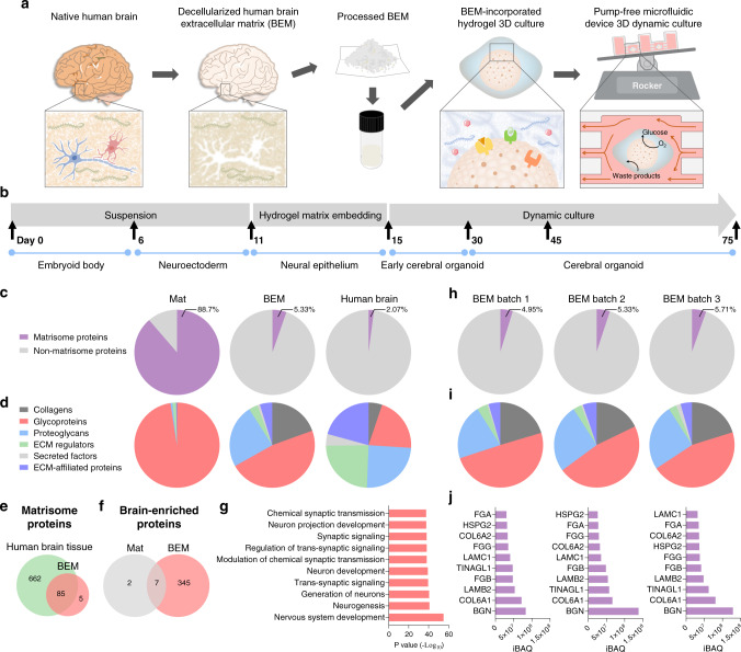
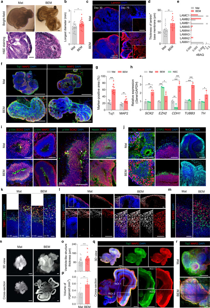
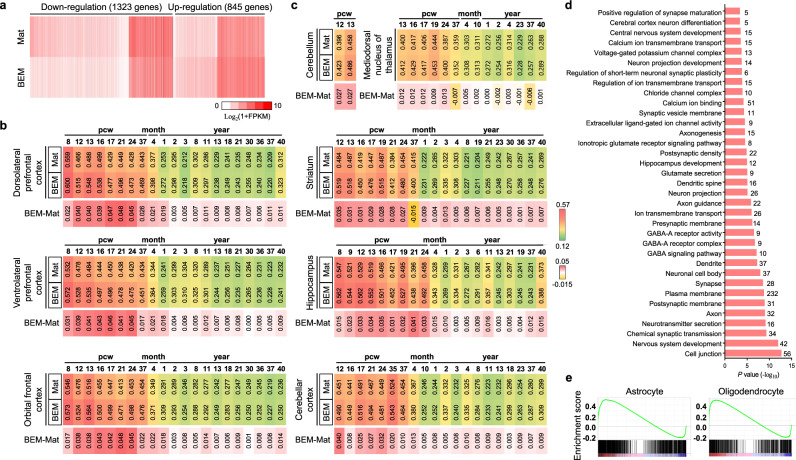
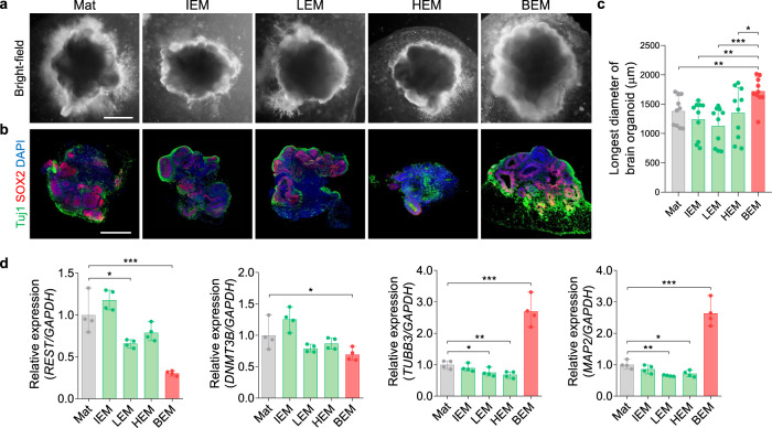
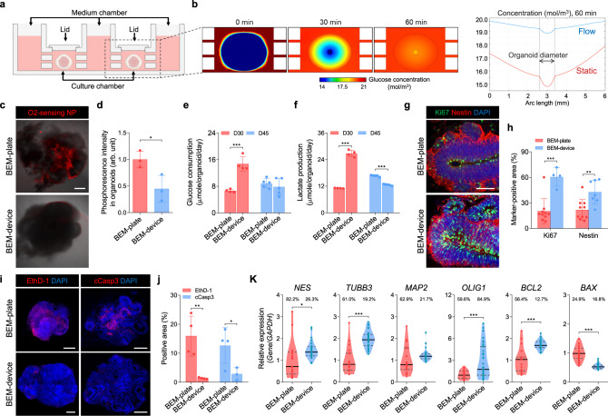
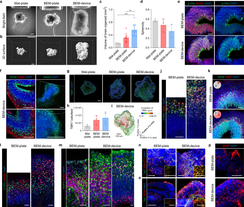
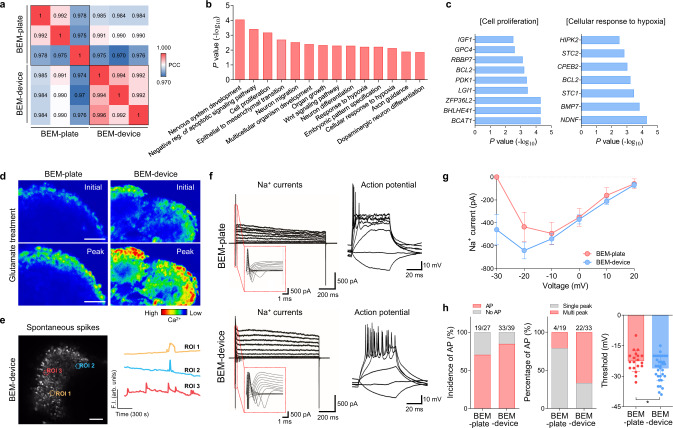
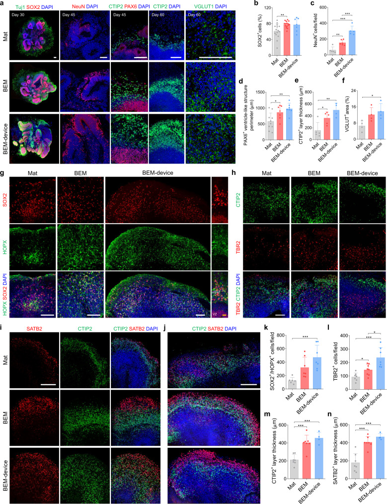
Brain organoids derived from human pluripotent stem cells provide a highly valuable in vitro model to recapitulate human brain development and neurological diseases. However, the current systems for brain organoid culture require further improvement for the reliable production of high-quality organoids. Here, we demonstrate two engineering elements to improve human brain organoid culture, (1) a human brain extracellular matrix to provide brain-specific cues and (2) a microfluidic device with periodic flow to improve the survival and reduce the variability of organoids. A three-dimensional culture modified with brain extracellular matrix significantly enhanced neurogenesis in developing brain organoids from human induced pluripotent stem cells. Cortical layer development, volumetric augmentation, and electrophysiological function of human brain organoids were further improved in a reproducible manner by dynamic culture in microfluidic chamber devices. Our engineering concept of reconstituting brain-mimetic microenvironments facilitates the development of a reliable culture platform for brain organoids, enabling effective modeling and drug development for human brain diseases.
Overview
The claims and statistics of this paper haven’t been checked yet.
Run the check — free, no account needed. It reads the open-access text and checks the sources it cites.
Integrity signals
Registry
Cites
Statistics
Figures
Verification
Claims the paper makes
The claims and statistics of this paper haven’t been checked yet.
Run the check — free, no account needed. It reads the open-access text and checks the sources it cites.
Claims other papers make about this one
Each is checked against this paper’s own text: does it say what the citing paper says it does?
The source backs part of the claim, not all of it — or backs it for a different population, condition or comparison.
In the citing paper
“While many other organoid models, including brain[13], intestinal [14], mammary gland [15], and retinal organoids [16], employ 3D ECM-based hydrogels to induce complex tissue architecture that cannot be achieved in ECM-free floating culture, the same has not been explored for skin yet.”
From Efficient Generation of Skin Organoids from Pluripotent Cells via Defined Extracellular Matrix Cues and Morphogen Gradients in a Spindle‐Shaped Microfluidic Device (2024) · cites this paper as [13]
Experimental — accuracy not yet measured
Self-check
Percentages, sums and reported differences re-computed from the paper’s own numbers.
The self-check needs the paper’s full text.
Citations
Cites
Gene Ontology: tool for the unification of biology
Ashburner, M., Ball, C. A., Blake, J. A., Botstein, D., Butler, H., Cherry, J. M., Davis, A. P., Dolinski, K., Dwight, S. S., Eppig, J. T., et al. (2000) · Nature Genetics · doi:10.1038/75556 · Cited by 43,206
Tissue-based map of the human proteome
Uhlén, M., Fagerberg, L., Hallström, B. M., Lindskog, C., Oksvold, P., Mardinoglu, A., Sivertsson, Å., Kampf, C., Sjöstedt, E., Asplund, A., et al. (2015) · Science · doi:10.1126/science.1260419 · Cited by 15,286
The Molecular Signatures Database Hallmark Gene Set Collection
Liberzon, A., Birger, C., Thorvaldsdóttir, H., Ghandi, M., Mesirov, J. P., & Tamayo, P. (2015) · Cell Systems · doi:10.1016/j.cels.2015.12.004 · Cited by 13,807
Shrestha, S., & Offer, S. M. (2016) · Medical Epigenetics · doi:10.1159/000444713 · Cited by 8,951
Pérez-Riverol, Y., Csordas, A., Bai, J.-W., Bernal-Llinares, M., Hewapathirana, S., Kundu, D. J., Inuganti, A., Griss, J., Mayer, G., Eisenacher, M., et al. (2018) · Nucleic Acids Res. · doi:10.1093/nar/gky1106 · Cited by 6,216
Cerebral organoids model human brain development and microcephaly
Lancaster, M. A., Renner, M., Martin, C.-A., Wenzel, D., Bicknell, L. S., Hurles, M. E., Homfray, T., Penninger, J. M., Jackson, A. P., & Knoblich, J. A. (2013) · Nature · doi:10.1038/nature12517 · Cited by 5,372
The Gene Ontology resource: enriching a GOld mine
The Gene Ontology Consortium, Carbon, S., Douglass, E., Good, B. M., Unni, D. R., Harris, N. L., Mungall, C. J., Basu, S., Chisholm, R. L., Dodson, R. J., et al. (2021) · Nucleic Acids Research · doi:10.1093/nar/gkaa1113 · Cited by 3,555
Organoid Models and Applications in Biomedical Research
Xinaris, C., Brizi, V., & Remuzzi, G. (2015) · The Nephron journals/Nephron journals · doi:10.1159/000433566 · Cited by 3,519
Mi, H., Muruganujan, A., Ebert, D., Huang, X., & Thomas, P. D. (2018) · Nucleic Acids Research · doi:10.1093/nar/gky1038 · Cited by 3,030
A draft map of the human proteome
Kim, M.-S., Pinto, S. M., Getnet, D., Nirujogi, R. S., Manda, S. S., Chaerkady, R., Madugundu, A. K., Kelkar, D. S., Isserlin, R., Jain, S., et al. (2014) · Nature · doi:10.1038/nature13302 · Cited by 2,198
Cited by
Sorted by citations
Organoids: The current status and biomedical applications
Yang, S., Hu, H., Kung, H., Zou, R.-Q., Dai, Y.-S., Hu, Y.-F., Wang, T., Lv, T., Yu, J., & Li, F. (2023) · MedComm · doi:10.1002/mco2.274 · Cited by 300
Three-Dimensional Cell Cultures: The Bridge between In Vitro and In Vivo Models
Urzì, O., Gasparro, R., Costanzo, E., Luca, A. D., Giavaresi, G., Fontana, S., & Alessandro, R. (2023) · International Journal of Molecular Sciences · doi:10.3390/ijms241512046 · Cited by 258
Kim, W., Gwon, Y., Park, S., Kim, H., & Kim, J. (2022) · Bioactive Materials · doi:10.1016/j.bioactmat.2022.03.039 · Cited by 216
Self-citationShell microelectrode arrays (MEAs) for brain organoids
Huang, Q., Tang, B.-H., Romero, J. C., Yang, Y., Elsayed, S. K., Pahapale, G. J., Lee, T.-J., Pantoja, I. E. M., Han, F., Berlinicke, C. J., et al. (2022) · bioRxiv · doi:10.1126/sciadv.abq5031 · Cited by 195
Microfluidic organoids-on-a-chip: The future of human models
Saorin, G., Caligiuri, I., & Rizzolio, F. (2022) · Seminars in Cell and Developmental Biology · doi:10.1016/j.semcdb.2022.10.001 · Cited by 213
Metadata
- Authors
- Cho, A.-N., Jin, Y., An, Y., Kim, J., Choi, Y. S., Lee, J. S., Kim, J., Choi, W.-Y., Koo, D.-J., Yu, W., Chang, G.-E., Kim, D.-Y., Jo, S.-H., Kim, J., Kim, S.-Y., Kim, Y.-G., Kim, J. Y., Choi, N., Cheong, E., Kim, Y.-J., Je, H. S., Kang, H.-C., & Cho, S.-W.
- Affiliations
- Yonsei University (KR)
- Kangwon National University (KR)
- Seoul National University (KR)
- Duke-NUS Medical School (SG)
- Soongsil University (KR)
- Korea Institute of Science and Technology (KR)
- Corresponding author
- Cho, Seung-Woo
- ORCID
- Venue
- Nature Communications
- Published
- 2021-08-05
- Volume
- 12
- Issue
- 1
- Pages
- 4730-4730
- ISSN
- 2041-1723 (ISSN-L)
- PMC
- PMC8342542
- OpenAlex
- W3188744629
- Cited by
- 397
- References
- 91
- Citations by year
- 2026: 77 · 2025: 83 · 2024: 96 · 2023: 66 · 2022: 69 · 2021: 6
- FWCI
- 18.60
- Type
- journal article
- Language
- en
- Open access
- gold
- doi.org
- Locations
- Nature Communications (publishedVersion, cc-by)
- PubMed Central (publishedVersion, cc-by)
- PubMed (publishedVersion)
- RePEc: Research Papers in Economics (submittedVersion)
- DOAJ (DOAJ: Directory of Open Access Journals) (submittedVersion, cc-by-sa)
- YUHSpace (Yonsei University Medical Library) (submittedVersion, cc-by-nc-nd)
- Funders
- National Research Foundation of Korea (2016M3C9A4921712, 2017M3C7A1047659, 2018M3A9H1021382)
- Institute for Basic Science (IBS-R026-D1)
- Samsung (SRFC-TC2003-03)
- Topics
- Life Sciences › Biochemistry, Genetics and Molecular Biology › Molecular Biology › Pluripotent Stem Cells Research
- Physical Sciences › Engineering › Biomedical Engineering › 3D Printing in Biomedical Research
- Life Sciences › Neuroscience › Developmental Neuroscience › Neurogenesis and neuroplasticity mechanisms
- Keywords
- brain organoids, microfluidics, brain extracellular matrix, induced pluripotent stem cells, neurogenesis, organoid maturation, dynamic culture, periodic flow, electrophysiological function, 3D cell culture
- MeSH terms
- Animals, Brain, Culture Media, Extracellular Matrix, Feasibility Studies, Humans, Models, Anatomic, Models, Neurological, Neuroglia, Organ Culture Techniques, Organoids, Swine, Hydrogels, Gene Expression Profiling, Neurogenesis, Electrophysiological Phenomena, Lab-On-A-Chip Devices, Induced Pluripotent Stem Cells
About these checks
Registry, retraction, Cites and statistics results are computed, not judged, with one exception: a printed reference that no index lists is read once by a model before it counts as a citation error. Claims are extracted by a model and kept only when their quote appears verbatim in the text; each cited source is then checked, with a model’s help, for whether it says what the claim relies on. Those verdicts can be wrong — read the claim’s quote and the cited source before relying on one.
Statistics and arithmetic are recomputed, not judged.
Confidence bands are shown only for fields calibrated on our annotated test set. None is calibrated yet, so no band is shown.
What counts as a claim ›How we measure confidence ›How we check this ›
Are you an author, or do you see a problem with this page? Request a review
