

The DOI matches its Crossref record; 91 of the 91 references it cites were found in a registry or an index.
Microfluidic device with brain extracellular matrix promotes structural and functional maturation of human brain organoids
Open accessAbstract
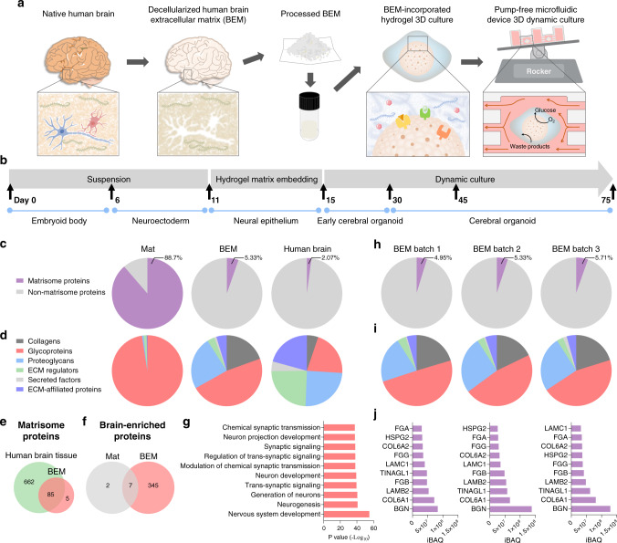
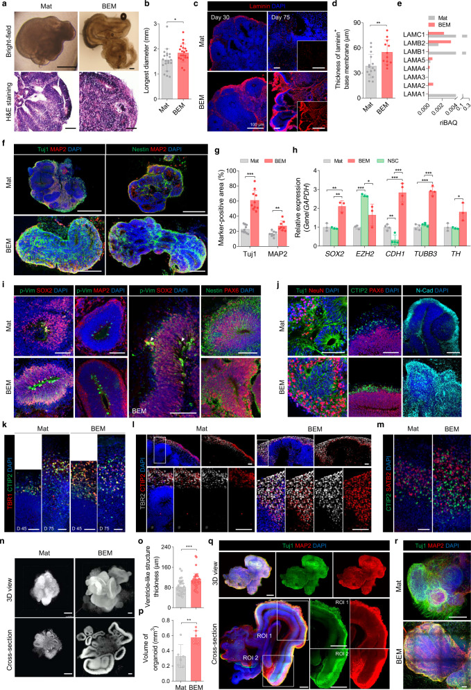
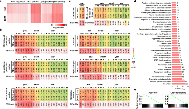
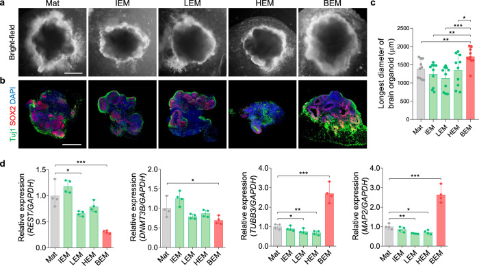
Brain organoids derived from human pluripotent stem cells provide a highly valuable in vitro model to recapitulate human brain development and neurological diseases. However, the current systems for brain organoid culture require further improvement for the reliable production of high-quality organoids. Here, we demonstrate two engineering elements to improve human brain organoid culture, (1) a human brain extracellular matrix to provide brain-specific cues and (2) a microfluidic device with periodic flow to improve the survival and reduce the variability of organoids. A three-dimensional culture modified with brain extracellular matrix significantly enhanced neurogenesis in developing brain organoids from human induced pluripotent stem cells. Cortical layer development, volumetric augmentation, and electrophysiological function of human brain organoids were further improved in a reproducible manner by dynamic culture in microfluidic chamber devices. Our engineering concept of reconstituting brain-mimetic microenvironments facilitates the development of a reliable culture platform for brain organoids, enabling effective modeling and drug development for human brain diseases.
Overview
The claims and statistics of this paper haven’t been checked yet.
Run the check — free, no account needed. It reads the open-access text and checks the sources it cites.
Integrity signals
Registry
Cites
Statistics
Figures
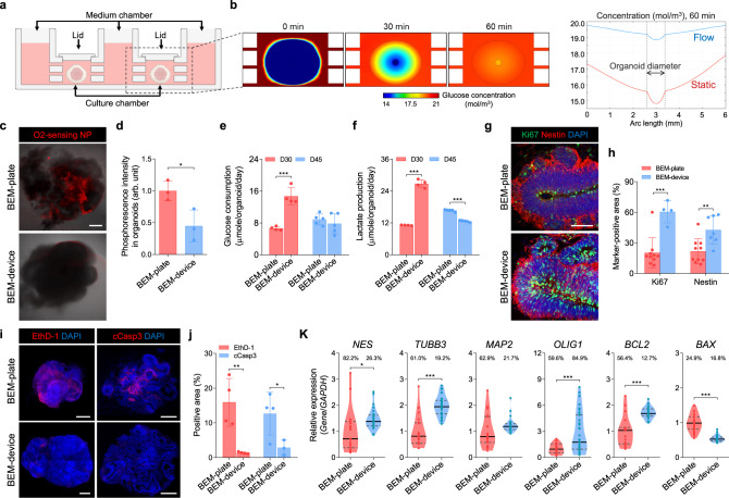
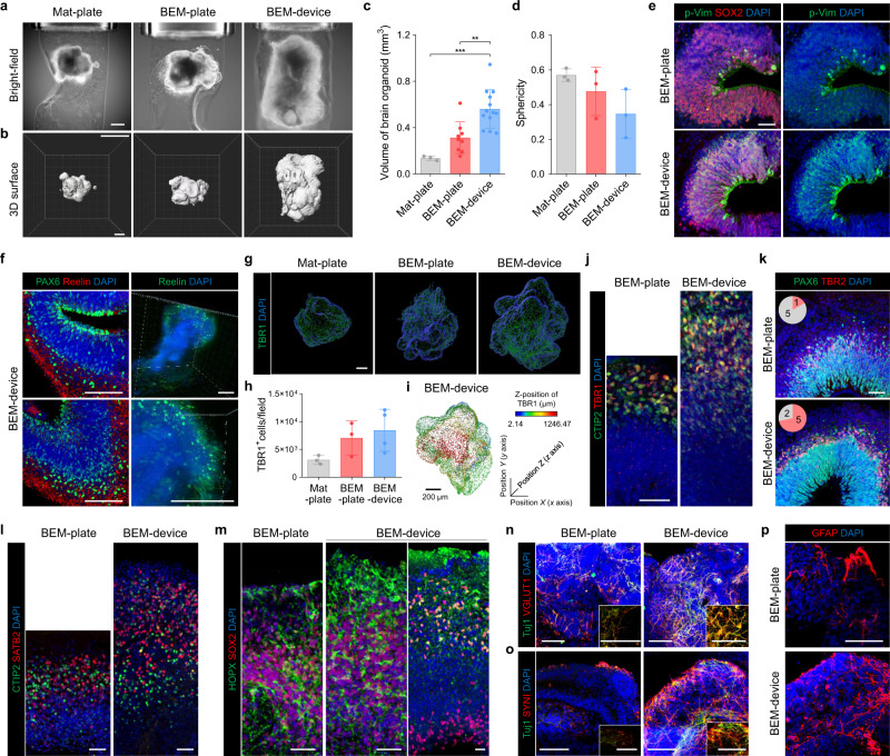
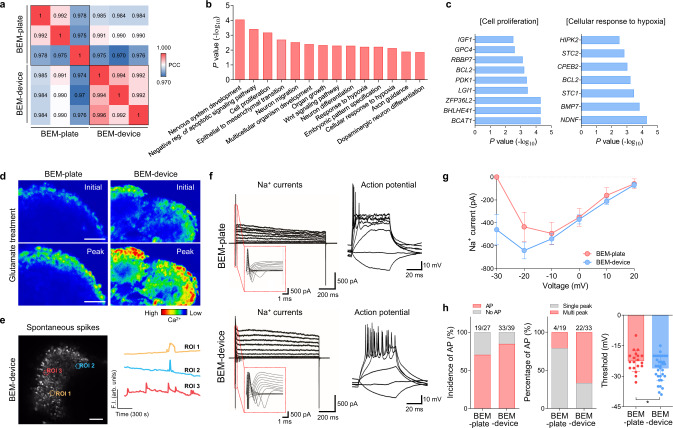
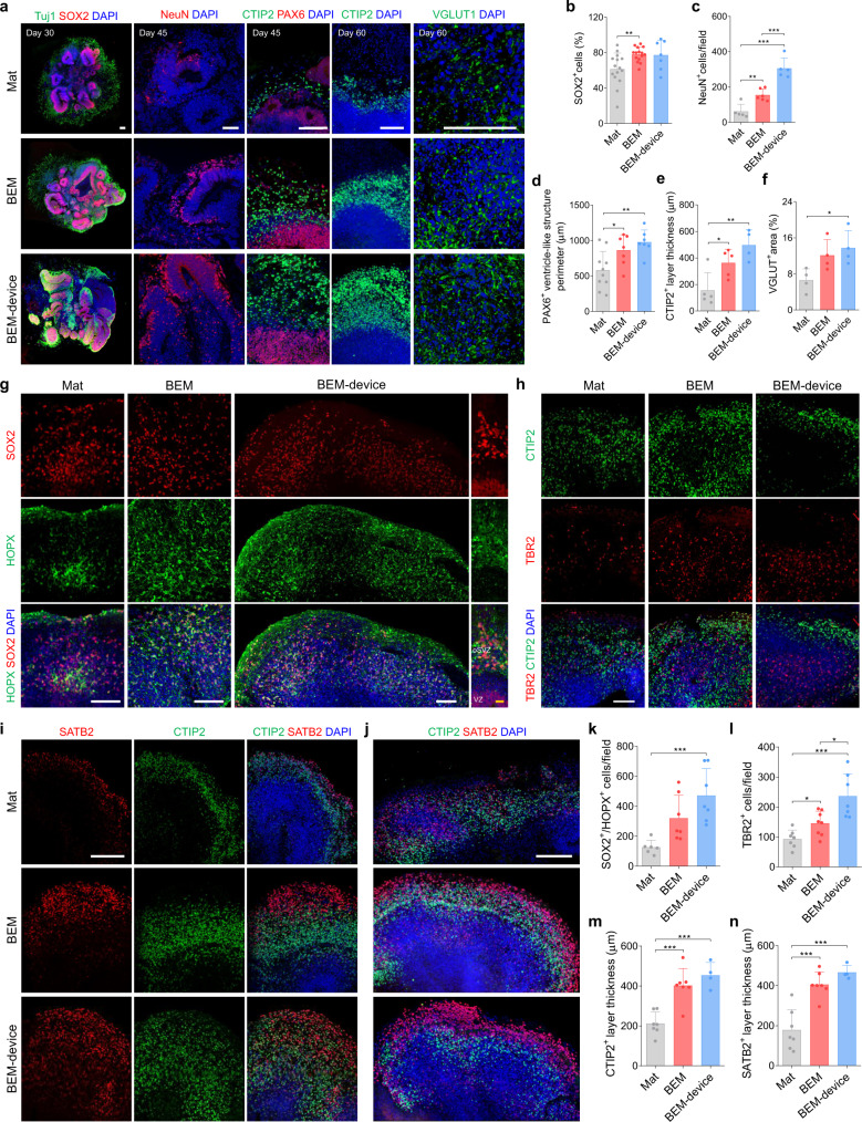
Verification
Claims the paper makes
The claims and statistics of this paper haven’t been checked yet.
Run the check — free, no account needed. It reads the open-access text and checks the sources it cites.
Claims other papers make about this one
Each is checked against this paper’s own text: does it say what the citing paper says it does?
The source backs part of the claim, not all of it — or backs it for a different population, condition or comparison.
In the citing paper
“While many other organoid models, including brain[13], intestinal [14], mammary gland [15], and retinal organoids [16], employ 3D ECM-based hydrogels to induce complex tissue architecture that cannot be achieved in ECM-free floating culture, the same has not been explored for skin yet.”
From Efficient Generation of Skin Organoids from Pluripotent Cells via Defined Extracellular Matrix Cues and Morphogen Gradients in a Spindle‐Shaped Microfluidic Device (2024) · cites this paper as [13]
Experimental — accuracy not yet measured
Self-check
Percentages, sums and reported differences re-computed from the paper’s own numbers.
The self-check needs the paper’s full text.
Citations
Cites
Gene Ontology: tool for the unification of biology
Ashburner, M., Ball, C. A., Blake, J. A., Botstein, D., Butler, H., Cherry, J. M., Davis, A. P., Dolinski, K., Dwight, S. S., Eppig, J. T., et al. (2000) · Nature Genetics · doi:10.1038/75556 · Cited by 43,206
Tissue-based map of the human proteome
Uhlén, M., Fagerberg, L., Hallström, B. M., Lindskog, C., Oksvold, P., Mardinoglu, A., Sivertsson, Å., Kampf, C., Sjöstedt, E., Asplund, A., et al. (2015) · Science · doi:10.1126/science.1260419 · Cited by 15,286
The Molecular Signatures Database Hallmark Gene Set Collection
Liberzon, A., Birger, C., Thorvaldsdóttir, H., Ghandi, M., Mesirov, J. P., & Tamayo, P. (2015) · Cell Systems · doi:10.1016/j.cels.2015.12.004 · Cited by 13,807
Shrestha, S., & Offer, S. M. (2016) · Medical Epigenetics · doi:10.1159/000444713 · Cited by 8,951
Pérez-Riverol, Y., Csordas, A., Bai, J.-W., Bernal-Llinares, M., Hewapathirana, S., Kundu, D. J., Inuganti, A., Griss, J., Mayer, G., Eisenacher, M., et al. (2018) · Nucleic Acids Res. · doi:10.1093/nar/gky1106 · Cited by 6,216
Cerebral organoids model human brain development and microcephaly
Lancaster, M. A., Renner, M., Martin, C.-A., Wenzel, D., Bicknell, L. S., Hurles, M. E., Homfray, T., Penninger, J. M., Jackson, A. P., & Knoblich, J. A. (2013) · Nature · doi:10.1038/nature12517 · Cited by 5,372
The Gene Ontology resource: enriching a GOld mine
The Gene Ontology Consortium, Carbon, S., Douglass, E., Good, B. M., Unni, D. R., Harris, N. L., Mungall, C. J., Basu, S., Chisholm, R. L., Dodson, R. J., et al. (2021) · Nucleic Acids Research · doi:10.1093/nar/gkaa1113 · Cited by 3,555
Organoid Models and Applications in Biomedical Research
Xinaris, C., Brizi, V., & Remuzzi, G. (2015) · The Nephron journals/Nephron journals · doi:10.1159/000433566 · Cited by 3,519
Mi, H., Muruganujan, A., Ebert, D., Huang, X., & Thomas, P. D. (2018) · Nucleic Acids Research · doi:10.1093/nar/gky1038 · Cited by 3,030
A draft map of the human proteome
Kim, M.-S., Pinto, S. M., Getnet, D., Nirujogi, R. S., Manda, S. S., Chaerkady, R., Madugundu, A. K., Kelkar, D. S., Isserlin, R., Jain, S., et al. (2014) · Nature · doi:10.1038/nature13302 · Cited by 2,198
Brain-Region-Specific Organoids Using Mini-bioreactors for Modeling ZIKV Exposure
Qian, X., Nguyen, H. N., Song, M. M., Hadiono, C., Ogden, S. C., Hammack, C., Yao, B., Hamersky, G. R., Jacob, F., Zhong, C., et al. (2016) · Cell · doi:10.1016/j.cell.2016.04.032 · Cited by 2,127
Generation of cerebral organoids from human pluripotent stem cells
Lancaster, M. A., & Knoblich, J. A. (2014) · Nature Protocols · doi:10.1038/nprot.2014.158 · Cited by 1,823
Functional cortical neurons and astrocytes from human pluripotent stem cells in 3D culture
Paşca, A. M., Sloan, S. A., Clarke, L. E., Tian, Y., Makinson, C. D., Huber, N., Kim, C. H., Park, J.-Y., O'Rourke, N. A., Nguyen, K. D., et al. (2015) · Nature Methods · doi:10.1038/nmeth.3415 · Cited by 1,609
Eiraku, M., Watanabe, K., Matsuo-Takasaki, M., Kawada, M., Yonemura, S., Matsumura, M., Wataya, T., Nishiyama, A., Muguruma, K., & Sasai, Y. (2008) · Cell Stem Cell · doi:10.1016/j.stem.2008.09.002 · Cited by 1,522
Assembly of functionally integrated human forebrain spheroids
Birey, F., Andersen, J., Makinson, C. D., Islam, S., Wei, W., Huber, N., Fan, H. C., Metzler, K. R. C., Panagiotakos, G., Thom, N., et al. (2017) · Nature · doi:10.1038/nature22330 · Cited by 1,365
Naba, A., Clauser, K. R., Hoersch, S., Liu, H., Carr, S. A., & Hynes, R. O. (2012) · Molecular & Cellular Proteomics · doi:10.1074/mcp.m111.014647 · Cited by 1,354
Whole-Brain Imaging with Single-Cell Resolution Using Chemical Cocktails and Computational Analysis
Susaki, E. A., Tainaka, K., Perrin, D., Kishino, F., Tawara, T., Watanabe, T. M., Yokoyama, C., Onoe, H., Eguchi, M., Yamaguchi, S., et al. (2014) · Cell · doi:10.1016/j.cell.2014.03.042 · Cited by 1,352
Cell diversity and network dynamics in photosensitive human brain organoids
Quadrato, G., Nguyen, T., Macosko, E. Z., Sherwood, J. L., Min Yang, S., Berger, D. R., Maria, N., Scholvin, J., Goldman, M., Kinney, J. P., et al. (2017) · Nature · doi:10.1038/nature22047 · Cited by 1,321
An in vivo model of functional and vascularized human brain organoids
Mansour, A. A., Gonçalves, J. T., Bloyd, C. W., Li, H., Fernandes, S., Quang, D., Johnston, S., Parylak, S. L., Jin, X., & Gage, F. H. (2018) · Nature Biotechnology · doi:10.1038/nbt.4127 · Cited by 1,211
An atlas of the protein-coding genes in the human, pig, and mouse brain
Sjöstedt, E., Zhong, W., Fagerberg, L., Karlsson, M., Mitsios, N., Adori, C., Oksvold, P., Edfors, F., Limiszewska, A., Hikmet, F., et al. (2020) · Science · doi:10.1126/science.aay5947 · Cited by 1,151
FOXG1-Dependent Dysregulation of GABA/Glutamate Neuron Differentiation in Autism Spectrum Disorders
Mariani, J., Coppola, G., Zhang, P., Abyzov, A., Provini, L., Tomasini, L., Amenduni, M., Szekely, A., Palejev, D., Wilson, M., et al. (2015) · Cell · doi:10.1016/j.cell.2015.06.034 · Cited by 1,149
Human cerebral organoids recapitulate gene expression programs of fetal neocortex development
Camp, J. G., Badsha, F., Florio, M., Kanton, S., Gerber, T., Wilsch-Bräuninger, M., Lewitus, E., Sykes, A., Hevers, W., Lancaster, M., et al. (2015) · Proceedings of the National Academy of Sciences · doi:10.1073/pnas.1520760112 · Cited by 1,130
Kadoshima, T., Sakaguchi, H., Nakano, T., Soen, M., Ando, S., Eiraku, M., & Sasai, Y. (2013) · Proceedings of the National Academy of Sciences · doi:10.1073/pnas.1315710110 · Cited by 999
Engineering of human brain organoids with a functional vascular-like system
Çakır, B., Xiang, Y., Tanaka, Y., Kural, M. H., Parent, M. J., Kang, Y., Chapeton, K., Patterson, B., Yuan, Y., He, C., et al. (2019) · Nature Methods · doi:10.1038/s41592-019-0586-5 · Cited by 989
Molecular Identity of Human Outer Radial Glia during Cortical Development
Pollen, A. A., Nowakowski, T. J., Chen, J., Retallack, H., Sandoval-Espinosa, C., Nicholas, C. R., Shuga, J., Liu, S. J., Oldham, M. C., Diaz, A., et al. (2015) · Cell · doi:10.1016/j.cell.2015.09.004 · Cited by 908
Engineering Stem Cell Organoids
Yin, X., Mead, B. E., Safaee, H., Langer, R., Karp, J. M., & Levy, O. (2016) · Cell Stem Cell · doi:10.1016/j.stem.2015.12.005 · Cited by 866
Jo, J., Xiao, Y., Sun, A. X., Cukuroglu, E., Tran, H.-D., Göke, J., Tan, Z. Y., Saw, T. Y., Tan, C.-P., Lokman, H., et al. (2016) · Cell Stem Cell · doi:10.1016/j.stem.2016.07.005 · Cited by 861
Self-citationOSVZ progenitors of human and ferret neocortex are epithelial-like and expand by integrin signaling
Fietz, S. A., Kelava, I., Vogt, J., Wilsch-Bräuninger, M., Stenzel, D., Fish, J. L., Corbeil, D., Riehn, A., Distler, W., Nitsch, R., et al. (2010) · Nature Neuroscience · doi:10.1038/nn.2553 · Cited by 830
Advanced CUBIC protocols for whole-brain and whole-body clearing and imaging
Susaki, E. A., Tainaka, K., Perrin, D., Yukinaga, H., Kuno, A., & Ueda, H. R. (2015) · Nature Protocols · doi:10.1038/nprot.2015.085 · Cited by 822
A new look at cerebrospinal fluid circulation
Brinker, T., Stopa, E., Morrison, J., & Klinge, P. (2014) · Fluids and Barriers of the CNS · doi:10.1186/2045-8118-11-10 · Cited by 819
Fused cerebral organoids model interactions between brain regions
Bagley, J. A., Reumann, D., Bian, S., Lévi-Strauss, J., & Knoblich, J. A. (2017) · Nature Methods · doi:10.1038/nmeth.4304 · Cited by 790
The use of brain organoids to investigate neural development and disease
Di Lullo, E., & Kriegstein, A. R. (2017) · Nature Reviews Neuroscience · doi:10.1038/nrn.2017.107 · Cited by 788
Guided self-organization and cortical plate formation in human brain organoids
Lancaster, M. A., Corsini, N. S., Wolfinger, S., Gustafson, E. H., Phillips, A. W., Burkard, T. R., Otani, T., Livesey, F. J., & Knoblich, J. A. (2017) · Nature Biotechnology · doi:10.1038/nbt.3906 · Cited by 783
Modes of neuronal migration in the developing cerebral cortex
Nadarajah, B., & Parnavelas, J. G. (2002) · Nature reviews. Neuroscience · doi:10.1038/nrn845 · Cited by 712
Xiang, Y., Tanaka, Y., Patterson, B., Kang, Y., Govindaiah, G., Roselaar, N., Çakır, B., Kim, K.-Y., Lombroso, A., Hwang, S., et al. (2017) · Cell stem cell · doi:10.1016/j.stem.2017.07.007 · Cited by 711
Self-Organization of Polarized Cerebellar Tissue in 3D Culture of Human Pluripotent Stem Cells
Muguruma, K., Nishiyama, A., Kawakami, H., Hashimoto, K., & Sasai, Y. (2015) · Cell Reports · doi:10.1016/j.celrep.2014.12.051 · Cited by 692
A single-cell RNA-seq survey of the developmental landscape of the human prefrontal cortex
Zhong, S., Zhang, S., Fan, X., Wu, Q., Yan, L., Dong, J., Zhang, H., Li, L., Sun, L., Pan, N., et al. (2018) · Nature · doi:10.1038/nature25980 · Cited by 689
Bilati, U., Allémann, É., & Doelker, É. (2004) · European Journal of Pharmaceutical Sciences · doi:10.1016/j.ejps.2004.09.011 · Cited by 687
Zheng, X., Boyer, L., Jin, M., Mertens, J., Kim, Y., Ma, L., Ma, L., Hamm, M., Gage, F. H., & Hunter, T. (2016) · eLife · doi:10.7554/elife.13374 · Cited by 671
Self-citationCerebral organoids at the air-liquid interface generate diverse nerve tracts with functional output
Giandomenico, S. L., Mierau, S. B., Gibbons, G. M., Wenger, L. M. D., Masullo, L., Sit, T., Sutcliffe, M., Boulanger, J., Tripodi, M., Derivery, E., et al. (2019) · Nature Neuroscience · doi:10.1038/s41593-019-0350-2 · Cited by 651
Sakaguchi, H., Kadoshima, T., Soen, M., Narii, N., Ishida, Y., Ohgushi, M., Takahashi, J., Eiraku, M., & Sasai, Y. (2015) · Nature Communications · doi:10.1038/ncomms9896 · Cited by 542
Modeling human cortical development in vitro using induced pluripotent stem cells
Mariani, J., Simonini, M. V., Palejev, D., Tomasini, L., Coppola, G., Szekely, A. M., Horvath, T. L., & Vaccarino, F. M. (2012) · Proceedings of the National Academy of Sciences · doi:10.1073/pnas.1202944109 · Cited by 526
Extracellular Matrix: Functions in the Nervous System
Barros, C. S., Franco, S. J., & Muller, U. (2011) · Cold Spring Harbor Perspectives in Biology · doi:10.1101/cshperspect.a005108 · Cited by 506
Self-formation of functional adenohypophysis in three-dimensional culture
Suga, H., Kadoshima, T., Minaguchi, M., Ohgushi, M., Soen, M., Nakano, T., Takata, N., Wataya, T., Muguruma, K., Miyoshi, H., et al. (2011) · Nature · doi:10.1038/nature10637 · Cited by 497
hESC-Derived Thalamic Organoids Form Reciprocal Projections When Fused with Cortical Organoids
Xiang, Y., Tanaka, Y., Cakir, B., Patterson, B., Kim, K.-Y., Sun, P., Kang, Y.-J., Zhong, M., Liu, X., Patra, P., et al. (2019) · Cell Stem Cell · doi:10.1016/j.stem.2018.12.015 · Cited by 485
Pereira, J. D., Sansom, S. N., Smith, J. C., Dobenecker, M., Tarakhovsky, A., & Livesey, F. J. (2010) · Proceedings of the National Academy of Sciences · doi:10.1073/pnas.1002530107 · Cited by 465
Induction of Expansion and Folding in Human Cerebral Organoids
Li, Y., Muffat, J., Omer, A., Bosch, I., Lancaster, M. A., Sur, M., Gehrke, L., Knoblich, J. A., & Jaenisch, R. (2017) · Cell Stem Cell · doi:10.1016/j.stem.2016.11.017 · Cited by 436
Human brain organoids on a chip reveal the physics of folding
Karzbrun, E., Kshirsagar, A., Cohen, S., Hanna, J. H., & Reiner, O. (2018) · Nature Physics · doi:10.1038/s41567-018-0046-7 · Cited by 421
Stem Cell Models of Human Brain Development
Kelava, I., & Lancaster, M. A. (2016) · Cell stem cell · doi:10.1016/j.stem.2016.05.022 · Cited by 385
Orešković, D., & Klarica, M. (2010) · Brain Research Reviews · doi:10.1016/j.brainresrev.2010.04.006 · Cited by 349
Specification of positional identity in forebrain organoids
Cederquist, G., Asciolla, J. J., Tchieu, J. H., Walsh, R. M., Cornacchia, D., Resh, M. D., & Studer, L. (2019) · Nature Biotechnology · doi:10.1038/s41587-019-0085-3 · Cited by 334
Reelin, Rap1 and N-cadherin orient the migration of multipolar neurons in the developing neocortex
Jossin, Y., & Cooper, J. A. (2011) · Nature Neuroscience · doi:10.1038/nn.2816 · Cited by 332
Human brain organoid-on-a-chip to model prenatal nicotine exposure
Wang, Y., Wang, L., Zhu, Y., & Qin, J. (2018) · Lab on a Chip · doi:10.1039/c7lc01084b · Cited by 330
N-cadherin mediates cortical organization in the mouse brain
Kadowaki, M., Nakamura, S., Machoň, O., Krauss, S. J. K., Radice, G. L., & Takeichi, M. (2006) · Developmental Biology · doi:10.1016/j.ydbio.2006.12.014 · Cited by 298
Giandomenico, S. L., Sutcliffe, M. A., & Lancaster, M. A. (2020) · Nature Protocols · doi:10.1038/s41596-020-00433-w · Cited by 293
Changes in cell-cycle kinetics during the development and evolution of primate neocortex
Kornack, D. R., & RAKIC, P. T. (1998) · Proceedings of the National Academy of Sciences · doi:10.1073/pnas.95.3.1242 · Cited by 292
Single-cell analysis reveals transcriptional heterogeneity of neural progenitors in human cortex
Johnson, M. B., Wang, P. P., Atabay, K. D., Murphy, E. A., Doan, R. N., Hecht, J. L., & Walsh, C. A. (2015) · Nature Neuroscience · doi:10.1038/nn.3980 · Cited by 288
Expression of tenascin in the developing and adult cerebellar cortex
Bartsch, S., Bartsch, U., Dörries, U., Faissner, A., Weller, A., Ekblom, P., & Schachner, M. (1992) · Journal of Neuroscience · doi:10.1523/jneurosci.12-03-00736.1992 · Cited by 271
Artegiani, B., Lyubimova, A., Muraro, M., van Es, J. H., van Oudenaarden, A., & Clevers, H. (2017) · Cell Reports · doi:10.1016/j.celrep.2017.11.050 · Cited by 259
Mitotic spindle orientation predicts outer radial glial cell generation in human neocortex
Ostrem, B. E. L., Lui, J. H., Hansen, D. V., & Kriegstein, A. R. (2013) · Nature Communications · doi:10.1038/ncomms2647 · Cited by 244
The Cells of Cajal-Retzius: Still a Mystery One Century After
Soriano, E., & del Rı́o, J. A. (2005) · Neuron · doi:10.1016/j.neuron.2005.04.019 · Cited by 243
Lee, J. S., Shin, J., Park, H.-M., Kim, Y.-G., Kim, B.-G., Oh, J.-W., & Cho, S.-W. (2014) · Biomacromolecules · doi:10.1021/bm4015039 · Cited by 236
Self-citationRole of radial glial cells in cerebral cortex folding
Borrell, V., & Götz, M. (2014) · Current Opinion in Neurobiology · doi:10.1016/j.conb.2014.02.007 · Cited by 230
Engineering stem cell-derived 3D brain organoids in a perfusable organ-on-a-chip system
Wang, Y., Wang, L., Guo, Y., Zhu, Y., & Qin, J. (2018) · RSC Advances · doi:10.1039/c7ra11714k · Cited by 201
Proteoglycans and neuronal migration in the cerebral cortex during development and disease
Maeda, N. (2015) · Frontiers in Neuroscience · doi:10.3389/fnins.2015.00098 · Cited by 188
Neonatal loss of γ–aminobutyric acid pathway expression after human perinatal brain injury
Robinson, S., Li, Q., DeChant, A. K., & Cohen, M. L. (2006) · Journal of Neurosurgery Pediatrics · doi:10.3171/ped.2006.104.6.396 · Cited by 172
Jin, Y., Kim, J., Lee, J. S., Min, S., Kim, S., Ahn, D., Kim, Y., & Cho, S. (2018) · Advanced Functional Materials · doi:10.1002/adfm.201801954 · Cited by 166
Self-citationDevelopment of the brain: a vital role for cerebrospinal fluid
Miyan, J. A., Nabiyouni, M., & Zendah, M. (2003) · Canadian Journal of Physiology and Pharmacology · doi:10.1139/y03-027 · Cited by 159
Advances in the Potential Biomarkers of Epilepsy
Kobylarek, D., Iwanowski, P. S., Lewandowska, Z., Limphaibool, N., Szafranek, S., Labrzycka, A., & Kozubski, W. P. (2019) · Frontiers in Neurology · doi:10.3389/fneur.2019.00685 · Cited by 157
Millifluidic culture improves human midbrain organoid vitality and differentiation
Berger, E., Magliaro, C., Paczia, N., Monzel, A. S., Antony, P. M. A., Linster, C. L., Bolognin, S., Ahluwalia, A. D., & Schwamborn, J. C. (2018) · Lab on a Chip · doi:10.1039/c8lc00206a · Cited by 153
Towards definition of an ECM parts list: An advance on GO categories
Naba, A., Hoersch, S., & Hynes, R. O. (2012) · Matrix Biology · doi:10.1016/j.matbio.2012.11.008 · Cited by 121
The extracellular matrix compartment of neural stem and glial progenitor cells
Faissner, A., & Reinhard, J. (2015) · Glia · doi:10.1002/glia.22839 · Cited by 116
Jin, Y., Lee, J. S., Kim, J., Min, S., Wi, S., Yu, J. H., Chang, G.-E., Cho, A., Choi, Y., Ahn, D.-H., et al. (2018) · Nature Biomedical Engineering · doi:10.1038/s41551-018-0260-8 · Cited by 116
Self-citationControl of Outer Radial Glial Stem Cell Mitosis in the Human Brain
Ostrem, B. E. L., Lui, J. H., Gertz, C. C., & Kriegstein, A. R. (2014) · Cell Reports · doi:10.1016/j.celrep.2014.06.058 · Cited by 112
Alcalá, S., Klee, M., Fernández, J., Fleischer, A., & Pimentel‐Muiños, F. X. (2007) · Oncogene · doi:10.1038/sj.onc.1210600 · Cited by 103
APC/C-Cdh1 coordinates neurogenesis and cortical size during development
Delgado‐Esteban, M., García-Higuera, I., Maestre, C., Moreno, S., & Almeida, Á. (2013) · Nature Communications · doi:10.1038/ncomms3879 · Cited by 97
Six-month cultured cerebral organoids from human ES cells contain matured neural cells
Matsui, T. K., Matsubayashi, M., Sakaguchi, Y. M., Hayashi, R. K., Zheng, C., Sugie, K., Hasegawa, M., Nakagawa, T., & Mori, E. (2018) · Neuroscience Letters · doi:10.1016/j.neulet.2018.01.040 · Cited by 80
Heck, N., Garwood, J., Loeffler, J., Larmet, Y., & Faissner, A. (2004) · Neuroscience · doi:10.1016/j.neuroscience.2004.06.078 · Cited by 73
Sood, D., Cairns, D. M., Dabbi, J. M., Ramakrishnan, C., Deisseroth, K. A., Black, L. D., Santaniello, S., & Kaplan, D. L. (2019) · Scientific Reports · doi:10.1038/s41598-019-54248-1 · Cited by 72
Sjöstedt, E., Fagerberg, L., Hallström, B. M., Månberg, A., Mitsios, N., Nilsson, P., Pontén, F., Hökfelt, T. G. M., Uhlén, M., & Mulder, J. (2015) · PLoS ONE · doi:10.1371/journal.pone.0130028 · Cited by 62
Phosphorescent nanoparticles for quantitative measurements of oxygen profiles in vitro and in vivo
Choi, N., Verbridge, S. S., Williams, R. M., Chen, J., Kim, J., Schmehl, R. H., Farnum, C. E., Zipfel, W. R., Fischbach, C., & Stroock, A. D. (2012) · Biomaterials · doi:10.1016/j.biomaterials.2011.11.048 · Cited by 56
Self-citationXu, J.-C., Xiao, M.-F., Jakovčevski, I., Sivukhina, E. V., Hargus, G., Cui, Y.-F., Irintchev, A., Schachner, M., & Bernreuther, C. (2013) · Journal of Cell Science · doi:10.1242/jcs.137612 · Cited by 51
Cho, A., Jin, Y., Kim, S., Kumar, S., Shin, H., Kang, H., & Cho, S. (2019) · ACS Applied Materials & Interfaces · doi:10.1021/acsami.9b03242 · Cited by 51
Self-citationSeo, H. I., Cho, A., Jang, J., Kim, D., Cho, S., & Chung, B. G. (2015) · Nanomedicine Nanotechnology Biology and Medicine · doi:10.1016/j.nano.2015.05.008 · Cited by 49
Self-citationLee, J. S., Roh, Y. H., Choi, Y. S., Jin, Y., Jeon, E. J., Bong, K. W., & Cho, S. (2019) · Advanced Functional Materials · doi:10.1002/adfm.201807803 · Cited by 48
Self-citationNeural ECM in laminar organization and connectivity development in healthy and diseased human brain
Milošević, N. J., Judáš, M., Aronica, E., & Kostović, I. (2014) · Progress in brain research · doi:10.1016/b978-0-444-63486-3.00007-4 · Cited by 43
Topical Review: Neuronal Migration in Cortical Development
Kanatani, S., Tabata, H., & Nakajima, K. (2005) · Journal of Child Neurology · doi:10.1177/08830738050200040201 · Cited by 34
Organoids for Advanced Therapeutics and Disease Models
Kim, S., Cho, A., Min, S., Kim, S., & Cho, S. (2018) · Advanced Therapeutics · doi:10.1002/adtp.201800087 · Cited by 33
Self-citationSun, B., Chang, E., Gerhartl, A., & Szele, F. G. (2018) · Cerebral Cortex · doi:10.1093/cercor/bhx289 · Cited by 30
Xu, J., Xiao, M., Jakovčevski, I., Sivukhina, E. V., Hargus, G., Cui, Y., Irintchev, A., Schachner, M., & Bernreuther, C. (2014) · Development · doi:10.1242/dev.108605 · Cited by 27
EZH2 Influences mdDA Neuronal Differentiation, Maintenance and Survival
Wever, I., von Oerthel, L., Wagemans, C. M. R. J., & Smidt, M. P. (2019) · Frontiers in Molecular Neuroscience · doi:10.3389/fnmol.2018.00491 · Cited by 14
Cited by
Sorted by citations
Organoids: The current status and biomedical applications
Yang, S., Hu, H., Kung, H., Zou, R.-Q., Dai, Y.-S., Hu, Y.-F., Wang, T., Lv, T., Yu, J., & Li, F. (2023) · MedComm · doi:10.1002/mco2.274 · Cited by 300
Three-Dimensional Cell Cultures: The Bridge between In Vitro and In Vivo Models
Urzì, O., Gasparro, R., Costanzo, E., Luca, A. D., Giavaresi, G., Fontana, S., & Alessandro, R. (2023) · International Journal of Molecular Sciences · doi:10.3390/ijms241512046 · Cited by 258
Kim, W., Gwon, Y., Park, S., Kim, H., & Kim, J. (2022) · Bioactive Materials · doi:10.1016/j.bioactmat.2022.03.039 · Cited by 216
Self-citationShell microelectrode arrays (MEAs) for brain organoids
Huang, Q., Tang, B.-H., Romero, J. C., Yang, Y., Elsayed, S. K., Pahapale, G. J., Lee, T.-J., Pantoja, I. E. M., Han, F., Berlinicke, C. J., et al. (2022) · bioRxiv · doi:10.1126/sciadv.abq5031 · Cited by 195
Microfluidic organoids-on-a-chip: The future of human models
Saorin, G., Caligiuri, I., & Rizzolio, F. (2022) · Seminars in Cell and Developmental Biology · doi:10.1016/j.semcdb.2022.10.001 · Cited by 213
Metadata
- Authors
- Cho, A.-N., Jin, Y., An, Y., Kim, J., Choi, Y. S., Lee, J. S., Kim, J., Choi, W.-Y., Koo, D.-J., Yu, W., Chang, G.-E., Kim, D.-Y., Jo, S.-H., Kim, J., Kim, S.-Y., Kim, Y.-G., Kim, J. Y., Choi, N., Cheong, E., Kim, Y.-J., Je, H. S., Kang, H.-C., & Cho, S.-W.
- Affiliations
- Yonsei University (KR)
- Kangwon National University (KR)
- Seoul National University (KR)
- Duke-NUS Medical School (SG)
- Soongsil University (KR)
- Korea Institute of Science and Technology (KR)
- Corresponding author
- Cho, Seung-Woo
- ORCID
- Venue
- Nature Communications
- Published
- 2021-08-05
- Volume
- 12
- Issue
- 1
- Pages
- 4730-4730
- ISSN
- 2041-1723 (ISSN-L)
- PMC
- PMC8342542
- OpenAlex
- W3188744629
- Cited by
- 397
- References
- 91
- Citations by year
- 2026: 77 · 2025: 83 · 2024: 96 · 2023: 66 · 2022: 69 · 2021: 6
- FWCI
- 18.60
- Type
- journal article
- Language
- en
- Open access
- gold
- doi.org
- Locations
- Nature Communications (publishedVersion, cc-by)
- PubMed Central (publishedVersion, cc-by)
- PubMed (publishedVersion)
- RePEc: Research Papers in Economics (submittedVersion)
- DOAJ (DOAJ: Directory of Open Access Journals) (submittedVersion, cc-by-sa)
- YUHSpace (Yonsei University Medical Library) (submittedVersion, cc-by-nc-nd)
- Funders
- National Research Foundation of Korea (2016M3C9A4921712, 2017M3C7A1047659, 2018M3A9H1021382)
- Institute for Basic Science (IBS-R026-D1)
- Samsung (SRFC-TC2003-03)
- Topics
- Life Sciences › Biochemistry, Genetics and Molecular Biology › Molecular Biology › Pluripotent Stem Cells Research
- Physical Sciences › Engineering › Biomedical Engineering › 3D Printing in Biomedical Research
- Life Sciences › Neuroscience › Developmental Neuroscience › Neurogenesis and neuroplasticity mechanisms
- Keywords
- brain organoids, microfluidics, brain extracellular matrix, induced pluripotent stem cells, neurogenesis, organoid maturation, dynamic culture, periodic flow, electrophysiological function, 3D cell culture
- MeSH terms
- Animals, Brain, Culture Media, Extracellular Matrix, Feasibility Studies, Humans, Models, Anatomic, Models, Neurological, Neuroglia, Organ Culture Techniques, Organoids, Swine, Hydrogels, Gene Expression Profiling, Neurogenesis, Electrophysiological Phenomena, Lab-On-A-Chip Devices, Induced Pluripotent Stem Cells
About these checks
Registry, retraction, Cites and statistics results are computed, not judged, with one exception: a printed reference that no index lists is read once by a model before it counts as a citation error. Claims are extracted by a model and kept only when their quote appears verbatim in the text; each cited source is then checked, with a model’s help, for whether it says what the claim relies on. Those verdicts can be wrong — read the claim’s quote and the cited source before relying on one.
Statistics and arithmetic are recomputed, not judged.
Confidence bands are shown only for fields calibrated on our annotated test set. None is calibrated yet, so no band is shown.
What counts as a claim ›How we measure confidence ›How we check this ›
Are you an author, or do you see a problem with this page? Request a review
