

The DOI matches its Crossref record; 58 of the 58 references it cites were found in a registry or an index.
Cryo-EM structure of the agonist-bound Hsp90-XAP2-AHR cytosolic complex
Open accessPublished version of a preprint
Abstract
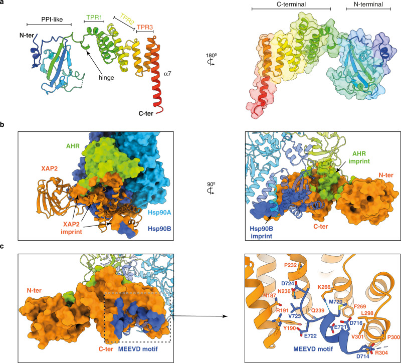
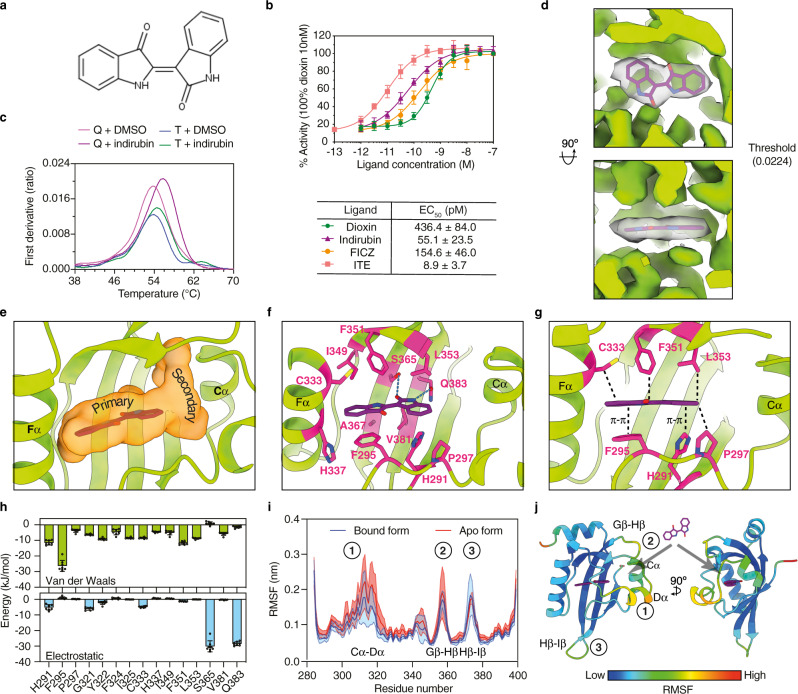
The aryl hydrocarbon receptor (AHR) is a ligand-dependent transcription factor that mediates a broad spectrum of (patho)physiological processes in response to numerous substances including pollutants, natural products and metabolites. However, the scarcity of structural data precludes understanding of how AHR is activated by such diverse compounds. Our 2.85 Å structure of the human indirubin-bound AHR complex with the chaperone Hsp90 and the co-chaperone XAP2, reported herein, reveals a closed conformation Hsp90 dimer with AHR threaded through its lumen and XAP2 serving as a brace. Importantly, we disclose the long-awaited structure of the AHR PAS-B domain revealing a unique organisation of the ligand-binding pocket and the structural determinants of ligand-binding specificity and promiscuity of the receptor. By providing structural details of the molecular initiating event leading to AHR activation, our study rationalises almost forty years of biochemical data and provides a framework for future mechanistic studies and structure-guided drug design.
Overview
The claims and statistics of this paper haven’t been checked yet.
Run the check — free, no account needed. It reads the open-access text and checks the sources it cites.
Integrity signals
Registry
Cites
Statistics
Figures
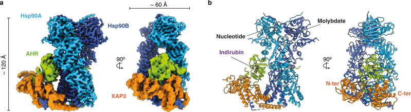
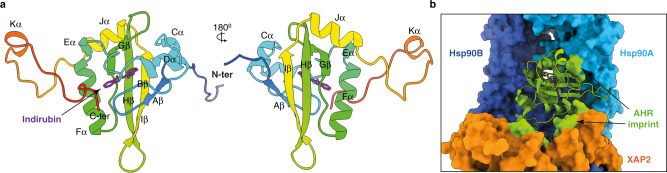
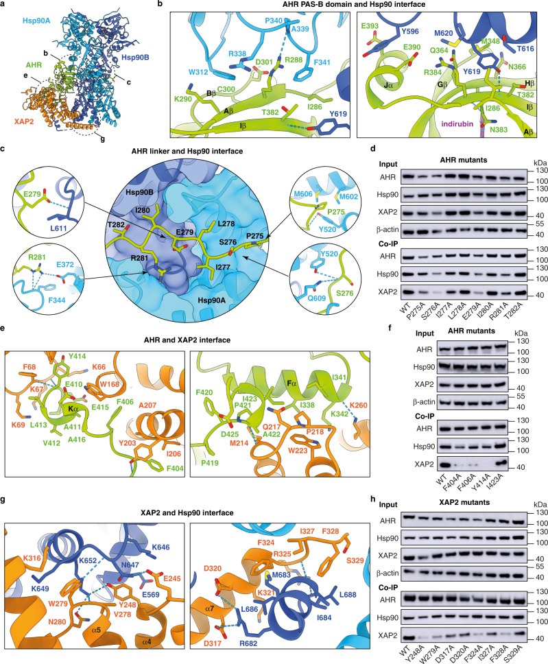
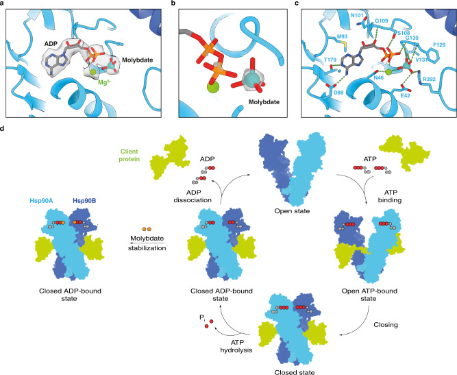
Verification
Claims the paper makes
The claims and statistics of this paper haven’t been checked yet.
Run the check — free, no account needed. It reads the open-access text and checks the sources it cites.
Claims other papers make about this one
Each is checked against this paper’s own text: does it say what the citing paper says it does?
The source backs part of the claim, not all of it — or backs it for a different population, condition or comparison.
In the citing paper
“Comparisons among these three states initially revealed that the DE-loop, situated between Dα and Eα of the AHR PAS-B domain, undergoes a conformational change towards the pocket upon ligand binding29,30 (Fig. 5a).”
From Structural basis for the ligand-dependent activation of heterodimeric AHR-ARNT complex (2025) · cites this paper as [29]
Experimental — accuracy not yet measuredThe cited source says what the claim attributes to it.
In the citing paper
“Notably, compared to two recently published structures of AHR cytosolic complexes bound to Indirubin and BaP29,40, these two ligands demonstrate similar binding patterns within the AHR-ARNT complexes (Supplementary Fig. 7g, h)”
From Structural basis for the ligand-dependent activation of heterodimeric AHR-ARNT complex (2025) · cites this paper as [29]
The cited source says what the claim attributes to it.
In the citing paper
“Previous studies have indicated that in the cytosolic AHR-HSP90-XAP2 complex29, this loop primarily interacts with XAP2 (Supplementary Fig. 9d).”
From Structural basis for the ligand-dependent activation of heterodimeric AHR-ARNT complex (2025) · cites this paper as [29]
Self-check
Percentages, sums and reported differences re-computed from the paper’s own numbers.
The self-check needs the paper’s full text.
Citations
Cites
UCSF Chimera—A visualization system for exploratory research and analysis
Pettersen, E. F., Goddard, T. D., Huang, C. C., Couch, G. S., Greenblatt, D. M., Meng, E. C., & Ferrin, T. E. (2004) · Journal of Computational Chemistry · doi:10.1002/jcc.20084 · Cited by 46,047
Features and development of Coot
Emsley, P., Lohkamp, B., Scott, W. G., & Cowtan, K. (2010) · Acta Crystallographica Section D Biological Crystallography · doi:10.1107/s0907444910007493 · Cited by 29,915
Abraham, M. J., Murtola, T., Schulz, R., Páll, S., Smith, J. C., Hess, B., & Lindahl, E. (2015) · SoftwareX · doi:10.1016/j.softx.2015.06.001 · Cited by 27,190
A smooth particle mesh Ewald method
Essmann, U., Perera, L., Berkowitz, M., Darden, T., Lee, H.-C., & Pedersen, L. (1995) · The Journal of Chemical Physics · doi:10.1063/1.470117 · Cited by 21,378
Polymorphic transitions in single crystals: A new molecular dynamics method
Parrinello, M., & Rahman, A. (1981) · Journal of Applied Physics · doi:10.1063/1.328693 · Cited by 20,009
Canonical sampling through velocity rescaling
Bussi, G., Donadio, D., & Parrinello, M. (2007) · The Journal of Chemical Physics · doi:10.1063/1.2408420 · Cited by 18,327
Development and testing of a general amber force field
Wang, J.-M., Wolf, R., Caldwell, J., Kollman, P., & Case, D. (2004) · Journal of Computational Chemistry · doi:10.1002/jcc.20035 · Cited by 18,318
The Amber biomolecular simulation programs
Case, D. A., Cheatham, T. E., Darden, T., Gohlke, H., Luo, R., Merz, K. M., Onufriev, A., Simmerling, C., Wang, B., & Woods, R. J. (2005) · Journal of Computational Chemistry · doi:10.1002/jcc.20290 · Cited by 9,535
Liebschner, D., Afonine, P. V., Baker, M. L., Bunkóczi, G., Chen, V. B., Croll, T. I., Hintze, B., Hung, L.-W., Jain, S., McCoy, A. J., et al. (2019) · Acta Crystallographica Section D Structural Biology · doi:10.1107/s2059798319011471 · Cited by 7,953
MotionCor2: anisotropic correction of beam-induced motion for improved cryo-electron microscopy
Zheng, S. Q., Palovcak, E. J., Armache, J., Verba, K. A., Cheng, Y., & Agard, D. A. (2017) · Nature Methods · doi:10.1038/nmeth.4193 · Cited by 7,496
Deciphering key features in protein structures with the new ENDscript server
Robert, X., & Gouet, P. (2014) · Nucleic Acids Research · doi:10.1093/nar/gku316 · Cited by 7,053
The I-TASSER Suite: protein structure and function prediction
Yang, J., Yan, R., Roy, A., Xu, D., Poisson, J., & Zhang, Y. (2015) · Nature Methods · doi:10.1038/nmeth.3213 · Cited by 5,596
RELION: Implementation of a Bayesian approach to cryo-EM structure determination
Scheres, S. H. W. (2012) · Journal of Structural Biology · doi:10.1016/j.jsb.2012.09.006 · Cited by 5,446
UCSF ChimeraX: Meeting modern challenges in visualization and analysis
Goddard, T. D., Huang, C. C., Meng, E. C., Pettersen, E. F., Couch, G. S., Morris, J. H., & Ferrin, T. E. (2018) · Protein Science · doi:10.1002/pro.3235 · Cited by 5,267
CTFFIND4: Fast and accurate defocus estimation from electron micrographs
Rohou, A. L., & Grigorieff, N. (2015) · Journal of Structural Biology · doi:10.1016/j.jsb.2015.08.008 · Cited by 4,759
MolProbity: More and better reference data for improved all‐atom structure validation
Williams, C. J., Headd, J. J., Moriarty, N. W., Prisant, M. G., Videau, L. L., Deis, L. N., Verma, V., Keedy, D. A., Hintze, B. J., Chen, V. B., et al. (2018) · Protein Science · doi:10.1002/pro.3330 · Cited by 4,599
The EMBL-EBI search and sequence analysis tools APIs in 2019
Madeira, F., Park, Y. M., Lee, J., Buso, N., Gur, T., Madhusoodanan, N., Basutkar, P., Tivey, A. R. N., Potter, S. C., Finn, R. D., et al. (2019) · Nucleic Acids Research · doi:10.1093/nar/gkz268 · Cited by 4,167
P-LINCS: A Parallel Linear Constraint Solver for Molecular Simulation
Hess, B. (2007) · Journal of Chemical Theory and Computation · doi:10.1021/ct700200b · Cited by 4,020
Tian, C., Kasavajhala, K., Belfon, K. A. A., Raguette, L. E., Huang, H., Migues, A. N., Bickel, J. D., Wang, Y., Pincay, J., Wu, Q., et al. (2019) · Journal of Chemical Theory and Computation · doi:10.1021/acs.jctc.9b00591 · Cited by 2,658
Rosenthal, P. B., & Henderson, R. (2003) · Journal of Molecular Biology · doi:10.1016/j.jmb.2003.07.013 · Cited by 2,416
CASTp 3.0: computed atlas of surface topography of proteins
Tian, W., Chen, C., Lei, X., Zhao, J., & Liang, J. (2018) · Nucleic Acids Research · doi:10.1093/nar/gky473 · Cited by 2,389
Denison, M. S., & Nagy, S. R. (2003) · Annual Review of Pharmacology and Toxicology · doi:10.1146/annurev.pharmtox.43.100901.135828 · Cited by 1,895
CASTp: Computed Atlas of Surface Topography of proteins
Binkowski, T. A. (2003) · Nucleic Acids Research · doi:10.1093/nar/gkg512 · Cited by 1,693
Building Water Models: A Different Approach
Izadi, S., Anandakrishnan, R., & Onufriev, A. V. (2014) · The Journal of Physical Chemistry Letters · doi:10.1021/jz501780a · Cited by 1,406
Rothhammer, V., & Quintana, F. J. (2019) · Nature Reviews Immunology · doi:10.1038/s41577-019-0125-8 · Cited by 1,197
The PAS Superfamily: Sensors of Environmental and Developmental Signals
Gu, Y.-Z., Hogenesch, J. B., & Bradfield, C. A. (2000) · The Annual Review of Pharmacology and Toxicology · doi:10.1146/annurev.pharmtox.40.1.519 · Cited by 1,044
Chen, S., McMullan, G., Faruqi, A. R., Murshudov, G. N., Short, J. M., Scheres, S. H. W., & Henderson, R. A. (2013) · Ultramicroscopy · doi:10.1016/j.ultramic.2013.06.004 · Cited by 1,005
Addressing Preferred Specimen Orientation in Single-Particle Cryo-EM through Tilting
Tan, Y.-Z., Baldwin, P. R., Davis, J., Williamson, J., Potter, C., Carragher, B., & Lyumkis, D. (2017) · Nature Methods · doi:10.1038/nmeth.4347 · Cited by 920
Aryl hydrocarbon receptor ligands in cancer: friend and foe
Murray, I. A., Patterson, A. D., & Perdew, G. H. (2014) · Nature reviews. Cancer · doi:10.1038/nrc3846 · Cited by 916
Denison, M. S., Soshilov, A. A., He, G., DeGroot, D. E., & Zhao, B. (2011) · Toxicological Sciences · doi:10.1093/toxsci/kfr218 · Cited by 748
The Aryl Hydrocarbon Receptor Complex and the Control of Gene Expression
Beischlag, T. V., Morales, J. L., Hollingshead, B. D., & Perdew, G. H. (2008) · Critical Reviews™ in Eukaryotic Gene Expression · doi:10.1615/critreveukargeneexpr.v18.i3.20 · Cited by 727
Indole and Tryptophan Metabolism: Endogenous and Dietary Routes to Ah Receptor Activation
Hubbard, T. D., Murray, I. A., & Perdew, G. H. (2015) · Drug Metabolism and Disposition · doi:10.1124/dmd.115.064246 · Cited by 670
UCSF Chimera, MODELLER, and IMP: An integrated modeling system
Yang, Z. R., Lasker, K., Schneidman‐Duhovny, D., Webb, B., Huang, C. C., Pettersen, E. F., Goddard, T. D., Meng, E. C., Šali, A., & Ferrin, T. E. (2011) · Journal of Structural Biology · doi:10.1016/j.jsb.2011.09.006 · Cited by 637
The Aryl Hydrocarbon Receptor in Barrier Organ Physiology, Immunology, and Toxicology
Esser, C., & Rannug, A. (2015) · Pharmacological Reviews · doi:10.1124/pr.114.009001 · Cited by 501
Atomic structure of Hsp90-Cdc37-Cdk4 reveals that Hsp90 traps and stabilizes an unfolded kinase
Verba, K. A., Wang, R. Y.-R., Arakawa, A., Liu, Y., Shirouzu, M., Yokoyama, S., & Agard, D. A. (2016) · Science · doi:10.1126/science.aaf5023 · Cited by 424
Adaptation of the human aryl hydrocarbon receptor to sense microbiota-derived indoles
Hubbard, T. D., Murray, I. A., Bisson, W. H., Lahoti, T. S., Gowda, K., Amin, S. G., Patterson, A. D., & Perdew, G. H. (2015) · Scientific Reports · doi:10.1038/srep12689 · Cited by 377
Schroeder, J. C., DiNatale, B. C., Murray, I. A., Flaveny, C. A., Liu, Q., Laurenzana, E. M., Lin, J.-M., Strom, S. C., Omiecinski, C. J., Amin, S., et al. (2009) · Biochemistry · doi:10.1021/bi901786x · Cited by 340
Nebert, D. W. (2017) · Progress in Lipid Research · doi:10.1016/j.plipres.2017.06.001 · Cited by 286
Okey, A. B. (2007) · Toxicological Sciences · doi:10.1093/toxsci/kfm096 · Cited by 264
The aryl hydrocarbon receptor as a mediator of host-microbiota interplay
Dong, F., & Perdew, G. H. (2020) · Gut Microbes · doi:10.1080/19490976.2020.1859812 · Cited by 202
Automatic generation of protein structure cartoons with Pro-origami
Stivala, A. D., Wybrow, M., Wirth, A. I., Whisstock, J. C., & Stuckey, P. J. (2011) · Bioinformatics · doi:10.1093/bioinformatics/btr575 · Cited by 172
Structure of Hsp90–p23–GR reveals the Hsp90 client-remodelling mechanism
Noddings, C. M., Wang, R. Y.-R., Johnson, J. L., & Agard, D. A. (2022) · Nature · doi:10.1038/s41586-021-04236-1 · Cited by 164
Structural Basis for Aryl Hydrocarbon Receptor-Mediated Gene Activation
Schulte, K. W., Green, E., Wilz, A., Platten, M., & Daumke, O. (2017) · Structure · doi:10.1016/j.str.2017.05.008 · Cited by 157
Seok, S., Lee, W., Jiang, L., Molugu, K., Zheng, A., Li, Y., Park, S., Bradfield, C. A., & Xing, Y. (2017) · Proceedings of the National Academy of Sciences · doi:10.1073/pnas.1617035114 · Cited by 135
Ligand Selectivity and Gene Regulation by the Human Aryl Hydrocarbon Receptor in Transgenic Mice
Flaveny, C. A., Murray, I. A., Chiaro, C. R., & Perdew, G. H. (2009) · Molecular Pharmacology · doi:10.1124/mol.109.054825 · Cited by 124
Structural characterization of mammalian bHLH-PAS transcription factors
Wu, D., & Rastinejad, F. (2017) · Current Opinion in Structural Biology · doi:10.1016/j.sbi.2016.09.011 · Cited by 122
Heid, S. E., Pollenz, R. S., & Swanson, H. I. (2000) · Molecular Pharmacology · doi:10.1016/s0026-895x(24)26445-2 · Cited by 120
Aryl Hydrocarbon Receptor and Its Diverse Ligands and Functions: An Exposome Receptor
Larigot, L., Benoit, L., Koual, M., Tomkiewicz, C., Barouki, R., & Coumoul, X. (2021) · The Annual Review of Pharmacology and Toxicology · doi:10.1146/annurev-pharmtox-052220-115707 · Cited by 117
Ramadoss, P., & Perdew, G. H. (2004) · Molecular Pharmacology · doi:10.1124/mol.66.1.129 · Cited by 114
The aryl hydrocarbon receptor (AhR) as a drug target for cancer chemotherapy
Safe, S. H., Cheng, Y., & Jin, U.-H. (2017) · Current Opinion in Toxicology · doi:10.1016/j.cotox.2017.01.012 · Cited by 99
Morgan, R. M. L., Hernández-Ramírez, L. C., Trivellin, G., Zhou, L., Roe, S. M., Korbonits, M., & Prodromou, C. (2012) · PLoS ONE · doi:10.1371/journal.pone.0053339 · Cited by 73
Esser, C., Lawrence, B. P., Sherr, D. H., Perdew, G. H., Puga, A., Barouki, R., & Coumoul, X. (2018) · International Journal of Molecular Sciences · doi:10.3390/ijms19113603 · Cited by 54
Soshilov, A., & Denison, M. S. (2011) · Journal of Biological Chemistry · doi:10.1074/jbc.m111.246439 · Cited by 49
Linnert, M., Lin, Y., Manns, A., Haupt, K., Paschke, A.-K., Fischer, G. S., Weiwad, M., & Lücke, C. (2013) · Biochemistry · doi:10.1021/bi301649m · Cited by 42
Faber, S. C., Soshilov, A. A., Giani Tagliabue, S., Bonati, L., & Denison, M. S. (2018) · International Journal of Molecular Sciences · doi:10.3390/ijms19092692 · Cited by 36
Soshilov, A. A., Motta, S., Bonati, L., & Denison, M. S. (2020) · International Journal of Molecular Sciences · doi:10.3390/ijms21072474 · Cited by 36
The aryl hydrocarbon receptor as a model PAS sensor
Vazquez-Rivera, E., Rojas, B. L., Parrott, J. C., Shen, A. L., Xing, Y., Carney, P. R., & Bradfield, C. A. (2021) · Toxicology Reports · doi:10.1016/j.toxrep.2021.11.017 · Cited by 22
Pham, T. H., Lecomte, S., Guével, R. L., Lardenois, A., Evrard, B., Chalmel, F., Ferrière, F., Balaguer, P., Efstathiou, T., & Pakdel, F. (2020) · International Journal of Molecular Sciences · doi:10.3390/ijms21041368 · Cited by 11
Self-citation
Cited by
Sorted by citations
The complex biology of aryl hydrocarbon receptor activation in cancer and beyond
Opitz, C. A., Holfelder, P., Prentzell, M. T., & Trump, S. (2023) · Biochemical Pharmacology · doi:10.1016/j.bcp.2023.115798 · Cited by 109
The aryl hydrocarbon receptor: a rehabilitated target for therapeutic immune modulation
Polonio, C. M., McHale, K. A., Sherr, D. H., Rubenstein, D., & Quintana, F. J. (2025) · Nature Reviews Drug Discovery · doi:10.1038/s41573-025-01172-x · Cited by 104
The aryl hydrocarbon receptor: structure, signaling, physiology and pathology
Coumoul, X., Barouki, R., Esser, C., Haarmann‐Stemmann, T., Lawrence, B. P., Lehmann, J., Moura‐Alves, P., Murray, I. A., Opitz, C. A., Perdew, G. H., et al. (2026) · Signal Transduction and Targeted Therapy · doi:10.1038/s41392-025-02500-8 · Cited by 111
“54 In this structure, AHR is bound to indirubin, a dietary-derived endogenous ligand produced from tryptophan by the intestinal microbiota.”
InfluentialSelf-citationTapinarof validates the aryl hydrocarbon receptor as a therapeutic target: A clinical review
Silverberg, J. I., Boguniewicz, M., Quintana, F. J., Clark, R. A., Gross, L., Hirano, I., Tallman, A. M., Brown, P. M., Fredericks, D., Rubenstein, D. S., et al. (2023) · Journal of Allergy and Clinical Immunology · doi:10.1016/j.jaci.2023.12.013 · Cited by 94
Cryo-EM reveals how Hsp90 and FKBP immunophilins co-regulate the glucocorticoid receptor
Noddings, C. M., Johnson, J. L., & Agard, D. A. (2023) · Nature Structural & Molecular Biology · doi:10.1038/s41594-023-01128-y · Cited by 75
“Interest-ingly, FKBP52 W259 is also conserved in the FKBP-like co-chaperone XAP2 and a recent structure reveals XAP2 engages with an Hsp90–client using the analogous XAP2 W168 , suggesting this residue is critical more broadly for FKBP co-chaperone:client engagement 42 .”
Background
Metadata
- Authors
- Gruszczyk, J., Grandvuillemin, L., Lai-Kee-Him, J., Paloni, M., Savva, C. G., Germain, P., Grimaldi, M., Boulahtouf, A., Kwong, H.-S., Bous, J., Ancelin, A., Bechara, C., Barducci, A., Balaguer, P., & Bourguet, W.
- Affiliations
- Centre National de la Recherche Scientifique (FR)
- University of Leicester (GB)
- Inserm (FR)
- Karolinska Institutet (SE)
- Corresponding author
- Gruszczyk, Jakub
- Bourguet, William
- ORCID
- Venue
- Nature Communications
- Published
- 2022-11-16
- Volume
- 13
- Issue
- 1
- Pages
- 7010-7010
- ISSN
- 2041-1723 (ISSN-L)
- PMC
- PMC9668932
- OpenAlex
- W4309473730
- Cited by
- 137
- References
- 58
- Citations by year
- 2026: 38 · 2025: 28 · 2024: 39 · 2023: 31 · 2022: 1
- FWCI
- 8.08
- Type
- journal article
- Language
- en
- Open access
- gold
- doi.org
- Locations
- Nature Communications (publishedVersion, cc-by)
- bioRxiv (Cold Spring Harbor Laboratory) (acceptedVersion, cc-by-nc-nd)
- PubMed Central (publishedVersion, cc-by)
- PubMed (publishedVersion)
- HAL (submittedVersion, cc-by)
- HAL (submittedVersion, cc-by-sa)
- HAL (submittedVersion)
- RePEc: Research Papers in Economics (submittedVersion)
- DOAJ (DOAJ: Directory of Open Access Journals) (submittedVersion, cc-by-sa)
- Funders
- European Commission (825489, HORIZON2020)
- Agence Nationale de la Recherche (ANR-16-IDEX-0006, ANR‐16‐IDEX‐, ANR-10-INBS-04-01, 10-INBS-04, 10-INBS-04-01, ANR‐10_INBS‐04, ANR-16, INBS-04-01, ANR-10, ANR-10-LABX-0012, ANR-10-INBS-0004)
- Université de Montpellier (ANR_16-IDEX-0 0 06)
- French Infrastructure for Integrated Structural Biology (ANR-10-INBS, ANR-10-INBS-04)
- Région Occitanie Pyrénées-Méditerranée
- Laboratoire d'Excellence EpiGenMed
- Medical Research Council (MC_PC_17136, ANR-10-INBS-04)
- Topics
- Life Sciences › Biochemistry, Genetics and Molecular Biology › Molecular Biology › Heat shock proteins research
- Life Sciences › Biochemistry, Genetics and Molecular Biology › Molecular Biology › Receptor Mechanisms and Signaling
- Life Sciences › Biochemistry, Genetics and Molecular Biology › Molecular Biology › Protein Structure and Dynamics
- Keywords
- aryl hydrocarbon receptor, HSP90, indirubin, ligand binding, PAS-B domain, ligand specificity, receptor activation, transcription factors, structure-based drug design
- MeSH terms
- Cytosol, Humans, Ligands, Receptors, Aryl Hydrocarbon, HSP90 Heat-Shock Proteins, Cryoelectron Microscopy, Intracellular Signaling Peptides and Proteins, Basic Helix-Loop-Helix Proteins
About these checks
Registry, retraction, Cites and statistics results are computed, not judged, with one exception: a printed reference that no index lists is read once by a model before it counts as a citation error. Claims are extracted by a model and kept only when their quote appears verbatim in the text; each cited source is then checked, with a model’s help, for whether it says what the claim relies on. Those verdicts can be wrong — read the claim’s quote and the cited source before relying on one.
Statistics and arithmetic are recomputed, not judged.
Confidence bands are shown only for fields calibrated on our annotated test set. None is calibrated yet, so no band is shown.
What counts as a claim ›How we measure confidence ›How we check this ›
Are you an author, or do you see a problem with this page? Request a review
