

The DOI matches its Crossref record; 76 of the 76 references it cites were found in a registry or an index.
The structure of a hibernating ribosome in a Lyme disease pathogen
Open accessPublished version of a preprint
Abstract
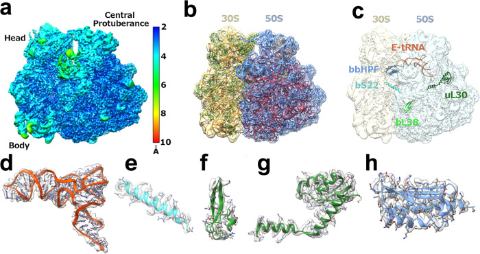
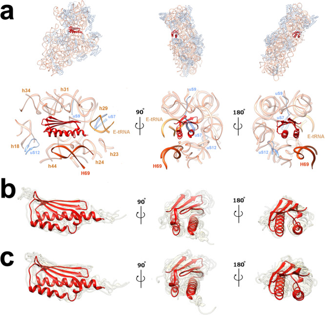
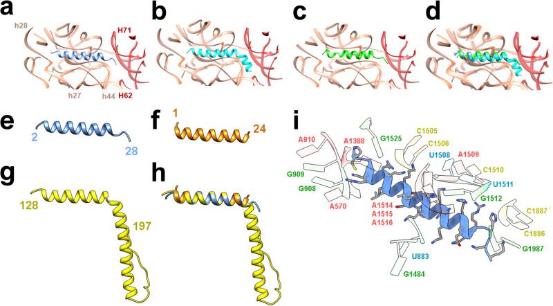
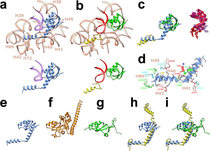
The spirochete bacterial pathogen Borrelia ( Borreliella) burgdorferi ( Bbu ) affects more than 10% of the world population and causes Lyme disease in about half a million people in the US annually. Therapy for Lyme disease includes antibiotics that target the Bbu ribosome. Here we present the structure of the Bbu 70S ribosome obtained by single particle cryo-electron microscopy at 2.9 Å resolution, revealing a bound hibernation promotion factor protein and two genetically non-annotated ribosomal proteins bS22 and bL38. The ribosomal protein uL30 in Bbu has an N-terminal α-helical extension, partly resembling the mycobacterial bL37 protein, suggesting evolution of bL37 and a shorter uL30 from a longer uL30 protein. Its analogy to proteins uL30m and mL63 in mammalian mitochondrial ribosomes also suggests a plausible evolutionary pathway for expansion of protein content in mammalian mitochondrial ribosomes. Computational binding free energy predictions for antibiotics reflect subtle distinctions in antibiotic-binding sites in the Bbu ribosome. Discovery of these features in the Bbu ribosome may enable better ribosome-targeted antibiotic design for Lyme disease treatment.
Overview
The claims and statistics of this paper haven’t been checked yet.
Run the check — free, no account needed. It reads the open-access text and checks the sources it cites.
Integrity signals
Registry
Cites
Statistics
Figures
Verification
Claims the paper makes
The claims and statistics of this paper haven’t been checked yet.
Run the check — free, no account needed. It reads the open-access text and checks the sources it cites.
Self-check
Percentages, sums and reported differences re-computed from the paper’s own numbers.
The self-check needs the paper’s full text.
Citations
Cites
UCSF Chimera—A visualization system for exploratory research and analysis
Pettersen, E. F., Goddard, T. D., Huang, C. C., Couch, G. S., Greenblatt, D. M., Meng, E. C., & Ferrin, T. E. (2004) · Journal of Computational Chemistry · doi:10.1002/jcc.20084 · Cited by 45,828
Highly accurate protein structure prediction with AlphaFold
Jumper, J., Evans, R., Pritzel, A., Green, T., Figurnov, M., Ronneberger, O., Tunyasuvunakool, K., Bates, R., Žídek, A., Potapenko, A., et al. (2021) · Nature · doi:10.1038/s41586-021-03819-2 · Cited by 43,764
Trott, O., & Olson, A. J. (2010) · Journal of Computational Chemistry · doi:10.1002/jcc.21334 · Cited by 38,412
PHENIX : a comprehensive Python-based system for macromolecular structure solution
Adams, P. D., Afonine, P. V., Bunkóczi, G., Chen, V. B., Davis, I. W., Echols, N., Headd, J. J., Hung, L.-W., Kapral, G. J., Grosse-Kunstleve, R. W., et al. (2010) · Acta Crystallographica Section D Biological Crystallography · doi:10.1107/s0907444909052925 · Cited by 23,716
cryoSPARC: algorithms for rapid unsupervised cryo-EM structure determination
Punjani, A., Rubinstein, J. L., Fleet, D. J., & Brubaker, M. A. (2017) · Nature Methods · doi:10.1038/nmeth.4169 · Cited by 11,626
UCSF ChimeraX: Structure visualization for researchers, educators, and developers
Pettersen, E. F., Goddard, T. D., Huang, C. C., Meng, E. C., Couch, G. S., Croll, T. I., Morris, J. H., & Ferrin, T. E. (2021) · Protein Science · doi:10.1002/pro.3943 · Cited by 9,514
KEGG: new perspectives on genomes, pathways, diseases and drugs
Kanehisa, M., Furumichi, M., Tanabe, M., Sato, Y., & Morishima, K. (2017) · Nucleic Acids Research · doi:10.1093/nar/gkw1092 · Cited by 8,865
UCSF ChimeraX: Meeting modern challenges in visualization and analysis
Goddard, T. D., Huang, C. C., Meng, E. C., Pettersen, E. F., Couch, G. S., Morris, J. H., & Ferrin, T. E. (2018) · Protein Science · doi:10.1002/pro.3235 · Cited by 5,267
Automated molecular microscopy: The new Leginon system
Suloway, C., Pulokas, J., Fellmann, D., Cheng, A., Guerra, F., Quispe, J., Stagg, S., Potter, C. S., & Carragher, B. (2005) · Journal of Structural Biology · doi:10.1016/j.jsb.2005.03.010 · Cited by 1,764
Non-uniform refinement: adaptive regularization improves single-particle cryo-EM reconstruction
Punjani, A., Zhang, H., & Fleet, D. J. (2020) · Nature Methods · doi:10.1038/s41592-020-00990-8 · Cited by 1,683
A ratchet-like inter-subunit reorganization of the ribosome during translocation
Frank, J., & Agrawal, R. K. (2000) · Nature · doi:10.1038/35018597 · Cited by 799
Self-citationEstimating the Frequency of Lyme Disease Diagnoses, United States, 2010–2018
Kugeler, K. J., Schwartz, A. M., Delorey, M. J., Mead, P. S., & Hinckley, A. F. (2021) · Emerging Infectious Diseases · doi:10.3201/eid2702.202731 · Cited by 583
The structure of the human mitochondrial ribosome
Amunts, A., Brown, A., Toots, J., Scheres, S. H. W., & Ramakrishnan, V. (2015) · Science · doi:10.1126/science.aaa1193 · Cited by 533
Crowell, H. L., Soneson, C., Germain, P.-L., Calini, D., Collin, L., Raposo, C., Malhotra, D., & Robinson, M. D. (2020) · Nature Communications · doi:10.1038/s41467-020-19894-4 · Cited by 515
The complete structure of the 55 S mammalian mitochondrial ribosome
Greber, B. J., Bieri, P., Leibundgut, M., Leitner, A., Aebersold, R., Boehringer, D., & Ban, N. (2015) · Science · doi:10.1126/science.aaa3872 · Cited by 395
Fast, accurate, and reliable molecular docking with QuickVina 2
Alhossary, A., Handoko, S. D., Mu, Y., & Kwoh, C.-K. (2015) · Bioinformatics · doi:10.1093/bioinformatics/btv082 · Cited by 375
Visualization of chemical modifications in the human 80S ribosome structure
Natchiar, S. K., Myasnikov, A. G., Kratzat, H., Hazemann, I., & Klaholz, B. P. (2017) · Nature · doi:10.1038/nature24482 · Cited by 362
Sharma, M. R., Koc, E. C., Datta, P. P., Booth, T. M., Spremulli, L. L., & Agrawal, R. K. (2003) · Cell · doi:10.1016/s0092-8674(03)00762-1 · Cited by 341
Self-citationElias, A. F., Stewart, P. E., Grimm, D., Caimano, M. J., Eggers, C. H., Tilly, K., Bono, J. L., Akins, D. R., Radolf, J. D., Schwan, T. G., et al. (2002) · Infection and Immunity · doi:10.1128/iai.70.4.2139-2150.2002 · Cited by 320
Rodnina, M. V. (2018) · Cold Spring Harbor Perspectives in Biology · doi:10.1101/cshperspect.a032664 · Cited by 278
ModeRNA: a tool for comparative modeling of RNA 3D structure
Rother, M., Rother, K., Puton, T., & Bujnicki, J. M. (2011) · Nucleic Acids Research · doi:10.1093/nar/gkq1320 · Cited by 273
Structural insights into the role of rRNA modifications in protein synthesis and ribosome assembly
Polikanov, Y. S., Melnikov, S. V., Söll, D., & Steitz, T. A. (2015) · Nature Structural & Molecular Biology · doi:10.1038/nsmb.2992 · Cited by 272
Roadblocks and resolutions in eukaryotic translation
Schuller, A. P., & Green, R. (2018) · Nature Reviews Molecular Cell Biology · doi:10.1038/s41580-018-0011-4 · Cited by 257
How Hibernation Factors RMF, HPF, and YfiA Turn Off Protein Synthesis
Polikanov, Y. S., Blaha, G. M., & Steitz, T. A. (2012) · Science · doi:10.1126/science.1218538 · Cited by 245
High-resolution cryo-EM structure of the human 80S ribosome
Natchiar, S. K., Myasnikov, A. G., Kratzat, H., Hazemann, I., & Klaholz, B. P. (2017) · Worldwide Protein Data Bank · doi:10.2210/pdb6ek0/pdb · Cited by 222
Modular Assembly of the Bacterial Large Ribosomal Subunit
Davis, J. H., Tan, Y. Z., Carragher, B. O., Potter, C. S., Lyumkis, D., & Williamson, J. R. (2016) · Cell · doi:10.1016/j.cell.2016.11.020 · Cited by 214
Marks, J., Kannan, K., Roncase, E. J., Klepacki, D., Kefi, A., Orelle, C., Vázquez-Laslop, N., & Mankin, A. S. (2016) · Proceedings of the National Academy of Sciences · doi:10.1073/pnas.1613055113 · Cited by 159
R2DT is a framework for predicting and visualising RNA secondary structure using templates
Sweeney, B., Hoksza, D., Nawrocki, E. P., Ribas, C. E., Madeira, F., Cannone, J. J., Gutell, R. R., Maddala, A., Meade, C. D., Williams, L. D., et al. (2021) · Nature Communications · doi:10.1038/s41467-021-23555-5 · Cited by 152
Mardirossian, M., Pérébaskine, N., Benincasa, M., Gambato, S., Hofmann, S., Huter, P., Müller, C., Hilpert, K., Innis, C. A., Tossi, A., et al. (2018) · Cell chemical biology · doi:10.1016/j.chembiol.2018.02.004 · Cited by 148
Structure of Erm-modified 70S ribosome reveals the mechanism of macrolide resistance
Svetlov, M. S., Syroegin, E. A., Aleksandrova, E. V., Atkinson, G. C., Gregory, S. T., Mankin, A. S., & Polikanov, Y. S. (2021) · Nature Chemical Biology · doi:10.1038/s41589-020-00715-0 · Cited by 148
Margos, G., Gofton, A. W., Wibberg, D., Dangel, A., Marosevic, D. V., Loh, S.-M., Oskam, C. L., & Fingerle, V. (2018) · PLoS ONE · doi:10.1371/journal.pone.0208432 · Cited by 143
Iyer, R., Caimano, M. J., Luthra, A., Axline, D., Corona, A., Iacobaş, D. A., Radolf, J. D., & Schwartz, I. S. (2014) · Molecular Microbiology · doi:10.1111/mmi.12882 · Cited by 140
Lin, T., Gao, L., Zhang, C., Odeh, E., Jacobs, M. B., Coutte, L., Chaconas, G., Philipp, M. T., & Norris, S. J. (2012) · PLoS ONE · doi:10.1371/journal.pone.0047532 · Cited by 140
Clinical spectrum of Lyme disease
la Garza, J. A. C., la Cruz-Valadez, E. D., Ocampo‐Candiani, J., et al. (2018) · European Journal of Clinical Microbiology & Infectious Diseases · doi:10.1007/s10096-018-3417-1 · Cited by 135
Seefeldt, A. C., Graf, M., Pérébaskine, N., Nguyen, F., Arenz, S., Mardirossian, M., Scocchi, M., Wilson, D. N., & Innis, C. A. (2016) · Nucleic Acids Research · doi:10.1093/nar/gkv1545 · Cited by 131
RNApdbee 2.0: multifunctional tool for RNA structure annotation
Żok, T., Antczak, M., Żurkowski, M., Popenda, M., Błażewicz, J., Adamiak, R. W., & Szachniuk, M. (2018) · Nucleic Acids Research · doi:10.1093/nar/gky314 · Cited by 130
Beckert, B., Turk, M., Czech, A., Berninghausen, O., Beckmann, R., Ignatova, Z., Plitzko, J. M., & Wilson, D. N. (2018) · Nature Microbiology · doi:10.1038/s41564-018-0237-0 · Cited by 128
The Ribosome Emerges from a Black Box
Ramakrishnan, V. (2014) · Cell · doi:10.1016/j.cell.2014.10.052 · Cited by 124
Mac, S., da Silva, S. R., & Sander, B. (2019) · PLoS ONE · doi:10.1371/journal.pone.0210280 · Cited by 111
Kowalski, T. J., Tata, S., Berth, W., Mathiason, M. A., & Agger, W. A. (2010) · Clinical Infectious Diseases · doi:10.1086/649920 · Cited by 106
The Complete Structure of the Mycobacterium smegmatis 70S Ribosome
Hentschel, J., Burnside, C., Mignot, I., Leibundgut, M., Boehringer, D., & Ban, N. (2017) · Cell Reports · doi:10.1016/j.celrep.2017.06.029 · Cited by 96
A Structural Model for the Large Subunit of the Mammalian Mitochondrial Ribosome
Mears, J. A., Sharma, M. R., Gutell, R. R., McCook, A. S., Richardson, P. E., Caulfield, T. R., Agrawal, R. K., & Harvey, S. C. (2006) · Journal of Molecular Biology · doi:10.1016/j.jmb.2006.01.094 · Cited by 94
Self-citationZinc depletion induces ribosome hibernation in mycobacteria
Li, Y., Sharma, M. R., Koripella, R. K., Yang, Y., Kaushal, P. S., Lin, Q., Wade, J. T., Gray, T. A., Derbyshire, K. M., Agrawal, R. K., et al. (2018) · Proceedings of the National Academy of Sciences · doi:10.1073/pnas.1804555115 · Cited by 94
Self-citationPolikanov, Y. S., Starosta, A. L., Juette, M. F., Altman, R. B., Terry, D. S., Lu, W., Burnett, B. J., Dinos, G. P., Reynolds, K. A., Blanchard, S. C., et al. (2015) · Molecular Cell · doi:10.1016/j.molcel.2015.04.014 · Cited by 93
Syroegin, E. A., Flemmich, L., Klepacki, D., Vázquez‐Laslop, N., Micura, R., & Polikanov, Y. S. (2022) · Nature Structural & Molecular Biology · doi:10.1038/s41594-022-00720-y · Cited by 93
Structure of the Bacillus subtilis hibernating 100S ribosome reveals the basis for 70S dimerization
Beckert, B., Abdelshahid, M., Schäfer, H., Steinchen, W., Arenz, S., Berninghausen, O., Beckmann, R., Bange, G., Turgay, K., & Wilson, D. N. (2017) · The EMBO Journal · doi:10.15252/embj.201696189 · Cited by 92
The cryo-EM structure of hibernating 100S ribosome dimer from pathogenic Staphylococcus aureus
Matzov, D., Aibara, S., Basu, A., Zimmerman, E., Bashan, A., Yap, M.-N. F., Amunts, A., & Yonath, A. (2017) · Nature Communications · doi:10.1038/s41467-017-00753-8 · Cited by 90
Datta, P. P., Sharma, M. R., Qi, L., Frank, J., & Agrawal, R. K. (2005) · Molecular Cell · doi:10.1016/j.molcel.2005.10.028 · Cited by 89
Self-citationKhusainov, I. S., Vicens, Q., Ayupov, R., Сергеевич, У. К., Myasnikov, A. G., Simonetti, A., Validov, S. Z., Kieffer, B., Yusupova, G. Z., Yusupov, M., et al. (2017) · The EMBO Journal · doi:10.15252/embj.201696105 · Cited by 89
Dong, Y., Zhou, G., Cao, W., Xu, X., Zhang, Y., Ji, Z., Yang, J., Chen, J., Liu, M., Fan, Y., et al. (2022) · BMJ Global Health · doi:10.1136/bmjgh-2021-007744 · Cited by 89
Context-Specific Action of Ribosomal Antibiotics
Vázquez‐Laslop, N., & Mankin, A. S. (2018) · Annual Review of Microbiology · doi:10.1146/annurev-micro-090817-062329 · Cited by 88
THE SYNTHESIS OF PROTEINS UPON RIBOSOMES.
Jd, W. (1964) · PubMed · Cited by 82
RNApdbee—a webserver to derive secondary structures from pdb files of knotted and unknotted RNAs
Antczak, M., Żok, T., Popenda, M., Łukasiak, P., Adamiak, R. W., Błażewicz, J., & Szachniuk, M. (2014) · Nucleic Acids Research · doi:10.1093/nar/gku330 · Cited by 78
Tetracenomycin X inhibits translation by binding within the ribosomal exit tunnel
Osterman, I. A., Wieland, M., Maviza, T. P., Lashkevich, K. A., Lukianov, D. A., Komarova, E. S., Zakalyukina, Y. V., Buschauer, R., Shiriaev, D. I., Leyn, S. A., et al. (2020) · Nature Chemical Biology · doi:10.1038/s41589-020-0578-x · Cited by 75
Advanced Data Acquisition From Electron Microscopes With SerialEM
Mastronarde, D. N. (2018) · Microscopy and Microanalysis · doi:10.1017/s1431927618004816 · Cited by 69
A selective antibiotic for Lyme disease
Leimer, N., Wu, X., Imai, Y., Morrissette, M., Pitt, N., Favre-Godal, Q., Iinishi, A., Jain, S., Caboni, M., Leus, I. V., et al. (2021) · Cell · doi:10.1016/j.cell.2021.09.011 · Cited by 66
Binding and Action of Amino Acid Analogs of Chloramphenicol upon the Bacterial Ribosome
Tereshchenkov, A. G., Dobosz-Bartoszek, M., Osterman, I. А., Marks, J., Sergeeva, V. A., Kasatsky, P. S., Komarova, E. S., Ставрианиди, А. Н., Родин, И. А., Konevega, A. L., et al. (2018) · Journal of Molecular Biology · doi:10.1016/j.jmb.2018.01.016 · Cited by 60
Ni, X., Davis, J. H., Jain, N., Razi, A., Benlekbir, S., McArthur, A. G., Rubinstein, J. L., Britton, R. A., Williamson, J. R., & Ortega, J. (2016) · Nucleic Acids Research · doi:10.1093/nar/gkw678 · Cited by 59
Cocozaki, A. I., Altman, R. B., Huang, J., Buurman, E. T., Kazmirski, S. L., Doig, P., Prince, D. B., Blanchard, S. C., Cate, J. H. D., & Ferguson, A. D. (2016) · Proceedings of the National Academy of Sciences · doi:10.1073/pnas.1605127113 · Cited by 56
Bhattacharjee, S., Feng, X., Maji, S., Dadhwal, P., Zhang, Z., Brown, Z. P., & Frank, J. (2024) · Cell · doi:10.1016/j.cell.2023.12.027 · Cited by 56
Exit tunnel modulation as resistance mechanism of S. aureus erythromycin resistant mutant
Halfon, Y., Matzov, D., Eyal, Z., Bashan, A., Zimmerman, E., Kjeldgaard, J. S., Ingmer, H., & Yonath, A. (2019) · Scientific Reports · doi:10.1038/s41598-019-48019-1 · Cited by 55
Structures of Mycobacterium smegmatis 70S ribosomes in complex with HPF, tmRNA, and P-tRNA
Mishra, S., Ahmed, T. U., Tyagi, A., Shi, J. R., & Bhushan, S. (2018) · Scientific Reports · doi:10.1038/s41598-018-31850-3 · Cited by 53
Khusainov, I. S., Fatkhullin, B. F., Pellegrino, S., Bikmullin, A. G., Liu, W.-T., Gabdulkhakov, A. G., Al‐Shebel, A., Golubev, A. A., Zeyer, D., Trachtmann, N., et al. (2020) · Nature Communications · doi:10.1038/s41467-020-15517-0 · Cited by 50
Koripella, R. K., Sharma, M. R., Bhargava, K., Datta, P. P., Kaushal, P. S., Keshavan, P., Spremulli, L. L., Banavali, N. K., & Agrawal, R. K. (2020) · Nature Communications · doi:10.1038/s41467-020-17715-2 · Cited by 47
Self-citationFlygaard, R. K., Boegholm, N., Yusupov, M., & Jenner, L. B. (2018) · Nature Communications · doi:10.1038/s41467-018-06724-x · Cited by 45
Kišonaitė, M., Wild, K., Lapouge, K., Ruppert, T., & Sinning, I. (2022) · Nature Communications · doi:10.1038/s41467-022-27967-9 · Cited by 38
Structural basis of sequestration of the anti-Shine-Dalgarno sequence in the Bacteroidetes ribosome
Jha, V., Roy, B., Jahagirdar, D., McNutt, Z., Shatoff, E., Boleratz, B. L., Watkins, D., Bundschuh, R. A., Basu, K., Ortega, J., et al. (2020) · Nucleic Acids Research · doi:10.1093/nar/gkaa1195 · Cited by 37
Chatellier, J., Rauffer‐Bruyère, N., Regenmortel, M. H. V. V., Altschuh, D., & Weiss, É. (1996) · Journal of Molecular Recognition · doi:10.1002/(sici)1099-1352(199601)9:1<1::aid-jmr241>3.0.co;2-6 · Cited by 35
Binding and Action of Triphenylphosphonium Analog of Chloramphenicol upon the Bacterial Ribosome
Chen, C., Pavlova, J. A., Lukianov, D. A., Tereshchenkov, A. G., Makarov, G. I., Khairullina, Z. Z., Tashlitsky, V. N., Paleskava, A., Konevega, A. L., Bogdanov, A. A., et al. (2021) · Antibiotics · doi:10.3390/antibiotics10040390 · Cited by 27
Distinct mechanisms of the human mitoribosome recycling and antibiotic resistance
Koripella, R. K., Deep, A., Agrawal, E. K., Keshavan, P., Banavali, N. K., & Agrawal, R. K. (2021) · Nature Communications · doi:10.1038/s41467-021-23726-4 · Cited by 25
Self-citationLi, Z., Ge, X., Zhang, Y., Zheng, L., Sanyal, S., & Gao, N. (2017) · Protein & Cell · doi:10.1007/s13238-017-0456-9 · Cited by 22
Predicting spatio‐temporal population patterns of Borrelia burgdorferi, the Lyme disease pathogen
Tran, T., Prusinski, M. A., White, J. L., Falco, R. C., Kokas, J., Vinci, V. C., Gall, W. K., Tober, K. J., Haight, J., Oliver, J., et al. (2022) · Journal of Applied Ecology · doi:10.1111/1365-2664.14274 · Cited by 19
Fazzino, L., Tilly, K., Dulebohn, D. P., & Rosa, P. A. (2015) · Infection and Immunity · doi:10.1128/iai.00925-15 · Cited by 15
Structural Studies Reveal the Role of Helix 68 in the Elongation Step of Protein Biosynthesis
Cimicata, G., Fridkin, G., Bose, T., Eyal, Z., Halfon, Y., Breiner-Goldstein, E., Fox, T. L., Zimmerman, E., Bashan, A., de Val, N., et al. (2022) · mBio · doi:10.1128/mbio.00306-22 · Cited by 14
Baid, P., & Sengupta, J. (2023) · International Journal of Biological Macromolecules · doi:10.1016/j.ijbiomac.2023.126876 · Cited by 13
Evolution: Mitochondrial Ribosomes Across Species
Agrawal, R. K., & Majumdar, S. (2023) · Methods in molecular biology · doi:10.1007/978-1-0716-3171-3_2 · Cited by 3
Self-citation
Research works printed but not linked: 1
- Bhattacharjee, S., Frank, J. & Feng, X. Mechanism of HflX-mediated recycling of E. coli ribosome studied by time-resolved cryo-EM. Preprint at bioRxiv https://doi.org/10.1101/2023.01.25.525430 (2023). · DOI
Metadata
- Authors
- Sharma, M. R., Manjari, S. R., Agrawal, E. K., Keshavan, P., Koripella, R. K., Majumdar, S., Marcinkiewicz, A. L., Lin, Y.-P., Agrawal, R. K., & Banavali, N. K.
- Affiliations
- New York State Department of Health (US)
- Corresponding author
- Agrawal, Rajendra K.
- Banavali, Nilesh K.
- ORCID
- Venue
- Nature Communications
- Published
- 2023-10-31
- Volume
- 14
- Issue
- 1
- Pages
- 6961-6961
- ISSN
- 2041-1723 (ISSN-L)
- PMC
- PMC10618245
- OpenAlex
- W4388040938
- Cited by
- 6
- References
- 76
- Citations by year
- 2026: 1 · 2025: 2 · 2024: 3
- FWCI
- 0.53
- Type
- journal article
- Language
- en
- Open access
- gold
- doi.org
- Licence
- cc-by
- creativecommons.org/licenses/by/4.0 (tdm)
- creativecommons.org/licenses/by/4.0 (vor)
- Locations
- Nature Communications (publishedVersion, cc-by)
- PubMed Central (publishedVersion, cc-by)
- PubMed (publishedVersion)
- DOAJ (DOAJ: Directory of Open Access Journals) (submittedVersion, cc-by-sa)
- Funders
- U.S. Department of Health & Human Services | NIH | National Institute of General Medical Sciences (GM061576, GM139277)
- Division of Intramural Research, National Institute of Allergy and Infectious Diseases (AI132422, AI155473)
- Topics
- Life Sciences › Biochemistry, Genetics and Molecular Biology › Molecular Biology › RNA and protein synthesis mechanisms
- Life Sciences › Biochemistry, Genetics and Molecular Biology › Structural Biology › Advanced Electron Microscopy Techniques and Applications
- Life Sciences › Biochemistry, Genetics and Molecular Biology › Molecular Biology › RNA modifications and cancer
- Keywords
- Borrelia burgdorferi, ribosome structure, ribosome hibernation, antibiotic binding sites, ribosome-targeted antibiotics, antibiotic design, Lyme disease
- MeSH terms
- Animals, Anti-Bacterial Agents, Bacterial Proteins, Humans, Lyme Disease, Mammals, Ribosomal Proteins, Ribosomes, Cryoelectron Microscopy
About these checks
Registry, retraction, Cites and statistics results are computed, not judged, with one exception: a printed reference that no index lists is read once by a model before it counts as a citation error. Claims are extracted by a model and kept only when their quote appears verbatim in the text; each cited source is then checked, with a model’s help, for whether it says what the claim relies on. Those verdicts can be wrong — read the claim’s quote and the cited source before relying on one.
Statistics and arithmetic are recomputed, not judged.
Confidence bands are shown only for fields calibrated on our annotated test set. None is calibrated yet, so no band is shown.
What counts as a claim ›How we measure confidence ›How we check this ›
Are you an author, or do you see a problem with this page? Request a review
Data sources
Bibliographic data: Semantic Scholar, OpenAlex, Crossref.
Contains information from the Semantic Scholar Academic Graph Datasets, made available under the ODC Attribution License (ODC-BY 1.0).
Semantic Scholar’s data is described in: Kinney, R., et al. (2023). The Semantic Scholar Open Data Platform. arXiv:2301.10140.
