

The DOI matches its Crossref record; 67 of the 67 references it cites were found in a registry or an index.
Omicron escapes the majority of existing SARS-CoV-2 neutralizing antibodies
Open accessPublished version of a preprint
Abstract
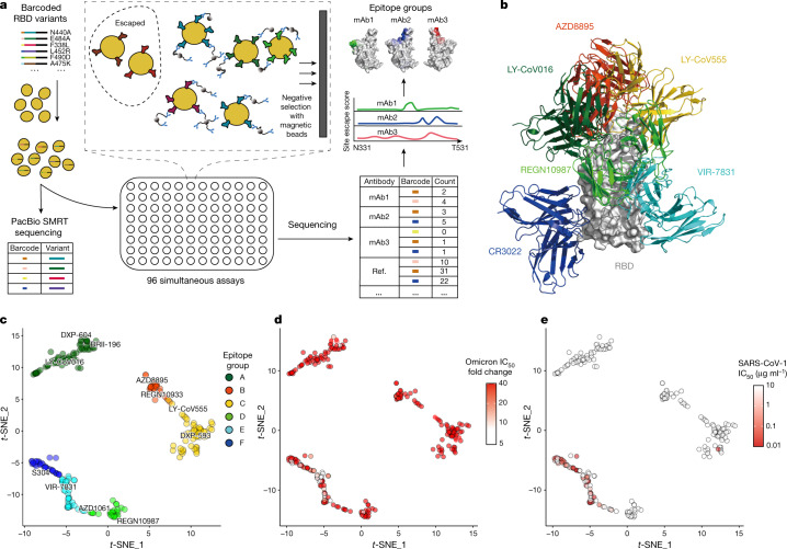
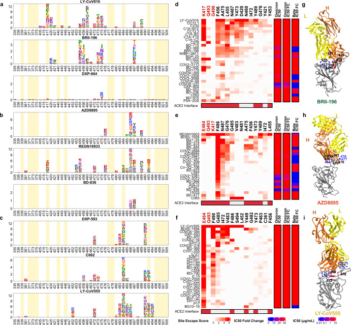
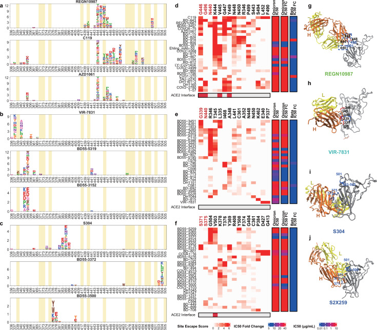
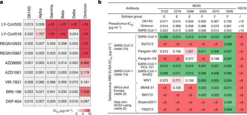
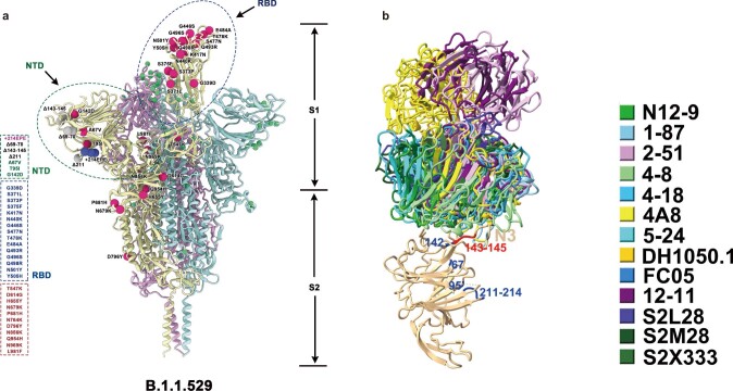
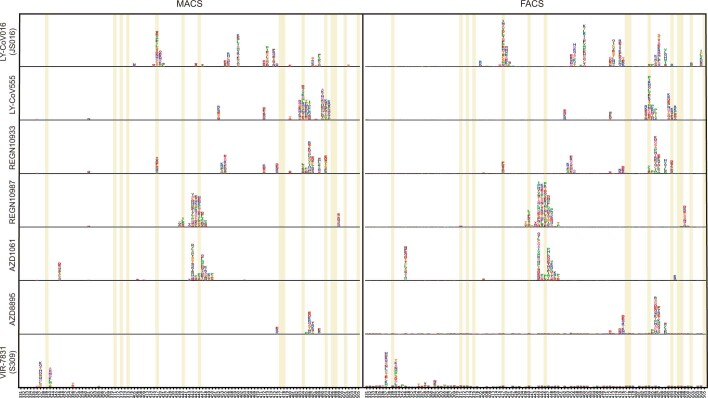
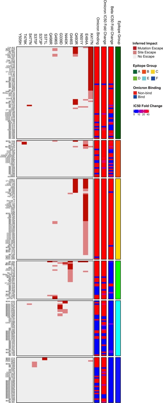
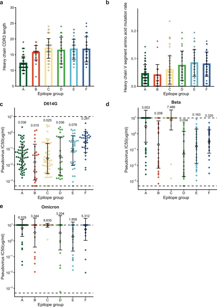
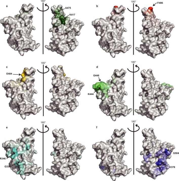
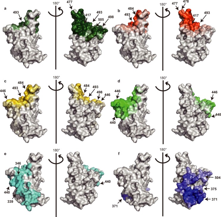
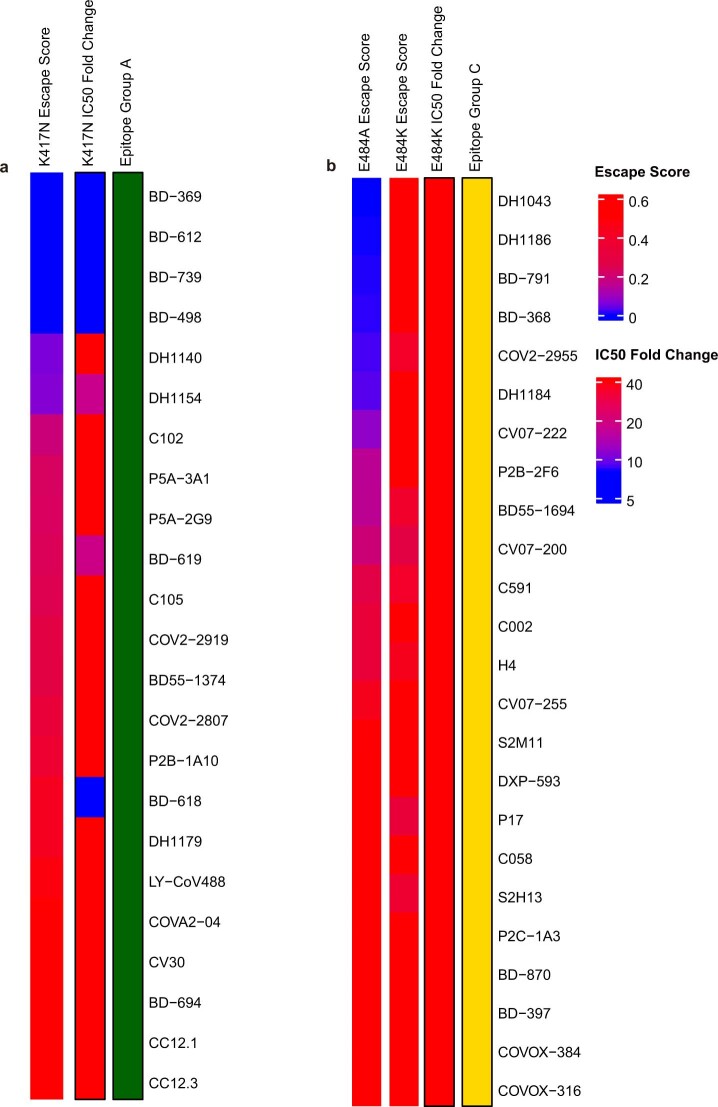
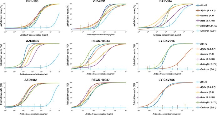
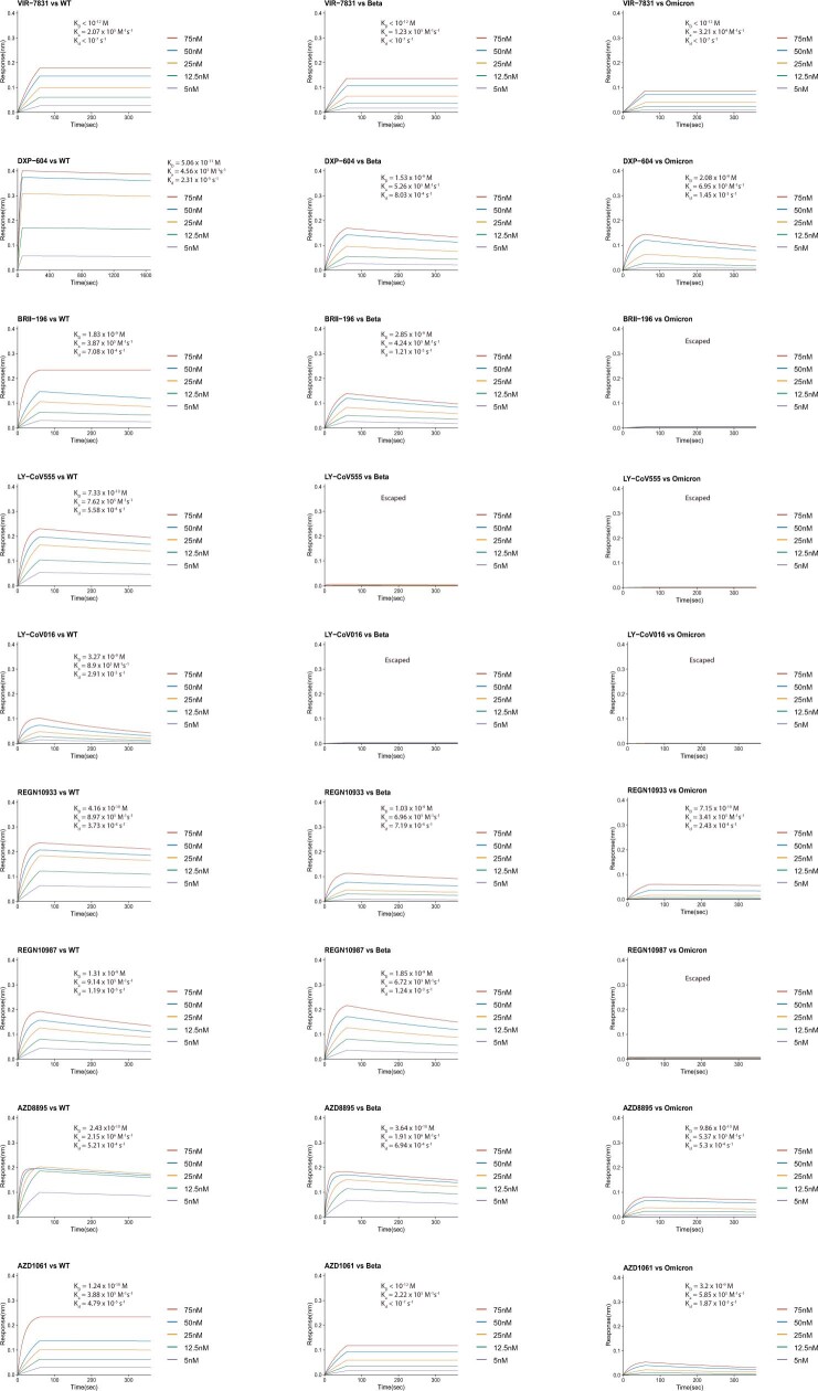
The SARS-CoV-2 B.1.1.529 (Omicron) variant contains 15 mutations of the receptor-binding domain (RBD). How Omicron evades RBD-targeted neutralizing antibodies requires immediate investigation. Here we use high-throughput yeast display screening 1,2 to determine the profiles of RBD escaping mutations for 247 human anti-RBD neutralizing antibodies and show that the neutralizing antibodies can be classified by unsupervised clustering into six epitope groups (A–F)—a grouping that is highly concordant with knowledge-based structural classifications 3–5 . Various single mutations of Omicron can impair neutralizing antibodies of different epitope groups. Specifically, neutralizing antibodies in groups A–D, the epitopes of which overlap with the ACE2-binding motif, are largely escaped by K417N, G446S, E484A and Q493R. Antibodies in group E (for example, S309) 6 and group F (for example, CR3022) 7 , which often exhibit broad sarbecovirus neutralizing activity, are less affected by Omicron, but a subset of neutralizing antibodies are still escaped by G339D, N440K and S371L. Furthermore, Omicron pseudovirus neutralization showed that neutralizing antibodies that sustained single mutations could also be escaped, owing to multiple synergetic mutations on their epitopes. In total, over 85% of the tested neutralizing antibodies were escaped by Omicron. With regard to neutralizing-antibody-based drugs, the neutralization potency of LY-CoV016, LY-CoV555, REGN10933, REGN10987, AZD1061, AZD8895 and BRII-196 was greatly undermined by Omicron, whereas VIR-7831 and DXP-604 still functioned at a reduced efficacy. Together, our data suggest that infection with Omicron would result in considerable humoral immune evasion, and that neutralizing antibodies targeting the sarbecovirus conserved region will remain most effective. Our results inform the development of antibody-based drugs and vaccines against Omicron and future variants.
Overview
The claims and statistics of this paper haven’t been checked yet.
Run the check — free, no account needed. It reads the open-access text and checks the sources it cites.
Integrity signals
Registry
Cites
Statistics
Figures
Verification
Claims the paper makes
The claims and statistics of this paper haven’t been checked yet.
Run the check — free, no account needed. It reads the open-access text and checks the sources it cites.
Claims other papers make about this one
Each is checked against this paper’s own text: does it say what the citing paper says it does?
In the citing paper
“Deep mutational scanning experiments of the SARS-CoV-2 receptor binding domain suggest that further mutations of Omicron BA.1/2 at sites 452 and 486 could lead to further immune evasion against antibodies isolated from individuals who had pre-Omicron and Omicron BA.1 breakthrough infections 21,28,29 .”
From Rapidly shifting immunologic landscape and severity of SARS-CoV-2 in the Omicron era in South Africa (2023) · cites this paper as [21]
In the citing paper
“Reinfections and vaccine breakthrough infections accounted for the majority of Omicron BA.1/2 infections, likely attributable to a minority of population remaining naïve prior to the Omicron BA.1/2 wave and to Omicron BA.1/2's immune-escape properties 16,21,22 .”
From Rapidly shifting immunologic landscape and severity of SARS-CoV-2 in the Omicron era in South Africa (2023) · cites this paper as [21]
In the citing paper
“Over reliance on immune memory may impact the breadth and strength of antibody and B-cell immunity as new antigenically drifted variants arise 15,21,32,33 .”
From Rapidly shifting immunologic landscape and severity of SARS-CoV-2 in the Omicron era in South Africa (2023) · cites this paper as [21]
Self-check
Percentages, sums and reported differences re-computed from the paper’s own numbers.
The self-check needs the paper’s full text.
Citations
Cites
SARS-CoV-2 variants, spike mutations and immune escape
Harvey, W. T., Carabelli, A. M., Jackson, B., Gupta, R. K., Thomson, E. C., Harrison, E. M., Ludden, C., Reeve, R., Rambaut, A., COVID-19 Genomics UK (COG-UK) Consortium, et al. (2021) · Nature Reviews Microbiology · doi:10.1038/s41579-021-00573-0 · Cited by 3,528
High-efficiency yeast transformation using the LiAc/SS carrier DNA/PEG method
Gietz, R. D., & Schiestl, R. H. (2007) · Nature Protocols · doi:10.1038/nprot.2007.13 · Cited by 2,785
Starr, T. N., Greaney, A. J., Hilton, S. K., Ellis, D., Crawford, K. H. D., Dingens, A. S., Navarro, M. J., Bowen, J. E., Tortorici, M. A., Walls, A. C., et al. (2020) · Cell · doi:10.1016/j.cell.2020.08.012 · Cited by 1,956
Cross-neutralization of SARS-CoV-2 by a human monoclonal SARS-CoV antibody
Pinto, D., Park, Y.-J., Beltramello, M., Walls, A. C., Tortorici, M. A., Bianchi, S., Jaconi, S., Culap, K., Zatta, F., De Marco, A., et al. (2020) · Nature · doi:10.1038/s41586-020-2349-y · Cited by 1,885
Convergent Antibody Responses to SARS-CoV-2 in Convalescent Individuals
Robbiani, D. F., Gaebler, C., Muecksch, F., Lorenzi, J. C. C., Wang, Z., Cho, A., Agudelo, M., Barnes, C. O., Gazumyan, A., Finkin, S., et al. (2020) · Nature · doi:10.1038/s41586-020-2456-9 · Cited by 1,853
Human neutralizing antibodies elicited by SARS-CoV-2 infection
Ju, B., Zhang, Q., Ge, J., Wang, R., Sun, J., Ge, X., Yu, J., Shan, S., Zhou, B., Song, S., et al. (2020) · Nature · doi:10.1038/s41586-020-2380-z · Cited by 1,667
Self-citationSARS-CoV-2 neutralizing antibody structures inform therapeutic strategies
Barnes, C. O., Jette, C. A., Abernathy, M. E., Dam, K.-M. A., Esswein, S. R., Gristick, H. B., Malyutin, A. G., Sharaf, N. G., Huey-Tubman, K. E., Lee, Y. E., et al. (2020) · Nature · doi:10.1038/s41586-020-2852-1 · Cited by 1,599
A highly conserved cryptic epitope in the receptor binding domains of SARS-CoV-2 and SARS-CoV
Yuan, M., Wu, N. C., Zhu, X., Lee, C.-C. D., So, R. T. Y., Lv, H., Mok, C. K. P., & Wilson, I. A. (2020) · Science · doi:10.1126/science.abb7269 · Cited by 1,429
Piccoli, L., Park, Y.-J., Tortorici, M. A., Czudnochowski, N., Walls, A. C., Beltramello, M., Silacci-Fregni, C., Pinto, D., Rosen, L. E., Bowen, J. E., et al. (2020) · Cell · doi:10.1016/j.cell.2020.09.037 · Cited by 1,395
Potent neutralizing antibodies against multiple epitopes on SARS-CoV-2 spike
Liu, L., Wang, P., Nair, M. S., Yu, J., Rapp, M., Wang, Q., Luo, Y., Chan, J. F.-W., Sahi, V., Figueroa, A., et al. (2020) · Nature · doi:10.1038/s41586-020-2571-7 · Cited by 1,349
Self-citation
Cited by
Sorted by citations
Items 801–900 of 1,000
Ren, P., Hu, Y., Peng, L., Yang, L., Suzuki, K., Fang, Z., Bai, M., Zhou, L., Feng, Y., Zou, Y., et al. (2023) · Signal Transduction and Targeted Therapy · doi:10.1038/s41392-023-01509-1 · Cited by 10
Hollingsworth, S. A., Noland, C. L., Vroom, K., Saha, A., Sam, M., Gao, Q., Zhou, H., Grandy, D. U., Singh, S., Wen, Z., et al. (2023) · Scientific Reports · doi:10.1038/s41598-023-40919-7 · Cited by 10
Sunagawa, J., Park, H., Kim, K. S., Komorizono, R., Choi, S., Torres, L. R., Woo, J., Jeong, Y. D., Hart, W. S., Thompson, R. N., et al. (2023) · Nature Communications · doi:10.1038/s41467-023-43043-2 · Cited by 11
O’Neill, A., Mantri, C. K., Tan, C. W., Saron, W. A. A., Nagaraj, S. K., Kala, M. P., Joy, C. M., Rathore, A. P. S., Tripathi, S., Wang, L., et al. (2023) · EBioMedicine · doi:10.1016/j.ebiom.2023.104924 · Cited by 12
Potent Anti‐SARS‐CoV‐2 Efficacy of COVID‐19 Hyperimmune Globulin from Vaccine‐Immunized Plasma
Yu, D., Li, Y., Hong, L., Wu, J., Hu, Y., Peng, Y., Li, T., Hou, J., Huang, W., Guan, L., et al. (2022) · Advanced Science · doi:10.1002/advs.202104333 · Cited by 11
Self-citationXu, Y., Li, Y., You, X., Pei, C., Wang, Z., Jiao, S., Zhao, X., Lin, X., Lü, Y., Jin, C., et al. (2022) · Frontiers in Chemistry · doi:10.3389/fchem.2022.871509 · Cited by 11
Dang, S., Ren, L., & Wang, J. (2022) · Chinese Medical Journal · doi:10.1097/cm9.0000000000002158 · Cited by 10
Self-citationSims, J. J., Lian, S., Meggersee, R., Kasimsetty, A., & Wilson, J. M. (2022) · PLoS ONE · doi:10.1371/journal.pone.0271359 · Cited by 10
Онищенко, Г. Г., Sizikova, T. E., Lebedev, V. N., & Borisevich, S. V. (2022) · Herald of the Russian Academy of Sciences · doi:10.1134/s1019331622040074 · Cited by 11
Schlör, A., Hirschberg, S., Amor, G. B., Meister, T. L., Arora, P., Pöhlmann, S., Hoffmann, M., Pfaender, S., Eddin, O. K., Kamhieh‐Milz, J., et al. (2022) · Frontiers in Immunology · doi:10.3389/fimmu.2022.930975 · Cited by 11
Dayuan, Z., Li, L., Luhua, X., Li, H., Dahao, C., Yumiao, L., Li, D.-X., & Deng, Y. (2022) · Heliyon · doi:10.1016/j.heliyon.2022.e11120 · Cited by 14
Oliveira, J. R., Remuzgo, C., Machado, R. R. G., Magawa, J. Y., Daher, I. P., Urbanski, A. H., Schmitz, G. J. H., Arcuri, H. A., Ferreira, M. A., Sasahara, G. L., et al. (2023) · Frontiers in Immunology · doi:10.3389/fimmu.2022.1010105 · Cited by 11
Lu, R., Liang, K.-H., Chiang, H.-L., Hsu, F., Lin, H.-T., Chen, W.-Y., Ke, F.-Y., Kumari, M., Chou, Y., Tao, M., et al. (2023) · Heliyon · doi:10.1016/j.heliyon.2023.e15587 · Cited by 12
IS IT TIME TO REFLECT, NOT ON THE “WHAT” BUT THE “WHY” IN EMERGING WILDLIFE DISEASE RESEARCH?
Kock, R. A. (2018) · Journal of Wildlife Diseases · doi:10.7589/2019-01-000 · Cited by 11
Engineered ACE2 counteracts vaccine-evading SARS-CoV-2 Omicron variantPreprint
Ikemura, N., Taminishi, S., Inaba, T., Arimori, T., Motooka, D., Katoh, K., Kirita, Y., Higuchi, Y., Li, S., Suzuki, T., et al. (2021) · bioRxiv (Cold Spring Harbor Laboratory) · doi:10.1101/2021.12.22.473804 · Cited by 11
Turelli, P., Fenwick, C. W., Raclot, C., Genet, V., Pantaleo, G., & Trono, D. (2022) · bioRxiv (Cold Spring Harbor Laboratory) · doi:10.1101/2022.07.28.501852 · Cited by 10
Muik, A., Lui, B. G., Diao, H., Fu, Y., Bacher, M., Toker, A., Grosser, J., Ozhelvaci, O., Grikscheit, K., Hoehl, S., et al. (2022) · bioRxiv (Cold Spring Harbor Laboratory) · doi:10.1101/2022.12.15.520569 · Cited by 11
Panorama of Breakthrough Infection Caused by SARS-CoV-2: A Review
Fan, Q., Nie, Z., & Xie, S. (2022) · Medicina · doi:10.3390/medicina58121733 · Cited by 10
Leveraging immunoliposomes as nanocarriers against SARS-CoV-2 and its emerging variants
Faizal, N. D. F. M., Ramli, N. A., Rani, N. N. I. M., Shaibie, N. A., Aarti, A., Poonsawas, P., Sharma, S. K., & Amin, M. C. I. M. (2023) · Asian Journal of Pharmaceutical Sciences · doi:10.1016/j.ajps.2023.100855 · Cited by 10
Deng, Y., Atyeo, C. G., Yuan, D., Chicz, T. M., Tibbitts, T. J., Gorman, M. J., Taylor, S., Lecouturier, V., Lauffenburger, D. A., Chicz, R. M., et al. (2023) · Cell Reports · doi:10.1016/j.celrep.2023.113292 · Cited by 10
Mei, S., Zou, Y., Jiang, S., Lü, X., Wang, Y., Han, J. D. J., Yang, P., Niu, M., Li, J., Yuan, K., et al. (2023) · European Journal of Medicinal Chemistry · doi:10.1016/j.ejmech.2023.115908 · Cited by 10
Self-citationEffect of the COVID-19 Pandemic on Respiratory Diseases and Their Economic Impacts
Jayaraman, A. S., Darekar, I., Dadhich, N. V., Tadepalli, L. S. M., Gongwang, Y., Singh, S. K., & Gavor, E. (2024) · Pathogens · doi:10.3390/pathogens13060491 · Cited by 10
Ferrucci, V., Miceli, M., Pagliuca, C., Bianco, O., Castaldo, L., Izzo, L., Cozzolino, M., Zannella, C., Oglio, F., Polcaro, A., et al. (2024) · Journal of Translational Medicine · doi:10.1186/s12967-024-05378-2 · Cited by 10
Su, C., He, J., Wang, L., Hu, Y., Cao, J., Bai, B., Qi, J., Gao, G. F., Yang, M., & Wang, Q. (2024) · mBio · doi:10.1128/mbio.01404-24 · Cited by 10
Dong, H., Zhou, R., Chen, J., Wei, J., Wei, Z., Yang, Z., Zhu, K., Yang, Y., Yang, Q., Liu, N., et al. (2024) · PLoS Pathogens · doi:10.1371/journal.ppat.1012625 · Cited by 10
Self-citationNan, X., Li, Y., Zhang, R., Wang, R., Lv, N., Li, J., Chen, Y., Zhou, B., Wang, Y., Wang, Z., et al. (2024) · Nature Communications · doi:10.1038/s41467-024-54746-5 · Cited by 11
Self-citationOluka, G. K., Sembera, J., Katende, J. S., Ankunda, V., Kato, L., Kurshan, A., Graham, C., Seow, J., Doores, K. J., Malim, M. H., et al. (2025) · Vaccines · doi:10.3390/vaccines13020143 · Cited by 9
Yilmaz, I. C., İpekoğlu, E. M., Saraydar, B., Bildik, T., Aksoy, A. G. B., Evcili, İ., Turay, N., Sürücü, N., Bulbul, A., Guvencli, N., et al. (2025) · Vaccine · doi:10.1016/j.vaccine.2025.126787 · Cited by 10
Dadonaite, B., Burrell, A. R., Logue, J., Chu, H. Y., Payne, D. C., Haslam, D. B., Staat, M. A., & Bloom, J. D. (2025) · Journal of Virology · doi:10.1128/jvi.00109-25 · Cited by 10
Nanovaccines: Antigen selection, stabilization, adjuvantation, formulation, and evaluation
Lu, L., Yang, M., Deng, D., Shi, X., Lovell, J. F., & Jin, H. (2025) · Coordination Chemistry Reviews · doi:10.1016/j.ccr.2025.216806 · Cited by 10
Ehling, R. A., Minot, M., Overath, M. D., Sheward, D. J., Han, J., Gao, B., Taft, J. M., Pertseva, M., Weber, C. R., Frei, L., et al. (2025) · Cell Systems · doi:10.1016/j.cels.2025.101391 · Cited by 8
Seekircher, L., Siller, A., Astl, M., Tschiderer, L., Wachter, G. A., Pfeifer, B. E., Huber, A., Gaber, M., Schennach, H., & Willeit, P. (2022) · Viruses · doi:10.3390/v14091877 · Cited by 10
Ríos‐Silva, M., Trujillo, X., Huerta, M., Benites-Godínez, V., Guzmán-Esquivel, J., Bricio‐Barrios, J. A., Mendoza‐Cano, O., Lugo-Radillo, A., & Murillo‐Zamora, E. (2022) · Pathogens · doi:10.3390/pathogens11101181 · Cited by 10
SARS-CoV-2 variants, immune escape, COVID-19 vaccine, and therapeutic strategies
Que, H., Chen, L., & Wei, X. (2022) · Science China Life Sciences · doi:10.1007/s11427-021-2164-6 · Cited by 10
On the Evolutionary Trajectory of SARS-CoV-2: Host Immunity as a Driver of Adaptation in RNA Viruses
Warger, J., & Gaudieri, S. (2022) · Viruses · doi:10.3390/v15010070 · Cited by 11
Farias, J. P., Pinheiro, J. R., Andreata‐Santos, R., Fogaça, M. M. C., Brito, R. D. D. S., da Cruz, E. F., de Castro‐Amarante, M. F., Pereira, S. S., Almeida, S. D. S., Moreira, L. M., et al. (2023) · Journal of Medical Virology · doi:10.1002/jmv.28481 · Cited by 10
Ito, K., Goto, K., Shirakawa, R., Horiuchi, H., & Ogasawara, K. (2023) · Biochemical and Biophysical Research Communications · doi:10.1016/j.bbrc.2023.03.057 · Cited by 10
Rodrigues‐da‐Silva, R. N., Conte, F. D. P., da Silva, G., Carneiro-Alencar, A. L., Gomes, P. R., Kuriyama, S. N., Neto, A. A. F., & Lima‐Junior, J. D. C. (2023) · Viruses · doi:10.3390/v15040923 · Cited by 11
Luo, Y., Liu, S., Xue, J., Yang, Y., Zhao, J., Sun, Y., Wang, B., Yin, S., Li, J., Xia, Y., et al. (2023) · Cell Discovery · doi:10.1038/s41421-023-00534-2 · Cited by 10
Self-citationAn RBD bispecific antibody effectively neutralizes a SARS-CoV-2 Omicron variant
Yuan, M., Zhu, Y., Liu, G., Wang, Y., Wang, G., Zhang, G., Ye, L., Qian, Z., & Liu, P. (2023) · One Health Advances · doi:10.1186/s44280-023-00012-0 · Cited by 10
Self-citationCryo-EM and cryo-ET of the spike, virion, and antibody neutralization of SARS-CoV-2 and VOCs
Xu, C., Han, W., & Cong, Y. (2023) · Current Opinion in Structural Biology · doi:10.1016/j.sbi.2023.102664 · Cited by 9
Kong, X., Gao, P., Jiang, Y., Lu, L., Zhao, M., Liu, Y., Deng, G., Zhu, H., Cao, Y., & Ma, L. (2023) · Virology Journal · doi:10.1186/s12985-023-02137-5 · Cited by 10
Self-citationEffectiveness of COVID-19 vaccination during pregnancy by circulating viral variant
Zerbo, O., Ray, G. T., FIREMAN, B. H., Layefsky, E., Goddard, K., Ross, P., Greenberg, M., & Klein, N. P. (2023) · AJOG Global Reports · doi:10.1016/j.xagr.2023.100264 · Cited by 10
Hybrid and SARS-CoV-2-vaccine immunity in kidney transplant recipients
Kared, H., Alirezaylavasani, A., Lund, K. P., Chopra, A., Tietze, L., Kasahara, T. D. M., Goll, G. L., Grødeland, G., Kaarbø, M., Reisæter, A. V., et al. (2023) · EBioMedicine · doi:10.1016/j.ebiom.2023.104833 · Cited by 12
Chen, J., Pan, L., Lu, Y. Z., Zhang, T., Xu, D., Yan, S., & Ouyang, Z. (2023) · Human Vaccines & Immunotherapeutics · doi:10.1080/21645515.2023.2276624 · Cited by 10
An enhanced broad-spectrum peptide inhibits Omicron variants in vivo
Bi, W., Tang, K., Chen, G., Xie, Y., Polizzi, N. F., DeGrado, W. F., Yuan, S., & Dang, B. (2024) · Cell Reports Medicine · doi:10.1016/j.xcrm.2024.101418 · Cited by 11
van den Dijssel, J., Duurland, M. C., Konijn, V. A. L., Kummer, L. Y. L., Hagen, R. R., Kuijper, L. H., Wieske, L., van Dam, K. P. J., Stalman, E. W., Steenhuis, M., et al. (2024) · Journal of Autoimmunity · doi:10.1016/j.jaut.2024.103175 · Cited by 11
Yu, P., Ran, J., Yang, R., Zhu, H., Song, L., Wu, Y., Zhao, T., & Xiong, T.-C. (2024) · Frontiers in Immunology · doi:10.3389/fimmu.2024.1374913 · Cited by 11
Hong, W., Hong, L., Peng, D., Huang, Y., He, C., Yang, H. J., Zhou, Y., Liu, J., Pan, X., Que, H., et al. (2024) · MedComm · doi:10.1002/mco2.539 · Cited by 10
Self-citationNutini, A., Zhang, J., Sohail, A., Arif, R., & Nofal, T. A. (2022) · Results in Physics · doi:10.1016/j.rinp.2022.105300 · Cited by 10
Boeckel, G. R., Hölscher, S. D., Bürger, C., Jacob, T., Krekeler, C., Shumilov, E., Reicherts, C., Bleckmann, A., Lenz, G., Vollenberg, R., et al. (2022) · Current Oncology · doi:10.3390/curroncol29040188 · Cited by 9
Tuells, J., Parra-Grande, M., Santos-Calle, F. J., Montagud, A. C., Egoavil, C. M., García-Rivera, C., Caballero, P., Bravo, E. M. G., Rodrı́guez, J. C., & Sánchez, J. A. H. (2022) · Vaccines · doi:10.3390/vaccines10040510 · Cited by 12
Li, M., Ye, Z., Tang, K., Guo, L., Bi, W., Zhang, Y., Tang, Y.-D., Rong, G., Sawan, M., Yin, X., et al. (2022) · Cell Research · doi:10.1038/s41422-022-00656-4 · Cited by 10
Yu, Y., Herrmann, A., Thonn, V., Cordsmeier, A., Neurath, M. F., Ensser, A., & Becker, C. (2022) · Cells · doi:10.3390/cells11081262 · Cited by 9
Self-citationOutpatient treatment options to address the SARS-CoV-2 variant Omicron
McCarthy, M. W. (2022) · Expert Review of Anti-infective Therapy · doi:10.1080/14787210.2022.2077191 · Cited by 10
Yang, S., Tong, Y., Chen, L., & Yu, W. (2022) · Molecular Biomedicine · doi:10.1186/s43556-022-00077-0 · Cited by 10
Self-citationLessons from SARS‑CoV‑2 and its variants (Review)
Qin, Z., Sun, Y., Zhang, J., Zhou, L., Chen, Y., & Huang, C. (2022) · Molecular Medicine Reports · doi:10.3892/mmr.2022.12779 · Cited by 10
Neutralizing Potency of Prototype and Omicron RBD mRNA Vaccines Against Omicron Variant
Zang, J., Yin, Y., Xu, S., Qiao, W., Liu, Q., Lavillette, D., Zhang, C., Wang, H., & Huang, Z. W. (2022) · Frontiers in Immunology · doi:10.3389/fimmu.2022.908478 · Cited by 11
Panahi, Y., Einollahi, B., Beiraghdar, F., Darvishi, M., Fathi, S., Javanbakht, M., Shafiee, S., & Akhavan‐Sigari, R. (2022) · Frontiers in Immunology · doi:10.3389/fimmu.2022.947602 · Cited by 10
Rodriguez, J. A., Gonzalez, J., Arboleda-Bustos, C. E., Mendoza, N., Martinez, C., & Pinzón, A. M. (2022) · Chemico-Biological Interactions · doi:10.1016/j.cbi.2022.110244 · Cited by 10
Roth, N., Gergen, J., Kovacikova, K., Mueller, S. O., Ulrich, L., Schön, J., Halwe, N. J., Fricke, C., Corleis, B., Dorhoi, A., et al. (2023) · Vaccines · doi:10.3390/vaccines11020318 · Cited by 10
Kuo, H., Kuo, K.-C., Du, P.-X., Keskin, B. B., Su, W., Ho, T., Tsai, P.-S., Pau, C. H., Shih, H.-C., Huang, Y., et al. (2023) · Molecular & Cellular Proteomics · doi:10.1016/j.mcpro.2023.100507 · Cited by 12
Structural Basis for the Enhanced Infectivity and Immune Evasion of Omicron Subvariants
Li, Y., Shen, Y., Zhang, Y., & Yan, R. (2023) · Viruses · doi:10.3390/v15061398 · Cited by 10
Khairnar, K., & Tomar, S. S. (2024) · Virus Research · doi:10.1016/j.virusres.2024.199365 · Cited by 10
Omicron breakthrough infection drives cross-variant neutralization and memory B cell formationPreprint
Quandt, J., Muik, A., Salisch, N. C., Lui, B. G., Lutz, S., Krüger, K., Wallisch, A.-K., Adams‐Quack, P., Bacher, M., Finlayson, A., et al. (2022) · bioRxiv (Cold Spring Harbor Laboratory) · doi:10.1101/2022.04.01.486695 · Cited by 10
Verkhivker, G. M., Agajanian, S., Kassab, R., & Krishnan, K. (2022) · Biomolecules · doi:10.3390/biom12070964 · Cited by 11
Thye, A. Y.-K., & Law, J. W. (2022) · Progress In Microbes & Molecular Biology · doi:10.36877/pmmb.a0000280 · Cited by 9
Awandu, S. S., Ochieng, A. O., Onyango, B., Magwanga, R. O., Were, P., Ochung’, A. A., Okumu, F. O., Oloo, M. A., Katieno, J. S., Lidechi, S., et al. (2022) · PLoS ONE · doi:10.1371/journal.pone.0272751 · Cited by 9
Detecting punctuated evolution in SARS-CoV-2 over the first year of the pandemic
Surya, K., Gardner, J. D., & Organ, C. L. (2023) · Frontiers in Virology · doi:10.3389/fviro.2023.1066147 · Cited by 9
Kim, W. S., Chae, H. D., Jung, I., Lee, W., Lee, W. J., Lee, J., Gong, Y., Lee, D., Kim, B.-W., Kim, J.-K., et al. (2023) · Fish & Shellfish Immunology · doi:10.1016/j.fsi.2023.108807 · Cited by 9
Surveillance for variants of SARS-CoV-2 to inform risk assessments
Cohen, H. A., Mesfin, S., Ikejezie, J., Kassamali, Z. A., Campbell, F., Adele, S., Guinko, N., Idoko, F., Mirembe, B. B., Mitri, M. E., et al. (2023) · Bulletin of the World Health Organization · doi:10.2471/blt.23.290093 · Cited by 9
Polyvalency: an emerging trend in the development of clinical antibodies
Shinde, S. H., Sandeep, & Pande, A. H. (2023) · Drug Discovery Today · doi:10.1016/j.drudis.2023.103846 · Cited by 9
Raisinghani, N., Alshahrani, M., Gupta, G., Xiao, S., Tao, P., & Verkhivker, G. M. (2024) · Journal of Chemical Information and Modeling · doi:10.1021/acs.jcim.3c01857 · Cited by 8
On-mask detection of SARS-CoV-2 related substances by surface enhanced Raman scattering
Liu, X., Xie, R., Li, K., Zhu, Z., Huang, X., Qian, H., Sun, Z., He, H., Ge, Y., Zhang, Q., et al. (2024) · Talanta · doi:10.1016/j.talanta.2024.126403 · Cited by 9
Self-citationHe, L., Wu, Q., Zhang, Z., Chen, L., Yu, K., Li, L., Jia, Q., Wang, Y., Ni, J., Wang, C., et al. (2024) · Molecular Pharmaceutics · doi:10.1021/acs.molpharmaceut.4c00165 · Cited by 9
Self-citationModulation of the pharmacokinetics of soluble ACE2 decoy receptors through glycosylation
Skeeters, S. S., Bagale, K., Stepanyuk, G. A., Thieker, D. F., Aguhob, A., Chan, K. K., Dutzar, B., Shalygin, S. S., Shajahan, A., Xu, Y., et al. (2024) · Molecular Therapy — Methods & Clinical Development · doi:10.1016/j.omtm.2024.101301 · Cited by 9
Yisimayi, A., Song, W., Wang, J., Jian, F., Yu, Y., Chen, X., Xu, Y., An, R., Wang, Y., Wang, J., et al. (2024) · Emerging Microbes & Infections · doi:10.1080/22221751.2024.2412623 · Cited by 9
Self-citationLemieux, G., Pérez‐Vargas, J., Désilets, A., Hassanzadeh, M., Thompson, C. A. H., Gravel-Trudeau, A., Joushomme, A., Ennis, S., Villanueva, I., Marouseau, É., et al. (2025) · Journal of Medicinal Chemistry · doi:10.1021/acs.jmedchem.4c02468 · Cited by 9
Wang, M., Li, M. J., Yan, G., Li, H., Zhou, J., & Yang, A. (2025) · Archives of Virology · doi:10.1007/s00705-025-06308-3 · Cited by 9
Fantin, R. F., Clark, J. J., Cohn, H., Jaiswal, D., Bozarth, B., Rao, V., Civljak, A., Lobo, I., Nardulli, J., Srivastava, K., et al. (2025) · The Lancet Microbe · doi:10.1016/j.lanmic.2025.101103 · Cited by 9
Alhashimi, M., Sayedahmed, E. E., Elkashif, A., Chothe, S. K., Wang, W.-C., Murala, M. S. T., Gairola, V., Wakamatsu, N., Gontu, A., Ramasamy, S., et al. (2025) · npj Vaccines · doi:10.1038/s41541-025-01198-7 · Cited by 9
Zhou, Y., Gammeltoft, K. A., Tjørnelund, H. D., Ryberg, L. A., Offersgaard, A., Czarnota, A., Duan, Z., Pham, L. V., Fahnøe, U., Peters, G. H. J., et al. (2025) · Emerging Microbes & Infections · doi:10.1080/22221751.2025.2552716 · Cited by 9
Qureshi, A., Sulaiman, S. A. S., Kumar, N., & Qureshi, P. A. A. A. (2022) · PLoS ONE · doi:10.1371/journal.pone.0270761 · Cited by 9
Kim, W. S., Kim, J. H., Lee, J., Ka, S. Y., Chae, H. D., Jung, I., Jung, S. T., & Na, J. (2022) · Bioengineering · doi:10.3390/bioengineering9110670 · Cited by 9
Fujita, S., Kosugi, Y., Kimura, I., Yamasoba, D., & Sato, K. (2022) · Viruses · doi:10.3390/v14122677 · Cited by 8
Zhang, Y., Zhang, H.-Q., Wang, G., Zhang, Z., Li, J., Chen, X., Hu, Y.-Y., Zeng, X., Shi, Y.-J., Wang, J., et al. (2022) · Antiviral Research · doi:10.1016/j.antiviral.2022.105507 · Cited by 10
Self-citationEngineered soluble ACE2 receptor: Responding to change with change
Li, G., Qian, K., Zhang, S., Fu, W., Zhao, J. L., Lei, C., & Hu, S. (2023) · Frontiers in Immunology · doi:10.3389/fimmu.2022.1084331 · Cited by 8
Wang, Z., Li, D., Chen, Y., Sun, Y., Jin, C., Hu, C., Feng, Y., Su, J., Ren, L., Hao, Y., et al. (2023) · Frontiers in Immunology · doi:10.3389/fimmu.2023.1160283 · Cited by 8
Khadela, A. D., Soni, S., Megha, K., Bhagat, S. U., & Chavda, V. P. (2023) · Biologics · doi:10.3390/biologics3020008 · Cited by 9
SARS-CoV-2 Prevalence and Variant Surveillance among Cats in Pittsburgh, Pennsylvania, USA
Ramasamy, S., Gontu, A., Neerukonda, S. N., Ruggiero, D., Morrow, B. L., Gupta, S., Amirthalingam, S., Hardham, J. M., Lizer, J. T., Yon, M., et al. (2023) · Viruses · doi:10.3390/v15071493 · Cited by 7
Dang, T. T. T., Anzurez, A., Nakayama‐Hosoya, K., Miki, S., Yamashita, K., de Souza, M. S., Matano, T., & Kawana‐Tachikawa, A. (2023) · Microbiology Spectrum · doi:10.1128/spectrum.02143-23 · Cited by 10
Xie, G., Wang, L., & Zhang, J. (2023) · Frontiers in Public Health · doi:10.3389/fpubh.2023.1190519 · Cited by 9
Ward, T., Morris, M., Gelman, A., Carpenter, B., Ferguson, W., Overton, C. E., & Fyles, M. (2023) · PLoS Computational Biology · doi:10.1371/journal.pcbi.1011580 · Cited by 9
Breakthrough SARS-COV-2 infection induces broad anti-viral T cell immunity
Lineburg, K. E., Crooks, P., Raju, J., Texier, L. L., Khaledi, P., Berry, K., Swaminathan, S., Panikkar, A., Rehan, S., Guppy-Coles, K., et al. (2023) · iScience · doi:10.1016/j.isci.2023.108474 · Cited by 9
A panel of multivalent nanobodies broadly neutralizing Omicron subvariants and recombinant
Zheng, L., Wang, H., Liu, X., Xu, C., Tian, M., Shi, G., Bai, C., Li, Z., Wang, J., & Liu, S. (2024) · Journal of Medical Virology · doi:10.1002/jmv.29528 · Cited by 9
Self-citationJaishwal, P., Jha, K., & Singh, S. P. (2024) · International Journal of Biological Macromolecules · doi:10.1016/j.ijbiomac.2024.134012 · Cited by 10
Cia, G., Pucci, F., & Rooman, M. J. (2022) · International Journal of Molecular Sciences · doi:10.3390/ijms23042078 · Cited by 9
Balandrán, J. C., Zamora-Herrera, G., Romo‐Rodríguez, R., & Pelayo, R. (2022) · Journal of Interferon & Cytokine Research · doi:10.1089/jir.2022.0028 · Cited by 11
Moving targets: COVID-19 vaccine efficacy against Omicron subvariants
da Silva, M. K., Fulco, U. L., da Silva, E. D., & Oliveira, J. I. N. (2022) · Molecular Therapy · doi:10.1016/j.ymthe.2022.07.004 · Cited by 9
Palladino, R., Mercogliano, M., Fiorilla, C., Frangiosa, A., Iodice, S., Zamparelli, S. S., Montella, E., Triassi, M., & Sanduzzi, A. (2022) · International Journal of Environmental Research and Public Health · doi:10.3390/ijerph19159670 · Cited by 9
Metadata
- Authors
- Cao, Y., Wang, J., Jian, F., Xiao, T., Song, W., Yisimayi, A., Huang, W., Li, Q., Wang, P., An, R., Wang, J., Wang, Y., Niu, X., Yang, S., Liang, H., Sun, H., Li, T., Yu, Y., Cui, Q., Liu, S., Yang, X., Du, S., Zhang, Z., Hao, X., Shao, F., Jin, R., Wang, X., Xiao, J., Wang, Y., & Xie, X. S.
- Affiliations
- Peking University (CN)
- National Institutes for Food and Drug Control (CN)
- Capital Medical University (CN)
- Chinese Academy of Sciences (CN)
- Corresponding author
- Cao, Yunlong
- Wang, Yao
- Wang, Xiangxi
- Xiao, Junyu
- Wang, Youchun
- Xie, Xiaoliang Sunney
- ORCID
- Venue
- Nature
- Published
- 2021-12-23
- Volume
- 602
- Issue
- 7898
- Pages
- 657-663
- ISSN
- 0028-0836 (ISSN-L)
- 1476-4687
- PMC
- PMC8866119
- OpenAlex
- W4220807057
- Cited by
- 2,006
- References
- 56
- Citations by year
- 2026: 143 · 2025: 242 · 2024: 329 · 2023: 525 · 2022: 735 · 2021: 20
- FWCI
- 127.72
- Type
- journal article
- Language
- en
- Open access
- hybrid
- doi.org
- Locations
- Nature (publishedVersion, cc-by)
- bioRxiv (Cold Spring Harbor Laboratory) (acceptedVersion)
- Research Square (acceptedVersion, cc-by)
- PubMed Central (publishedVersion, cc-by)
- PubMed (publishedVersion)
- Funders
- Peking University
- Topics
- Health Sciences › Medicine › Infectious Diseases › SARS-CoV-2 and COVID-19 Research
- Life Sciences › Biochemistry, Genetics and Molecular Biology › Molecular Biology › vaccines and immunoinformatics approaches
- Life Sciences › Immunology and Microbiology › Immunology › Monoclonal and Polyclonal Antibodies Research
- Keywords
- Omicron variant, SARS-CoV-2, receptor binding domain, neutralizing antibodies, antibody escape, K417N, vaccine design
- MeSH terms
- Angiotensin-Converting Enzyme 2, COVID-19, SARS-CoV-2, COVID-19 Vaccines, Antibodies, Monoclonal, Antibodies, Viral, Cells, Cultured, Convalescence, Humans, Immune Sera, Models, Molecular, Mutation, Neutralization Tests, Epitopes, B-Lymphocyte, Immune Evasion, Antibodies, Neutralizing, Spike Glycoprotein, Coronavirus
About these checks
Registry, retraction, Cites and statistics results are computed, not judged, with one exception: a printed reference that no index lists is read once by a model before it counts as a citation error. Claims are extracted by a model and kept only when their quote appears verbatim in the text; each cited source is then checked, with a model’s help, for whether it says what the claim relies on. Those verdicts can be wrong — read the claim’s quote and the cited source before relying on one.
Statistics and arithmetic are recomputed, not judged.
Confidence bands are shown only for fields calibrated on our annotated test set. None is calibrated yet, so no band is shown.
What counts as a claim ›How we measure confidence ›How we check this ›
Are you an author, or do you see a problem with this page? Request a review
