

The DOI matches its Crossref record; 53 of the 53 references it cites were found in a registry or an index.
TDP-43 represses cryptic exon inclusion in the FTD-ALS gene UNC13A
Open accessPublished version of a preprint
Abstract
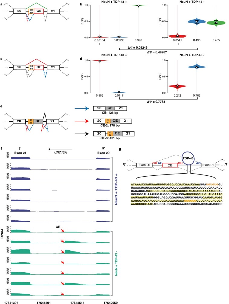
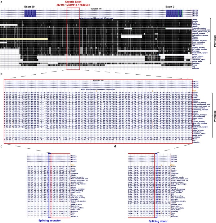
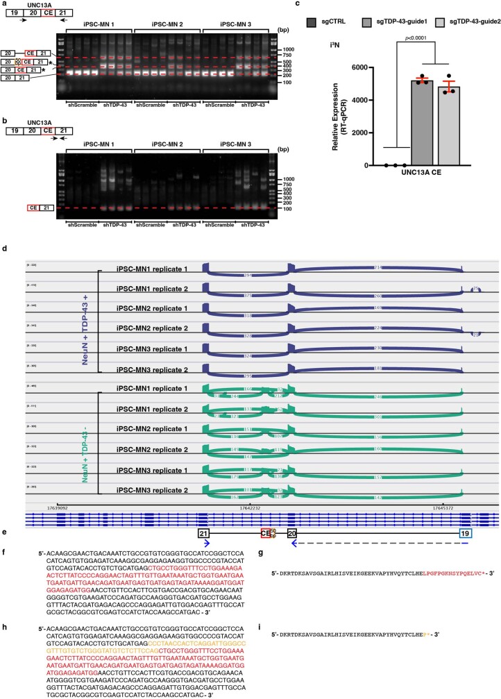
A hallmark pathological feature of the neurodegenerative diseases amyotrophic lateral sclerosis (ALS) and frontotemporal dementia (FTD) is the depletion of RNA-binding protein TDP-43 from the nucleus of neurons in the brain and spinal cord 1 . A major function of TDP-43 is as a repressor of cryptic exon inclusion during RNA splicing 2–4 . Single nucleotide polymorphisms in UNC13A are among the strongest hits associated with FTD and ALS in human genome-wide association studies 5,6 , but how those variants increase risk for disease is unknown. Here we show that TDP-43 represses a cryptic exon-splicing event in UNC13A . Loss of TDP-43 from the nucleus in human brain, neuronal cell lines and motor neurons derived from induced pluripotent stem cells resulted in the inclusion of a cryptic exon in UNC13A mRNA and reduced UNC13A protein expression. The top variants associated with FTD or ALS risk in humans are located in the intron harbouring the cryptic exon, and we show that they increase UNC13A cryptic exon splicing in the face of TDP-43 dysfunction. Together, our data provide a direct functional link between one of the strongest genetic risk factors for FTD and ALS ( UNC13A genetic variants), and loss of TDP-43 function.
Overview
The claims and statistics of this paper haven’t been checked yet.
Run the check — free, no account needed. It reads the open-access text and checks the sources it cites.
Integrity signals
Registry
Cites
Statistics
Figures
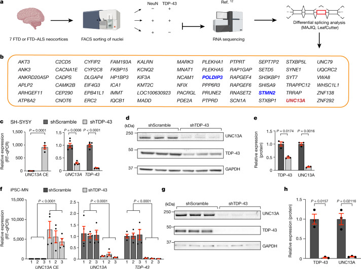
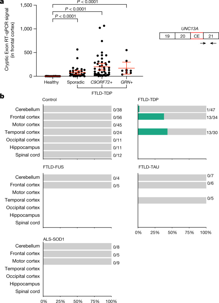
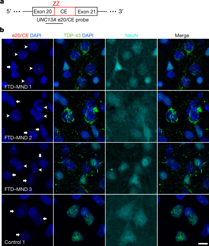
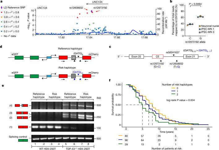
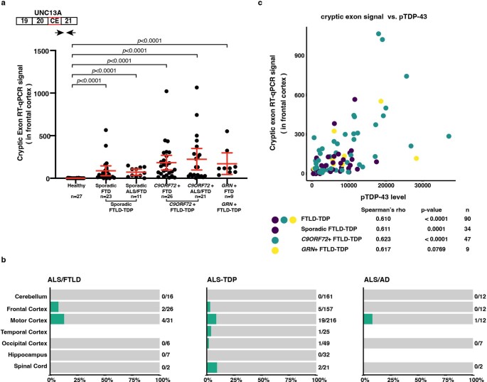
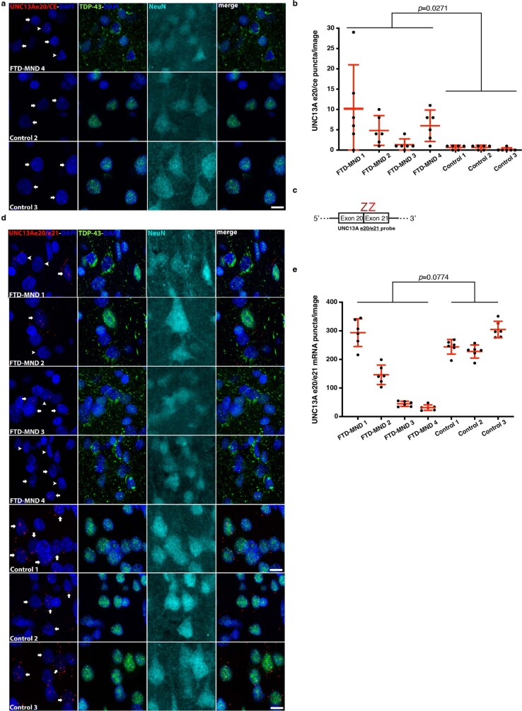
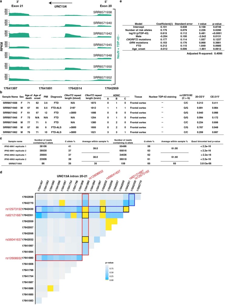
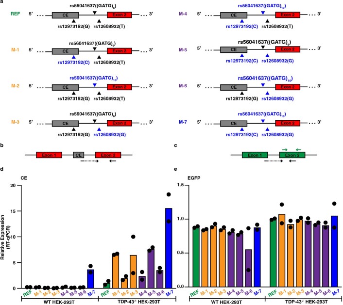
Verification
Claims the paper makes
The claims and statistics of this paper haven’t been checked yet.
Run the check — free, no account needed. It reads the open-access text and checks the sources it cites.
Claims other papers make about this one
Each is checked against this paper’s own text: does it say what the citing paper says it does?
In the citing paper
“The most common genes identified in clinical practice include SOD1, C9ORF72, TARDBP, FUS, and OPTNTD-P43 (55–57, 73, 74).”
From Modulation of the gut–brain axis via the gut microbiota: a new era in treatment of amyotrophic lateral sclerosis (2023) · cites this paper as [73]
Self-check
Percentages, sums and reported differences re-computed from the paper’s own numbers.
The self-check needs the paper’s full text.
Citations
Cites
Moderated estimation of fold change and dispersion for RNA-seq data with DESeq2
Love, M. I., Huber, W., & Anders, S. (2014) · Genome Biology · doi:10.1186/s13059-014-0550-8 · Cited by 95,106
A global reference for human genetic variation
The 1000 Genomes Project Consortium, authors, C., Auton, A., Abecasis, G. R., Steering committee, Altshuler, D. M., Durbin, R. M., Abecasis, G. R., Bentley, D. R., Chakravarti, A., et al. (2015) · Nature · doi:10.1038/nature15393 · Cited by 18,247
Self-citationTHE GENETICS OF CAENORHABDITIS ELEGANS
Brenner, S. (1974) · Genetics · doi:10.1093/genetics/77.1.71 · Cited by 15,220
The Human Genome Browser at UCSC
Kent, W. J., Sugnet, C. W., Furey, T. S., Roskin, K. M., Pringle, T. H., Zahler, A. M., & Haussler, A. D. (2002) · Genome Research · doi:10.1101/gr.229102 · Cited by 10,400
Ubiquitinated TDP-43 in Frontotemporal Lobar Degeneration and Amyotrophic Lateral Sclerosis
Neumann, M., Sampathu, D. M., Kwong, L. K., Truax, A. C., Micsenyi, M. C., Chou, T. T., Bruce, J., Schuck, T., Grossman, M., Clark, C. M., et al. (2006) · Science · doi:10.1126/science.1134108 · Cited by 6,576
LocusZoom: regional visualization of genome-wide association scan results
Pruim, R. J., Welch, R. P., Sanna, S., Teslovich, T. M., Chines, P. S., Gliedt, T. P., Boehnke, M., Abecasis, G. R., & Willer, C. J. (2010) · Bioinformatics · doi:10.1093/bioinformatics/btq419 · Cited by 2,707
Limbic-predominant age-related TDP-43 encephalopathy (LATE): consensus working group report
Nelson, P. T., Dickson, D. W., Trojanowski, J. Q., Jack, C. R., Boyle, P. A., Arfanakis, K., Rademakers, R., Alafuzoff, I., Attems, J., Brayne, C., et al. (2019) · Brain · doi:10.1093/brain/awz099 · Cited by 1,547
Self-citationLong pre-mRNA depletion and RNA missplicing contribute to neuronal vulnerability from loss of TDP-43
Polymenidou, M., Lagier-Tourenne, C., Hutt, K. R., Huelga, S. C., Moran, J., Liang, T. Y., Ling, S.-C., Sun, E., Wancewicz, E., Mazur, C., et al. (2011) · Nature Neuroscience · doi:10.1038/nn.2779 · Cited by 1,335
Characterizing the RNA targets and position-dependent splicing regulation by TDP-43
Tollervey, J. R., Curk, T., Rogelj, B., Briese, M., Cereda, M., Kayikci, M., König, J., Hortobágyi, T., Nishimura, A. L., Župunski, V., et al. (2011) · Nature Neuroscience · doi:10.1038/nn.2778 · Cited by 1,165
TDP-43 and FUS/TLS: emerging roles in RNA processing and neurodegeneration
Lagier-Tourenne, C., Polymenidou, M., & Cleveland, D. W. (2010) · Human Molecular Genetics · doi:10.1093/hmg/ddq137 · Cited by 1,021
Cited by
Sorted by citations
Items 501–576 of 576
Cao, M. C., Swanson, M. E. V., Basak, I., McDonald, K., Arnold, F. J., Stockford, C. M., Guo, G., Curtis, M. A., Faull, R. L. M., Hughes, S. M., et al. (2026) · Acta Neuropathologica Communications · doi:10.1186/s40478-026-02237-7
TDP-43 pathology is linked to motor neuron loss and is independent of stress granules in vivoPreprint
Dubinski, A. F., Ferdi, A., Choughari, M., Spence, H., Adhikary, A., Fauchon, C., Touati, M., Gagné, M., Liu, M., Peyrard, S., et al. (2026) · bioRxiv (Cold Spring Harbor Laboratory) · doi:10.64898/2026.02.11.705439 · Cited by 1
Benchmarking foundation models for splice site and exon annotationPreprint
He, Z., & Florea, L. (2026) · bioRxiv (Cold Spring Harbor Laboratory) · doi:10.64898/2026.02.22.707219
Neuronal TDP-43 regulates myelin formation via neurexin 1 mRNA stabilization
Li, J., Iguchi, Y., Yoshida, K., Kato, D., Araki, K., Kobayashi, K., Yokoi, S., Yoshimoto, R., Iida, M., Amakusa, Y., et al. (2026) · Proceedings of the National Academy of Sciences · doi:10.1073/pnas.2513642123
TDP-43-driven alternative splicing of UQCRC2 modulates mitochondrial bioenergetics
Xue, X., Hou, J., Zhang, Z., Yang, Z., Chang, L., Yang, Z., Liu, S., Huang, H., Mei, L., Mi, Y., et al. (2026) · Biology Direct · doi:10.1186/s13062-026-00745-z
Cabrera-Rodríguez, R., Pons-Grífols, A., Pernas, M., Casado, C., Trujillo‐González, R., Lorenzo-Sánchez, I., Reyes-Castañeda, I., Trinité, B., Blanco, J., & Valenzuela-Fernández, A. (2026) · Cell Communication and Signaling · doi:10.1186/s12964-026-02762-x
Cellular modifiers of TDP-43 phase transition and cytoplasmic aggregationPreprint
Chin, N. Y., Zhang, Q., Zou, J., Cheng, K. C.-C., Zheng, W., & Ye, Y. (2026) · eLife · doi:10.7554/elife.110172
Nuclear export governs TDP-43 phase transitions and cytoplasmic aggregationPreprint
Chin, N. Y., Zhang, Q., Zou, J., Cheng, K. C.-C., Zheng, W., & Ye, Y. (2026) · eLife · doi:10.7554/elife.110172.1
Zhang, L., Huang, Y., Huang, W., Huang, Q., Lin, Y., & Gu, J. (2026) · Journal of Alzheimer s Disease · doi:10.1177/13872877261424284
TYK2 mediates neuroinflammation in Alzheimer’s disease brains with TDP-43 pathology
König, L. E., Rodriguez, S., Hug, C. B., Daneshvari, S., Chung, A., Appleman, M., Tsai, M., Bradshaw, G. A., Şahin, A., Song, Y., et al. (2026) · Nature Communications · doi:10.1038/s41467-026-70243-3 · Cited by 7
McLellan, C., Campos‐Melo, D., Hammond, R. R., & Strong, M. J. (2026) · Acta Neuropathologica · doi:10.1007/s00401-026-03005-6
Shouman, S., Saif, N. A., Ahmed, S. M., Eltaher, S. S., Shehata, M., Abdelrahman, K., Elhadidy, M., & El‐Badri, N. (2026) · Stem cell biology and regenerative medicine · doi:10.1007/978-3-032-12705-1_1
Selective, genetically induced increase in synaptic vesicle priming
Aldahabi, M., Bálint, F., Lőrincz, A., Lipstein, N., Brose, N., & Nusser, Z. (2026) · Science Advances · doi:10.1126/sciadv.aee6848
Ramzan, F., Mishra, G. G., Alshehabi, Y., Perry, C. M., Jadon, L. M. Q., Kumar, A., Kim, H., Uy, R. C., Dang, A., Fayyad, M., et al. (2026) · bioRxiv (Cold Spring Harbor Laboratory) · doi:10.64898/2026.04.05.716148 · Cited by 3
Youssef, H., Gatto, R. G., Ghayal, N. B., Ayuso, V. E., Jansen‐West, K. R., Dunmore, J. A., Song, Y., Yue, M., Cook, C. N., Deture, M. A., et al. (2026) · Annals of Neurology · doi:10.1002/ana.78223
Self-citationAhmed, M., Volkening, K., McLellan, C., Shoesmith, C. L., Balci, T. B., & Strong, M. J. (2026) · Amyotrophic Lateral Sclerosis and Frontotemporal Degeneration · doi:10.1080/21678421.2026.2655734 · Cited by 1
Qi, Y., Wang, C., & Liu, J. (2026) · Intelligent Decision Technologies · doi:10.1177/18724981251397520
Generation and validation of a human iPSC-derived TDP-43 knockout model for ALS disease modelingPreprint
Gurumurthy, S., Bhargava, A., Huynh, N. P., Krzystek, T. J., Vieira, F. G., & Denton, K. R. (2026) · bioRxiv (Cold Spring Harbor Laboratory) · doi:10.64898/2026.04.29.720127 · Cited by 1
Pulst, S. M., Paul, S., Nguyen, H. T., Dansithong, W., Figueroa, K. P., Gandelman, M., Bonini, N. M., & Scoles, D. R. (2026) · Cell Death and Disease · doi:10.1038/s41419-026-08830-x
Ganssauge, J., Chandrashekar, H. B., Zhang, Y. H., Fusciardi, A. R., Namboori, S. C., & Bhinge, A. (2026) · bioRxiv (Cold Spring Harbor Laboratory) · doi:10.64898/2026.05.10.724071
Chauhan, B. S., Brennan, M. A., Forstmeier, P. C., Han, Y., Godfrey, R. K., Keuren‐Jensen, K. R. V., Sattler, R., Ichida, J. K., & Zarnescu, D. C. (2026) · Acta Neuropathologica Communications · doi:10.64898/2026.05.08.723849
Yokoi, S., Iguchi, Y., & Katsuno, M. (2026) · Frontiers in Molecular Neuroscience · doi:10.3389/fnmol.2026.1864964
Condorelli, G. A., Iozzia, A., Bonifacio, D., & Pelin, A.-M. (2026) · Neurochemical Research · doi:10.1007/s11064-026-04770-2
Caspase-4 transgenic mice exhibit cytoplasmic TDP-43 accumulation and age-dependent neuropathology
Jia, Q., Zhu, L., Li, D., Nan, Z., Hou, J., Zhao, Y., Zhu, G., Ou, K., Guo, M., Dui, H., et al. (2026) · Nature Communications · doi:10.1038/s41467-026-73724-7
Zhou, Q., Gong, T., Liu, J., Ma, J., Zheng, M., Zhong, Z., Deng, H., Lin, H., Feng, N., Lei, X., et al. (2026) · Neural Regeneration Research · doi:10.4103/nrr.nrr-d-25-00859
Molecular Dissection of TDP-43 as a Leading Cause of ALS/FTLD
漆谷, 真., Tamaki, Y., & URUSHITANI, M. (n.d.) · Institutional Repositories DataBase (IRDB)
He, C., Liu, Z., Yuan, Y., Tang, L., Wang, M., Li, Y., Zhao, Q., Weng, L., Du, J., Wu, H., et al. (2026) · European Journal of Neurology · doi:10.1111/ene.70653
A cytoplasmic turn in TDP-43 toxicity
Donnelly, C. J. (2026) · Neuron · doi:10.1016/j.neuron.2026.05.018
Novel Potential Risk Loci for Migraine in the Portuguese Population
Marco, R. C. D., Pucci, K., Santos, M., Gil‐Gouveia, R., Cavadas, B., Sousa, A., Alves‐Ferreira, M., Azevedo, L., Lemos, C., & Dias, A. (2026) · International Journal of Molecular Sciences · doi:10.3390/ijms27125165 · Cited by 1
Loss of TDP-43 Drives Innate Immune Activation Through Relish in Drosophila
Romano, G., Klima, R., & Feiguin, F. M. (2026) · International Journal of Molecular Sciences · doi:10.3390/ijms27125359
Kaur, H., Kaur, M., Sethi, G. K., Kaur, A. S., Mishra, A., & Sharma, N. (2026) · CNS & Neurological Disorders - Drug Targets · doi:10.2174/0118715273440191260226063811
Diagnosis and management of frontotemporal dementia: a narrative review.
Cenacchi, V., Lombardo, T., Palacino, F., Cattaruzza, T., Manganotti, P., & Benussi, A. (2026) · Neurodegenerative Disease Management · doi:10.1080/17582024.2026.2687481
Splicing kinases in neuronal health and neurodegeneration
El‐Gamil, D. S., Emad, M., Darwish, S. S., Hwang, T., Abadi, A. H., Engel, M., & Abdel‐Halim, M. (2026) · Elsevier eBooks · doi:10.1016/b978-0-443-44173-8.00022-x
Corti, S. P., Alberti, C., Ottoboni, L., Magni, G., Gagliardi, D., Marcotti, F., Zanotti, S., Moggio, M. G., & Comi, G. P. (2026) · Brain Communications · doi:10.1093/braincomms/fcag241
Neurotropic viruses and the TDP-43 axis: Reframing sporadic ALS through a viral-proteinopathy lens
Guan, T., Kong, J., & Lombard, J. (2026) · Cell Death · doi:10.1016/j.celld.2026.100018 · Cited by 1
Are patient-derived models of amyotrophic lateral sclerosis a game changer for novel drug discovery?
Morimoto, S., & Okano, H. (2026) · Expert Opinion on Drug Discovery · doi:10.1080/17460441.2026.2689746
TDP-43 proteinopathy as a biomarker and therapeutic target in amyotrophic lateral sclerosis
Christoforidou, E., McFagan, E., McLaughlin, M., & Hafezparast, M. (2026) · Biochemical Society Transactions · doi:10.1042/bst20260896
Nuclear export modulates TDP-43 phase transition and cytoplasmic aggregationPreprint
Chin, N. Y., Zhang, Q., Zou, J., Cheng, K. C.-C., Zheng, W., & Ye, Y. (2026) · eLife · doi:10.7554/elife.110172.2
Xie, X., Jiao, X.-Q., Yang, K., & Zhang, Q. (2026) · Molecular Biology Reports · doi:10.1007/s11033-026-12221-y
Xavier, J., Singh, M., Tamhankar, S. P., Griffett, K., & Amin, R. (2026) · ACS Pharmacology & Translational Science · doi:10.1021/acsptsci.6c00171
Guth, M. A. S. (2026) · Drug Discovery Today · doi:10.1016/j.drudis.2026.104752
Are Signal Peptides Hidden Regulators of Neurodegenerative Disease?
Karbownik, M., Fidura, M., & Perlikowska, R. (2026) · Biomedicines · doi:10.3390/biomedicines14081781
The UNC13A cryptic exon associates with cognitive impairment in Alzheimer’s disease
Reeves, M. M., Carpintero, S. A., Zanovello, M., Jansen‐West, K. R., Yue, M., Calliari, A., Song, Y., Dunmore, J. A., Cidfuentes, C. M., Ball, S., et al. (2026) · Alzheimer s Research & Therapy · doi:10.1186/s13195-026-02157-7
Self-citationCellular modifiers of TDP-43 phase transition and cytoplasmic aggregationPreprint
Chin, N. Y., Zhang, Q., Zou, J., Cheng, K. C.-C., Zheng, W., & Ye, Y. (2026) · eLife · doi:10.7554/elife.110172.3
Fragola, G., Weeks, R. D., Wolter, J., Bryan, A. F., Kapfer, K. N., Tian, X., Necarsulmer, J., Evangelista, B., Bhat, V., Arooji, O. K., et al. (2026) · bioRxiv (Cold Spring Harbor Laboratory) · doi:10.64898/2026.08.14.741835
TDP-43–Associated Neurodegenerative Disease Conceptualization and Integrated Staging
Benatar, M., Barmada, S., Jicha, G. A., Lagier‐Tourenne, C., Gregory, J., Al‐Chalabi, A., Boeve, B. F., Jack, C. R., Seeley, W. W., Maragakis, N. J., et al. (2026) · JAMA Neurology · doi:10.1001/jamaneurol.2026.2812 · Cited by 1
Self-citationBreakdown in the synaptic vesicle cycle defines early and reversible cortical pathogenesis in ALSPreprint
László, Z. I., Avila, A. S., McFarlane, A., van der Hoorn, D., Gil, R. S., Spires‐Jones, T. L., Gillingwater, T. H., Walker, A. K., & Henstridge, C. M. (2026) · bioRxiv (Cold Spring Harbor Laboratory) · doi:10.64898/2026.08.21.746168
Lindeboom, J. J., Gutierrez, R., Kirik, V., & Ehrhardt, D. W. (2026) · The Plant Cell · doi:10.1093/plcell/koag222
Wolozin, B. L., Best, M. N., Ellini, M., Ma, Y., Bok, S., Hwang, D., & Filipponi, C. (2026) · Cells · doi:10.3390/cells15171596
ALS-Associated Exitron Splicing Produces UBQLN2 Isoforms with Distinct PropertiesPreprint
Koo, A. S.-H., Zhou, J., Méndez-Albelo, N. M., Wei, Y., Xu, W., Zhao, X., Dewey, C. N., & Tibbetts, R. S. (2026) · bioRxiv (Cold Spring Harbor Laboratory) · doi:10.2139/ssrn.7435655
The Role of cGAS-STING-Driven PANoptosis in Neurodegenerative Diseases and Therapeutic Prospects
Hou, X., & Yu, W. (2026) · Cells · doi:10.3390/cells15181631
Weerakkody, J. S., Bokstaller, F., Sthanu, U., & Pitt, D. (2026) · bioRxiv (Cold Spring Harbor Laboratory) · doi:10.64898/2026.09.08.749915
Laureyssen, C., Thal, D. R., & Sleegers, K. (2026) · Acta Neuropathologica · doi:10.1007/s00401-026-03085-4
Prevention of Unc13a cryptic splicing is sufficient to preserve memoryPreprint
Cao, T., Baghel, M. S., Thapa, R., Gautam, S., Mallika, A. P., Wei, Y., Chen, X. K., Sinha, I. R., Burns, G. D., Renganathan, S., et al. (2026) · bioRxiv (Cold Spring Harbor Laboratory) · doi:10.64898/2026.09.11.751031
Loss of TDP-43 function drives cryptic circular RNAs in neurodegenerative diseasesPreprint
Zhu, C., Zhao, Z., Song, B., Chen, Y.-M., Xiao, Y., Zhang, Z., Ye, Y., Xu, N., Zhang, R., Huang, Y., et al. (2026) · bioRxiv (Cold Spring Harbor Laboratory) · doi:10.64898/2026.09.17.752169
Zhu, R., & Li, H. (2026) · Frontiers in Aging Neuroscience · doi:10.3389/fnagi.2026.1967205
Spillman, A. M., Alsop, E. B., Gittings, L. M., Garcia‐Mansfield, K., Piras, I. S., Bonfitto, A., Martinez, M. N., Sharma, R., Preller, K. R., Huentelman, M. J., et al. (2026) · Acta Neuropathologica · doi:10.1007/s00401-026-03084-5
Huang, S., Fuscà, D., Moron, V., Escher, M., Ramesh, N., Toppe, D., Zhao, Z., Turrel, O., Wu, Y., Liu, S., et al. (2026) · Current Biology · doi:10.1016/j.cub.2026.09.007
Hatano, Y., Ishihara, T., Tada, M., Li, Y., Hirokawa, S., Uemura, M., Ando, S., Yokoseki, A., Kakita, A., & Onodera, O. (2026) · European Journal of Human Genetics · doi:10.1038/s41431-026-02249-w
New treatments on the horizon for Alzheimer’s and other neurodegenerative diseases
Bailey, H., Layfield, R., & Žerovnik, E. (2026) · Elsevier eBooks · doi:10.1016/b978-0-443-34176-2.00003-1
Venkatesan, A., Sinha, P., Basak, J., & Bahadur, R. P. (2026) · Computational Biology and Chemistry · doi:10.1016/j.compbiolchem.2026.109462
Sakai, S., Oiwa, K., Iguchi, Y., Akçay, İ. M., Watanabe, S., Komine, O., Nagata, N., Horiuchi, M., Sreedharan, J., Katsuno, M., et al. (2026) · EMBO Reports · doi:10.1038/s44319-026-00953-8
Direct and Indirect Modulators of TDP-43 Aggregation in vitro
Caicedo, K. M. R. (2026) · Digital Commons - DU (University of Denver) · doi:10.56902/etdcrp.2026.187
Ohara, M., Ishigaki, S., Watts, J. K., Yamada, K., Ikezu, T., & Ikezu, S. (2026) · Molecular Neurodegeneration Advances · doi:10.1186/s44477-026-00044-x
Disease-associated variants alter a novel physiological role of hnRNP H2 in neuronal developmentPreprint
Lourinho, A., Silva, M. S., Tahiri, E., D'Ambrosio, M., Pinto, A. D., Calvo-Parra, S., Pietro, G. D., Galvao, G., Caniceiro, A. B., de Almeida, L. P., et al. (2026) · bioRxiv (Cold Spring Harbor Laboratory) · doi:10.64898/2026.10.02.756225
Temporal RNA splicing dysregulation in a human multicellular model of C9orf72 ALSPreprint
Aliya, B., Liu, S., Mansfield, A., Yao, S. X., Mouhi, S., Wariyar, S. S., Tenkorang, J. S., Smith, N. E., Chung, M., Yu, A. J. R., et al. (2026) · bioRxiv (Cold Spring Harbor Laboratory) · doi:10.64898/2026.10.13.755334
Wang, T., Ji, C., & Zhao, T. (2026) · Frontiers in Medicine · doi:10.3389/fmed.2026.1967704
Swindell, W. R., Ali, A. T., Iacoangeli, A., & Bhai, S. F. (2026) · Brain Sciences · doi:10.3390/brainsci16101120
Figlia, G., & Bremer, J. (2026) · Signal Transduction and Targeted Therapy · doi:10.1038/s41392-026-03002-x
Beyond Monogenic Inheritance: Emerging Genetic Architecture of Amyotrophic Lateral Sclerosis
Ramadan, S., Panza, E., & Orlacchio, A. (2026) · International Journal of Molecular Sciences · doi:10.3390/ijms27209350
Engineered proteins to restore and enhance neurotransmitter releasePreprint
Chin, M., Yang, K., Ricardo López, C., Tan, C., & Kaeser, P. S. (2026) · bioRxiv (Cold Spring Harbor Laboratory) · doi:10.64898/2026.10.23.761409
Okoshi, K., & Aoki, Y. (2026) · Journal of Human Genetics · doi:10.1038/s10038-026-01522-6
Motor Neuron Induction and Differentiation
Flores, A. F., Haghi, G., Chaineau, M., Chen, X., Rocha, C., Rao, T., & Durcan, T. M. (2019) · Zenodo (CERN European Organization for Nuclear Research) · doi:10.5281/zenodo.3367775
TDP-43 dysfunction destabilizes glial homeostatic identity in ALS/FTD
Dale, A. E., & Sattler, R. (2026) · Brain Research · doi:10.1016/j.brainres.2026.150618
Orsulik, D., & Niccoli, T. (2026) · Frontiers in Cellular Neuroscience · doi:10.3389/fncel.2026.1911064
Jiménez-Villegas, J., Carnicero-Senabre, D., Sicart, A., Bosch, L. V. D., Cuadrado, A., & Rojo, A. I. (2026) · Redox Biology · doi:10.1016/j.redox.2026.104462
Metadata
- Authors
- Ma, X. R., Prudencio, M., Koike, Y., Vatsavayai, S. C., Kim, G., Harbinski, F., Briner, A., Rodriguez, C. M., Guo, C., Akiyama, T., Schmidt, H. B., Cummings, B. B., Wyatt, D. W., Kurylo, K., Miller, G., Mekhoubad, S., Sallee, N., Mekonnen, G., Ganser, L., Rubien, J. D., Jansen-West, K., Cook, C. N., Pickles, S., Oskarsson, B., Graff-Radford, N. R., Boeve, B. F., Knopman, D. S., Petersen, R. C., Dickson, D. W., Shorter, J., Myong, S., Green, E. M., Seeley, W. W., Petrucelli, L., & Gitler, A. D.
- Affiliations
- Stanford Medicine (US)
- Mayo Clinic (US)
- University of California, San Francisco (US)
- Maze (United States) (US)
- The University of Queensland (AU)
- Johns Hopkins University (US)
- University of Pennsylvania (US)
- Mayo Clinic in Florida (US)
- Corresponding author
- Petrucelli, Leonard
- Gitler, Aaron D.
- ORCID
- Ma, X. Rosa
- Prudencio, Mercedes
- Koike, Yuka
- Vatsavayai, Sarat C.
- Kim, Garam
- Briner, Adam
- Rodriguez, Caitlin M.
- Guo, Caiwei
- Akiyama, Tetsuya
- Schmidt, H. Broder
- Cummings, Beryl B.
- Wyatt, David W.
- Kurylo, Katherine
- Miller, Georgiana
- Ganser, Laura
- Rubien, Jack D.
- Cook, Casey N.
- Pickles, Sarah
- Oskarsson, Björn
- Boeve, Bradley F.
- Knopman, David S.
- Petersen, Ronald C.
- Dickson, Dennis W.
- Shorter, James
- Myong, Sua
- Green, Eric M.
- Seeley, William W.
- Petrucelli, Leonard
- Gitler, Aaron D.
- Venue
- Nature
- Published
- 2022-02-23
- Volume
- 603
- Issue
- 7899
- Pages
- 124-130
- ISSN
- 0028-0836 (ISSN-L)
- 1476-4687
- PMC
- PMC8891019
- OpenAlex
- W4213441627
- Cited by
- 574
- References
- 53
- Citations by year
- 2026: 126 · 2025: 151 · 2024: 121 · 2023: 107 · 2022: 57
- FWCI
- 57.66
- Type
- journal article
- Language
- en
- Open access
- hybrid
- doi.org
- Locations
- Nature (publishedVersion, cc-by)
- bioRxiv (Cold Spring Harbor Laboratory) (acceptedVersion, cc-by-nc)
- PubMed Central (publishedVersion, cc-by)
- PubMed (publishedVersion)
- eScholarship (California Digital Library) (submittedVersion, cc-by)
- RePEc: Research Papers in Economics (submittedVersion)
- Funders
- ALS Association
- BrightFocus Foundation (A2020279F)
- Takeda Science Foundation
- Johns Hopkins University
- Target ALS
- Rainwater Charitable Foundation
- National Institutes of Health (W81XWH)
- University of California, San Francisco (P30AG062422, P01AG019724)
- Wu Tsai Neurosciences Institute, Stanford University
- Stanford Research Computing Center, Stanford University
- National Institute on Aging (RF1AG064690, R01AG063780, U01AG057195, U01AG006786, RF1AG071326, T32AG047126, U19AG063911, P01AG019724, P30AG062422, P30AG062677)
- National Human Genome Research Institute (T32HG000044)
- National Institute of General Medical Sciences (F32GM139268, T32GM007231)
- National Institute of Neurological Disorders and Stroke (F32NS116208, P01NS084974, R01NS104437, RF1NS120992, RF1NS113636, R35NS097273, R35NS097263, U54NS123743)
- Robert Packard Center for ALS Research, Johns Hopkins University
- Topics
- Health Sciences › Medicine › Clinical Neurology › Amyotrophic Lateral Sclerosis Research
- Life Sciences › Biochemistry, Genetics and Molecular Biology › Molecular Biology › RNA Research and Splicing
- Life Sciences › Neuroscience › Cellular and Molecular Neuroscience › Alzheimer's disease research and treatments
- Keywords
- TDP-43, UNC13A, RNA splicing, amyotrophic lateral sclerosis, frontotemporal dementia, single nucleotide polymorphisms, genetic risk factors, RNA-binding proteins, motor neurons, intronic variants, induced pluripotent stem cells
- MeSH terms
- Amyotrophic Lateral Sclerosis, DNA-Binding Proteins, Exons, Humans, Motor Neurons, Nerve Tissue Proteins, Genome-Wide Association Study, Frontotemporal Dementia
About these checks
Registry, retraction, Cites and statistics results are computed, not judged, with one exception: a printed reference that no index lists is read once by a model before it counts as a citation error. Claims are extracted by a model and kept only when their quote appears verbatim in the text; each cited source is then checked, with a model’s help, for whether it says what the claim relies on. Those verdicts can be wrong — read the claim’s quote and the cited source before relying on one.
Statistics and arithmetic are recomputed, not judged.
Confidence bands are shown only for fields calibrated on our annotated test set. None is calibrated yet, so no band is shown.
What counts as a claim ›How we measure confidence ›How we check this ›
Are you an author, or do you see a problem with this page? Request a review
