

The DOI matches its Crossref record; 107 of the 108 references it cites were found in a registry or an index.
A prenatal skin atlas reveals immune regulation of human skin morphogenesis
Open accessAbstract
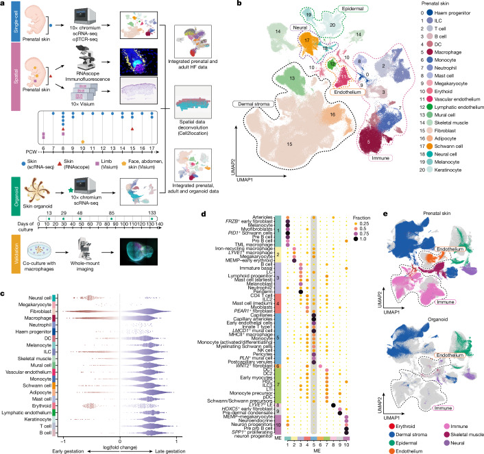
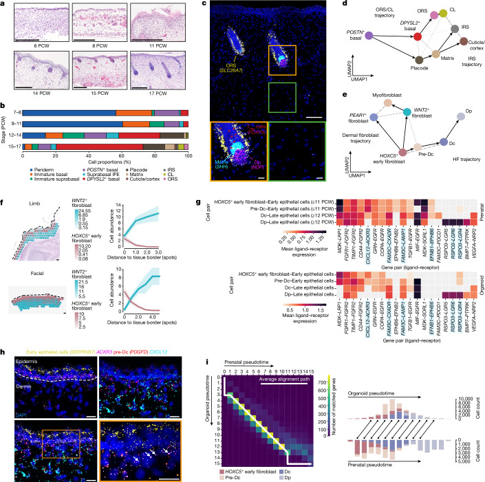
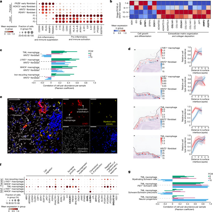
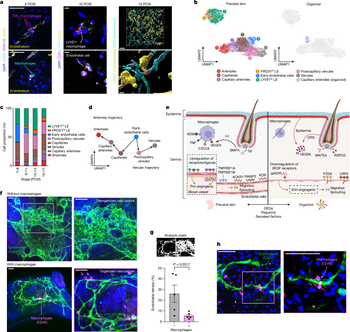
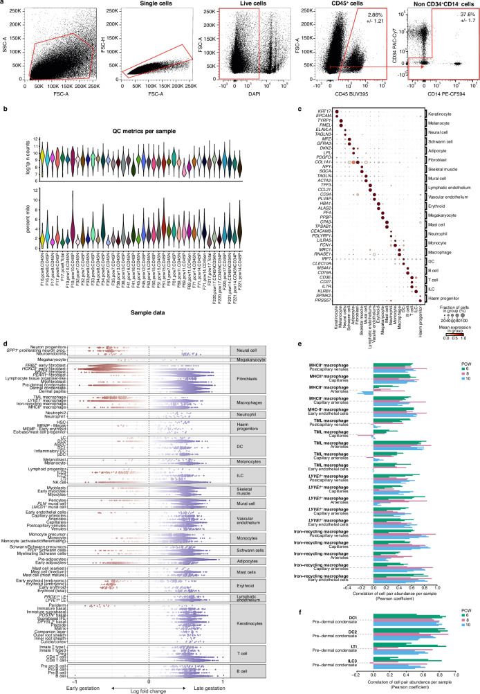
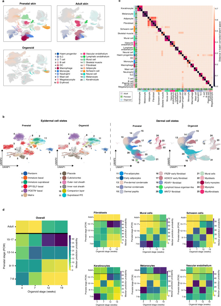
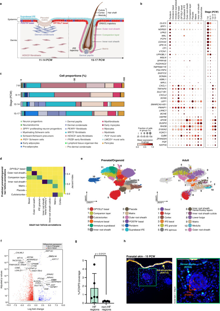
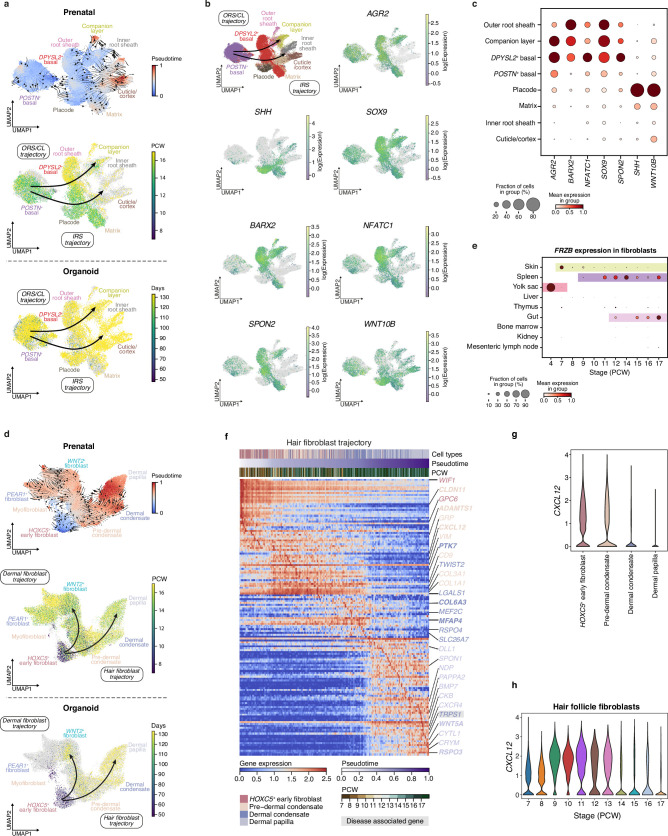
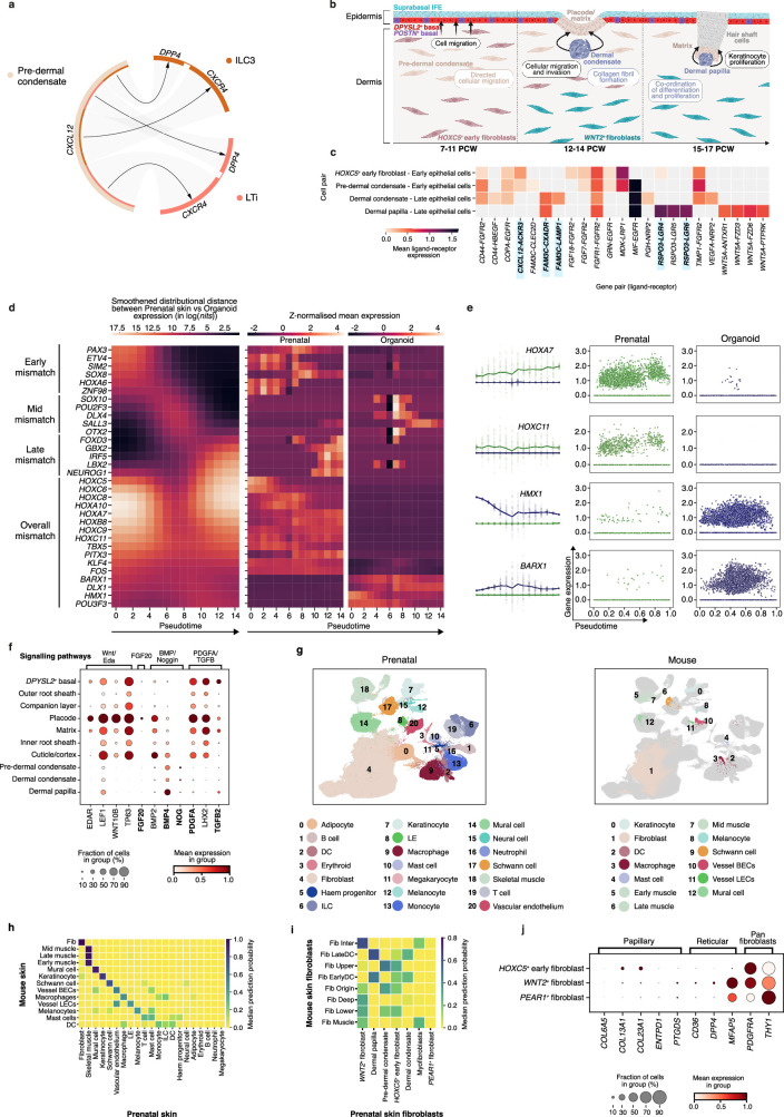
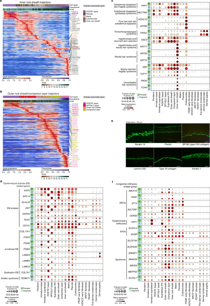
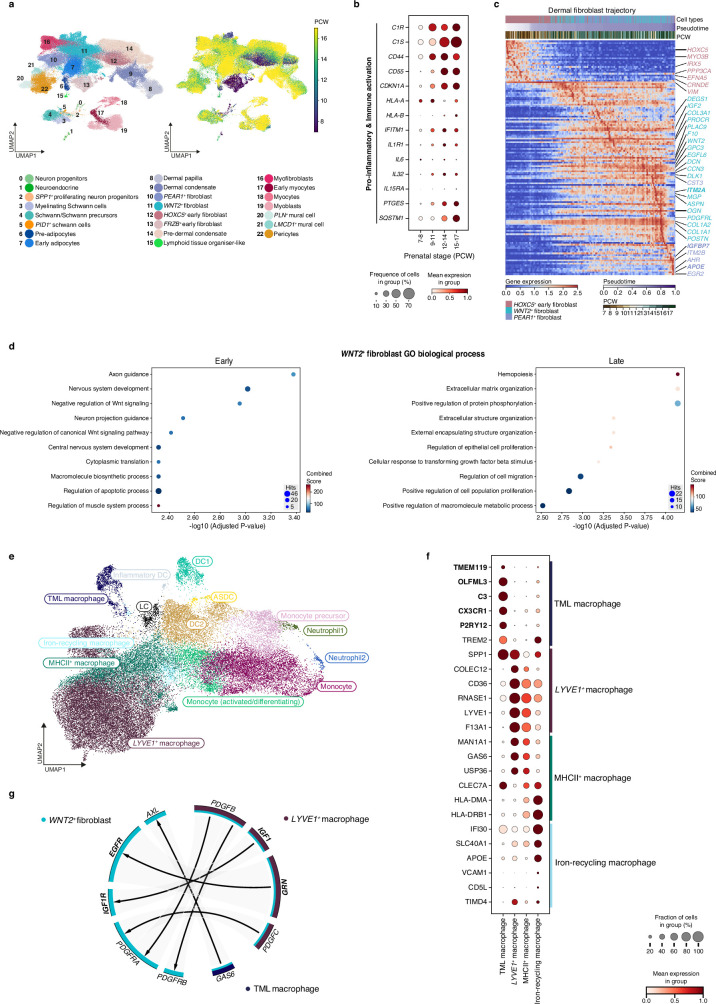
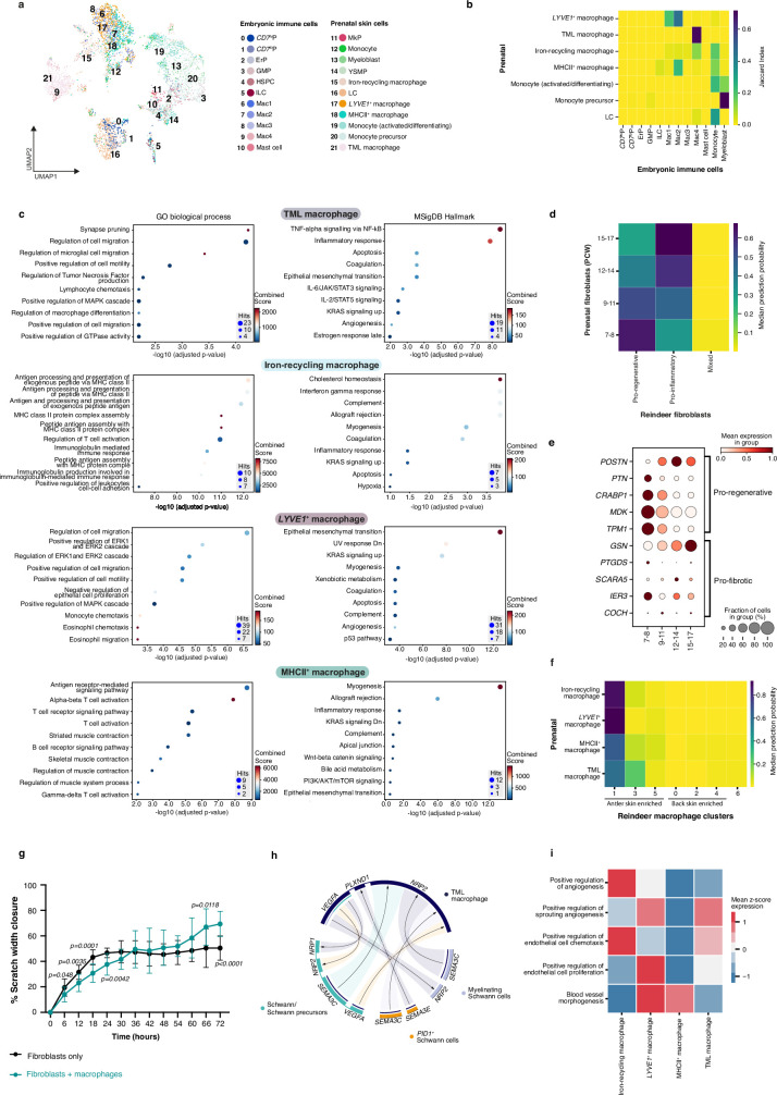
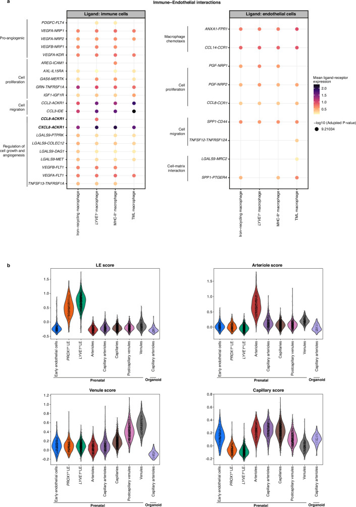
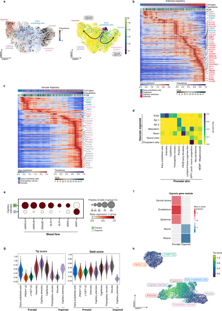
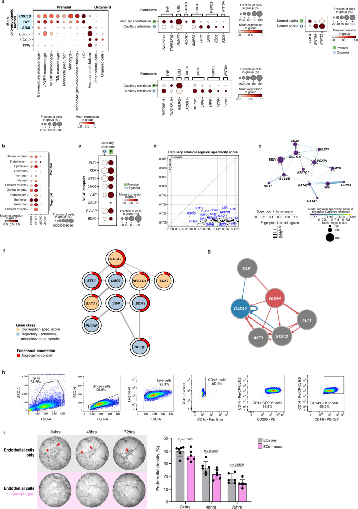
. The SkO model closely recapitulated in vivo skin epidermal and dermal cell types during hair follicle development and expression of genes implicated in the pathogenesis of genetic hair and skin disorders. However, the SkO model lacked immune cells and had markedly reduced endothelial cell heterogeneity and quantity. Our in vivo prenatal skin cell atlas indicated that macrophages and macrophage-derived growth factors have a role in driving endothelial development. Indeed, vascular network remodelling was enhanced following transfer of autologous macrophages derived from induced pluripotent stem cells into SkO cultures. Innate immune cells are therefore key players in skin morphogenesis beyond their conventional role in immunity, a function they achieve through crosstalk with non-immune cells.
Overview
The claims and statistics of this paper haven’t been checked yet.
Run the check — free, no account needed. It reads the open-access text and checks the sources it cites.
Integrity signals
Registry
Cites
Statistics
Figures
Verification
Claims the paper makes
The claims and statistics of this paper haven’t been checked yet.
Run the check — free, no account needed. It reads the open-access text and checks the sources it cites.
Claims other papers make about this one
Each is checked against this paper’s own text: does it say what the citing paper says it does?
The cited source doesn’t support the claim as written.
In the citing paper
“The dataset, collected by Jin et al.,8 consists of scratch assays performed on PC-3 prostate cancer cells with varying initial confluence prior to the scratch.”
From Inference of weak-form partial differential equations describing migration and proliferation mechanisms in wound healing experiments on cancer cells (2025) · cites this paper as [8]
Checked by a reasoning model — may be wrong.
Experimental — accuracy not yet measuredThe source backs part of the claim, not all of it — or backs it for a different population, condition or comparison.
In the citing paper
“By estimating parameters for these models, past work has helped identify quantitative effects of biological perturbations in scratch assays8,10,11.”
From Inference of weak-form partial differential equations describing migration and proliferation mechanisms in wound healing experiments on cancer cells (2025) · cites this paper as [8]
Experimental — accuracy not yet measuredThe source backs part of the claim, not all of it — or backs it for a different population, condition or comparison.
In the citing paper
“Moreover, vascular-immune interactions were primarily inferred from histological proximity, flow cytometry and gene-expression patterns; spatial transcriptomics and single-cell multiomics approaches will be valuable for elucidating cell-type-specific crosstalk at single-cell resolution 54,55 .”
From Vascularized human skin assembloids model compartmental skin organization and immune responsiveness in vitro (2026) · cites this paper as [55]
Experimental — accuracy not yet measuredThe source backs part of the claim, not all of it — or backs it for a different population, condition or comparison.
In the citing paper
“Recent advances in human induced pluripotent stem cell (hiPSC) technology have enabled the generation of skin organoids (SKO) with hair follicles, pigmentation, dermal stratification, appendage patterning, and innervation.[ 25 , 26 , 28 , 29 , 30 , 31 , 32 , 33 , 34 , 35 ]”
From Coordinated Development of Immune Cell Populations in Vascularized Skin Organoids from Human Induced Pluripotent Stem Cells (2025) · cites this paper as [35]
Experimental — accuracy not yet measuredThe source backs part of the claim, not all of it — or backs it for a different population, condition or comparison.
In the citing paper
“Previous human SKO models were devoid of vascularization and immune cells.[ 25 , 26 , 28 , 29 , 30 , 31 , 32 , 33 , 34 , 35 , 36 , 37 , 38 , 59 ]”
From Coordinated Development of Immune Cell Populations in Vascularized Skin Organoids from Human Induced Pluripotent Stem Cells (2025) · cites this paper as [35]
Experimental — accuracy not yet measured
The cited source says what the claim attributes to it.
In the citing paper
“Although macrophage co‐culture with SKOs has been reported,[ 35 ] a physiologically relevant vascularized hiPSC‐derived SKO has yet to be achieved.[ 36 , 37 , 38 ]”
From Coordinated Development of Immune Cell Populations in Vascularized Skin Organoids from Human Induced Pluripotent Stem Cells (2025) · cites this paper as [35]
The cited source says what the claim attributes to it.
In the citing paper
“Currently, SKOs lack vasculature and immune cells in a physiologically relevant context.[ 25 , 35 , 36 , 37 , 38 ]”
From Coordinated Development of Immune Cell Populations in Vascularized Skin Organoids from Human Induced Pluripotent Stem Cells (2025) · cites this paper as [35]
The check wasn’t confident either way, so it gives no verdict — never disputed.
In the citing paper
“First-principles modeling has established a variety of partial differential equation (PDE) models that accurately represent the behavior of scratch assays with a high density of cells and under a variety of conditions8,9.”
From Inference of weak-form partial differential equations describing migration and proliferation mechanisms in wound healing experiments on cancer cells (2025) · cites this paper as [8]
Experimental — accuracy not yet measuredThe check wasn’t confident either way, so it gives no verdict — never disputed.
In the citing paper
“There exist models that have considered density-dependent effects on migration, for instance representing cells interacting with each other, and on cell proliferation, for instance, modeling its saturation at high cell density8,27.”
From Inference of weak-form partial differential equations describing migration and proliferation mechanisms in wound healing experiments on cancer cells (2025) · cites this paper as [8]
Experimental — accuracy not yet measuredThe check wasn’t confident either way, so it gives no verdict — never disputed.
In the citing paper
“With it, we were able to rapidly identify an accurate model describing the evolution of cell concentration over time for a variety of initial seeding densities with accuracy that matched or improved upon results in the literature8.”
From Inference of weak-form partial differential equations describing migration and proliferation mechanisms in wound healing experiments on cancer cells (2025) · cites this paper as [8]
Experimental — accuracy not yet measuredThe check wasn’t confident either way, so it gives no verdict — never disputed.
In the citing paper
“As seen in Fig. 6A, the cell densities achieved are comparable to those observed by Jin et al.,8 and admit treatment of the problem using continuum advection-diffusion-reaction PDEs.”
From Inference of weak-form partial differential equations describing migration and proliferation mechanisms in wound healing experiments on cancer cells (2025) · cites this paper as [8]
Experimental — accuracy not yet measuredThe check wasn’t confident either way, so it gives no verdict — never disputed.
In the citing paper
“Lagergren et al. demonstrated the power of this approach on the Jin et al. dataset8 (also analyzed by Jin et al.), when they inferred density-dependent reaction-diffusion equations for cell migration10. They identified non-linear functions of concentration for diffusivity and reaction terms, and found that these functions vary with initial seeding concentration, consistent with our findings.”
From Inference of weak-form partial differential equations describing migration and proliferation mechanisms in wound healing experiments on cancer cells (2025) · cites this paper as [8]
Experimental — accuracy not yet measured
Self-check
Percentages, sums and reported differences re-computed from the paper’s own numbers.
The self-check needs the paper’s full text.
Citations
Cites
Fast, sensitive and accurate integration of single-cell data with Harmony
Korsunsky, I., Millard, N., Fan, J., Slowikowski, K., Zhang, F., Wei, K., Baglaenko, Y., Brenner, M., Loh, P.-R., & Raychaudhuri, S. (2019) · Nature Methods · doi:10.1038/s41592-019-0619-0 · Cited by 10,488
QuPath: Open source software for digital pathology image analysis
Bankhead, P., Loughrey, M. B., Fernández, J. A., Dombrowski, Y., McArt, D. G., Dunne, P. D., McQuaid, S., Gray, R. T., Murray, L. J., Coleman, H. G., et al. (2017) · Scientific Reports · doi:10.1038/s41598-017-17204-5 · Cited by 8,908
SCANPY: large-scale single-cell gene expression data analysis
Wolf, F. A., Angerer, P., & Theis, F. J. (2018) · Genome Biology · doi:10.1186/s13059-017-1382-0 · Cited by 8,881
Enrichr: interactive and collaborative HTML5 gene list enrichment analysis tool
Chen, E. Y., Tan, C. M., Kou, Y., Duan, Q., Wang, Z., Meirelles, G. V., Clark, N. R., & Ma’ayan, A. (2013) · BMC Bioinformatics · doi:10.1186/1471-2105-14-128 · Cited by 7,814
ImageJ2: ImageJ for the next generation of scientific image data
Rueden, C., Schindelin, J., Hiner, M., DeZonia, B. E., Walter, A., Arena, E. T., & Eliceiri, K. W. (2017) · BMC Bioinformatics · doi:10.1186/s12859-017-1934-z · Cited by 6,087
Macrophages in Tissue Repair, Regeneration, and Fibrosis
Wynn, T. A., & Vannella, K. M. (2016) · Immunity · doi:10.1016/j.immuni.2016.02.015 · Cited by 4,481
circlize implements and enhances circular visualization in R
Gu, Z., Gu, L., Eils, R., Schlesner, M., & Brors, B. (2014) · Bioinformatics · doi:10.1093/bioinformatics/btu393 · Cited by 4,290
Efremova, M., Vento-Tormo, M., Teichmann, S. A., & Vento-Tormo, R. (2020) · Nature Protocols · doi:10.1038/s41596-020-0292-x · Cited by 3,449
Self-citationThe Human Transcription Factors
Lambert, S. A., Jolma, A., Campitelli, L. F., Das, P. K., Yin, Y., Albu, M., Chen, X., Taipale, J., Hughes, T. R., & Weirauch, M. T. (2018) · Cell · doi:10.1016/j.cell.2018.01.029 · Cited by 3,399
Scrublet: computational identification of cell doublets in single-cell transcriptomic data
Wolock, S. L., Lopez, R., & Klein, A. M. (2018) · doi:10.1101/357368 · Cited by 2,758
Research works printed but not linked: 4
- Sadler, T. W. & Langman, J. Langman’s Medical Embryology (Lippincott Raven, 2000).
- Grubbs, H., Nassereddin, A. & Morrison, M. Embryology, Hair. StatPearls [Internet] https://www.ncbi.nlm.nih.gov/books/NBK534794/ (2022).
- Darby, I. A. & Desmoulière, A. in Textbook on Scar Management: State of the Art Management and Emerging Technologies (eds Téot, L. et al.) (Springer, 2020).
- Wallace, C. S. Statistical and Inductive Inference by Minimum Message Length (Springer Science & Business Media, 2005).
Cited by
Sorted by citations
The Human Cell Atlas from a cell census to a unified foundation model
Rood, J., Wynne, S., Robson, L., Hupalowska, A., Randell, J., Teichmann, S. A., & Regev, A. (2024) · Nature · doi:10.1038/s41586-024-08338-4 · Cited by 173
Self-citationHuman Wound and Its Burden: Updated 2025 Compendium of Estimates
Sen, C. K. (2025) · Advances in Wound Care · doi:10.1177/21621918251359554 · Cited by 115
Steele, L., Olabi, B., Roberts, K., Mazin, P., Koplev, S., Tudor, C. L., Rumney, B. M., Admane, C., Jiang, T., Correa‐Gallegos, D., et al. (2025) · Nature Immunology · doi:10.1038/s41590-025-02267-8 · Cited by 95
Self-citationThe spatial and temporal activation of macrophages during fibrosis
Behmoaras, J. V., Mulder, K., Ginhoux, F., & Petretto, E. G. (2025) · Nature reviews. Immunology · doi:10.1038/s41577-025-01186-x · Cited by 75
Sui, J., Hou, Y., Ding, C., Zheng, Z., Chen, M., Yu, H., Liu, L., Liu, R., Zhang, X., Xu, S., et al. (2025) · Advanced Materials · doi:10.1002/adma.202505061 · Cited by 38
Spatial architecture of development and disease
Lázár, E., & Lundeberg, J. (2025) · Nature Reviews Genetics · doi:10.1038/s41576-025-00892-5 · Cited by 38
Kan, L., Yu, Y., Wang, Y., Shi, L., Fan, T., Chen, H., & Ren, C. (2025) · Molecular Cancer · doi:10.1186/s12943-025-02328-4 · Cited by 32
Maternal stress triggers early-life eczema through fetal mast cell programming
Serhan, N., Abdullah, N. S., Ghéziel, N., Loste, A., Ekren, R., Labit, E., Gonzalez, A., Oliva, G., Tarot, P., Petitfils, C., et al. (2025) · Nature · doi:10.1038/s41586-025-09419-8 · Cited by 30
Wu, Y., Mao, J., Zhou, Y., Hong, G., Wu, H., Hu, Z., Huang, X., Shi, J., Xie, Z., & Lan, Y. (2025) · Research · doi:10.34133/research.0644 · Cited by 28
Qiu, S., Wang, Z., Guo, S., Xie, D., Cai, Y., Wang, X., Lin, C., Tang, S., Xie, Y., & Zhang, A. (2025) · Cell Insight · doi:10.1016/j.cellin.2025.100252 · Cited by 22
Single-cell transcriptomics identifies chondrocyte differentiation dynamics in vivo and in vitro
Lawrence, J. E. G., Woods Jon, S., Roberts, K., Sumanaweera, D., Balogh, P., Li, T., Predeus, A. V., He, P., Polański, K., Prigmore, E., et al. (2025) · Developmental Cell · doi:10.1016/j.devcel.2025.06.031 · Cited by 19
Self-citationPiezoelectric-immunomodulatory electrospun membrane for enhanced repair of refractory wounds
Liu, S. Y., Yuan, J., Nie, M.-D., Lin, X., Li, X., Luo, K., Huo, S., Bai, Y., & Mao, N. (2025) · Journal of Nanobiotechnology · doi:10.1186/s12951-025-03393-z · Cited by 18
Mostina, M., Sun, J., Sim, S. L., Ahmed, I. A., Souza‐Fonesca‐Guimaraes, F., Wolvetang, E. J., Brown, J., Kumari, S., Khosrotehrani, K., & Shafiee, A. (2025) · Advanced Healthcare Materials · doi:10.1002/adhm.202502108 · Cited by 17
Advances in engineered organoid models of skin for biomedical research
Zeng, D., Li, S., Du, F., Xia, Y., Zhang, J., Yu, S., & Qin, J. (2025) · Burns & Trauma · doi:10.1093/burnst/tkaf016 · Cited by 22
Microglia and neuroinflammation: function, heterogeneity, and crosstalk
Zong, S., Cui, X., Wu, S., & Lu, Z. (2026) · Cellular and Molecular Immunology · doi:10.1038/s41423-026-01438-3 · Cited by 28
Cytotoxic IgG: Mechanisms, functions, and applications
Wöhner, M., & Nimmerjahn, F. (2025) · Immunity · doi:10.1016/j.immuni.2025.05.008 · Cited by 18
Yan, Y., Li, M., Guo, L., Zhang, W., Wu, R., Guan, T., Ling, J., Yang, Y., Liu, M., Gu, X., et al. (2025) · Materials Today Bio · doi:10.1016/j.mtbio.2025.101957 · Cited by 14
A peripheral glial niche orchestrates the early stages of skin wound healing
Stierli, S., Salas-Bastos, A., Micheli, S., Ballwein, I., Kelemen, A., Lehmann, J., Loos, B., Jordaan, W., Stifter, S. A., Gwerder, M., et al. (2026) · Cell stem cell · doi:10.1016/j.stem.2025.12.015 · Cited by 16
Role of Fibroblast‐Immune Crosstalk in Kidney, Lung, and Skin Tertiary Lymphoid Structures
Cross, A. R., Shelley, J. R., Newman, R., Strid, J., & Denton, A. E. (2025) · Immunological Reviews · doi:10.1111/imr.70059 · Cited by 13
The role of multiomics in revealing the mechanism of skin repair and regeneration
Shi, S., Ou, X., Long, J., Lu, X., Xu, S., & Li, G. (2025) · Frontiers in Pharmacology · doi:10.3389/fphar.2025.1497988 · Cited by 14
Diversity of human skin three-dimensional organotypic cultures
Jia, Y. Y., & Atwood, S. X. (2024) · Current Opinion in Genetics & Development · doi:10.1016/j.gde.2024.102275 · Cited by 12
Singh, K., Koroma, A. K., Pandey, R., Wang, Y., Cheng, J., Haas, P., Aghapour, M., Krug, L., Hainzl, A., Wlaschek, M., et al. (2025) · Advanced Science · doi:10.1002/advs.202504920 · Cited by 14
Li, Z., Ou, F., Zhang, Z., Ning, C., Wang, F., Xie, T., Pan, W., Zhang, Z., Liang, Q., Zhao, S., et al. (2025) · Composites Science and Technology · doi:10.1016/j.compscitech.2025.111061 · Cited by 10
Cellular atlases are unlocking the mysteries of the human body
Yanai, I., Haas, S. F., Lippert, C., & Kretzmer, H. (2024) · Nature · doi:10.1038/d41586-024-03552-6 · Cited by 10
Zheng, M., An, S., Park, I. G., Kim, J., Kim, W.-S., Noh, M., & Sung, J.-H. (2024) · International Journal of Molecular Sciences · doi:10.3390/ijms26010095 · Cited by 9
Self-citationFetal Fibroblast Heterogeneity Defines Dermal Architecture during Human Embryonic Skin Development
Morioka, N., Gânier, C., & Watt, F. M. (2025) · Journal of Investigative Dermatology · doi:10.1016/j.jid.2024.12.027 · Cited by 10
Decoding Macrophage Dynamics: A Pathway to Understanding and Treating Inflammatory Skin Diseases
Gu, S., Xu, L., Bin, H., Xiong, K., Yang, X., & Ye, J. (2025) · International Journal of Molecular Sciences · doi:10.3390/ijms26094287 · Cited by 9
The microglial lineage: present and beyond
Chen, W., Wu, Z., & Li, H. (2025) · Trends in Immunology · doi:10.1016/j.it.2025.04.001 · Cited by 9
Generation and characterization of vestibular inner ear organoids from human pluripotent stem cells
van der Valk, W. H., Nist-Lund, C. A., Zhang, J., Perea, C., Jin, J., Gim, K. Y., Steinhart, M. R., Lee, J., & Koehler, K. R. (2025) · Nature Protocols · doi:10.1038/s41596-025-01191-3 · Cited by 12
Self-citationZhou, H., Zhang, Y.-X., Lei, Q.-K., Li, Y., & Zheng, Y. (2025) · Stem Cell Research & Therapy · doi:10.1186/s13287-025-04447-7 · Cited by 9
Luo, J., Zheng, D., Lu, B., Ding, X., Wang, M., Fang, B., Fang, S., & Qiu, H. (2025) · Materials & Design · doi:10.1016/j.matdes.2025.114247 · Cited by 7
Kageyama, T., Anakama, R., Hamano, S., Tu, S., Migita, Y., Asaba, T., Nanmo, A., Ishikawa, K., Yan, L., Seo, J., et al. (2026) · ACS Biomaterials Science & Engineering · doi:10.1021/acsbiomaterials.5c01780 · Cited by 6
An integrated single cell and spatial omics atlas of human prenatal developmentPreprint
Webb, S., Rose, A., Xu, C., Steele, L., Kuri, M. A., Stephenson, E., İnecik, K., Jafree, D. J., Foster, A. R., Basurto-Lozada, D., et al. (2026) · bioRxiv (Cold Spring Harbor Laboratory) · doi:10.64898/2026.03.30.714220 · Cited by 7
Self-citationSrivastava, S., Kinnunen, P. C., Wang, Z., Ho, K. K. Y., Humphries, B. A., Chen, S., Linderman, J. J., Luker, G. D., Luker, K. E., & Garikipati, K. (2025) · PLOS Computational Biology · doi:10.1371/journal.pcbi.1013607 · Cited by 5
Ghosh, S. K., Ghosh, S., Yeung, K., Xu, C., Wu, J., Mao, C., Ling, Z., Okuro, K., & Qiao, W. (2025) · Materials Today Bio · doi:10.1016/j.mtbio.2025.102219 · Cited by 5
Luo, Z., Huang, D., & Zhao, Y. (2025) · Small Science · doi:10.1002/smsc.202500322 · Cited by 5
Luminescent nanofibers for human skin textures photocopying
Tian, T., Han, H., Lin, X., Kang, H. D., Yang, M., Chen, Z., Wu, C., Jiang, Y., Li, W., Chang, X., et al. (2025) · Nature Communications · doi:10.1038/s41467-025-64703-5 · Cited by 5
Peter, S. E., Riaz, F., Li, Y., Zhao, X., & Pan, F. (2026) · Frontiers in Immunology · doi:10.3389/fimmu.2026.1737438 · Cited by 7
He, C., Wang, Y., Fang, X., Jiang, W., Liu, S., Yi, X., Zhang, K., Lin, H., Zeng, Q., Zhu, X., et al. (2025) · Journal of Materials Chemistry B · doi:10.1039/d5tb00683j · Cited by 5
Li, J., Tang, Y., Chen, P., Ding, Z., & Zhou, M. (2025) · Radiation Medicine and Protection · doi:10.1016/j.radmp.2025.09.005 · Cited by 5
Man, F., Wang, K., Yang, Z., An, L., You, C., Li, R., Cai, Y., Li, Y., Chen, L., Liu, Y., et al. (2026) · Interdisciplinary medicine · doi:10.1002/inmd.70076 · Cited by 4
Spatiotemporal decoding of skin biology: development, aging, disease, and regeneration
Xi, D., Huang, R. S., Li, Y., Xia, Z., & Ji, S. (2026) · Burns & Trauma · doi:10.1093/burnst/tkag019 · Cited by 5
Tang, Y., Ye, J., Xu, T., Sun, Y., Meng, W., Zhu, L., Jia, Z., Wu, Q., Chen, D., & Wu, F. (2025) · Journal of Cellular and Molecular Medicine · doi:10.1111/jcmm.70590 · Cited by 5
Hybrid living electronic skin for proactive scarless wound healing
Ma, Y., Xiong, C., Guo, F., Huang, M., Xie, X., Guo, Q., & Shi, P. (2025) · Biomaterials · doi:10.1016/j.biomaterials.2025.123721 · Cited by 4
Shi, R., Hu, C., Zhang, W., Wan, L., Shi, Y., Zhen, N., Mao, Z., Zhou, B., Luo, G., & Deng, J. (2026) · Advanced Materials · doi:10.1002/adma.202522987 · Cited by 6
Xu, Z., Chen, R., Lin, W., Wang, W., Tan, X., Huo, S., Liu, J., & Xu, G. (2025) · Colloids and Surfaces B Biointerfaces · doi:10.1016/j.colsurfb.2025.115162 · Cited by 3
He, H., Xiao, M., Song, L., Tian, Y., & Jia, Y. (2025) · British Journal of Dermatology · doi:10.1093/bjd/ljaf353 · Cited by 3
Immunological control of skin development: from homeostasis to developmental pathologies
Wei, Y., Gu, J., Zhao, Z., & Gu, B. (2025) · Frontiers in Immunology · doi:10.3389/fimmu.2025.1685633 · Cited by 4
A comparative review of single-cell atlases: mapping cellular diversity across species and tissues
Look, K., Goh, P. E., Ming, L. C., Law, S. H., Tan, S. L., Tan, E. W., Ser, H. L., & Goh, B. H. (2025) · Cellular and Molecular Life Sciences · doi:10.1007/s00018-025-05952-x · Cited by 3
Yue, L., Liang, Y., Zhong, P., Fang, L., Yun, Z., Su, Q., Li, C., Xiang, M., Zhang, J., Wang, J., et al. (2025) · Nature Communications · doi:10.1038/s41467-025-65848-z · Cited by 4
Zhang, H., Xiang, W., Tong, Y., Jiang, X., & Guo, L. (2025) · Clinical Reviews in Allergy & Immunology · doi:10.1007/s12016-025-09115-5 · Cited by 4
CellScope: high-performance cell atlas workflow with tree-structured representation
Li, B., Lin, R., Ni, T., Yan, G., Burns, M. J., Li, J. J., & Yao, Z. (2025) · Nature Communications · doi:10.1038/s41467-025-67890-3 · Cited by 3
Zhang, R., Chen, L., Xu, W., Ye, J., Wang, Z., Xu, W., & Wu, B. (2026) · International Endodontic Journal · doi:10.1111/iej.70098 · Cited by 3
Cohen, E., Xu, Y., Ghodke, S., Orosco, A., Wang, D., Johnson, C. N., Steen, K. A., Sarkar, M. K., Özlü, N., Tsoi, L. C., et al. (2026) · Science Translational Medicine · doi:10.1126/scitranslmed.adx9123 · Cited by 7
The Spectrum of Fibroblast Immune Biology
Guarnieri, A., & Turley, S. J. (2026) · Annual Review of Immunology · doi:10.1146/annurev-immunol-083024-035801 · Cited by 3
Molecular basis of variations in facial soft tissues: insights from cichlid fishes
Ahi, E. P., Sturmbauer, C., Albertson, R. C., & Singh, P. (2025) · Developmental Biology · doi:10.1016/j.ydbio.2025.10.014 · Cited by 3
Su, C., Pan, R. L., Li, H., Liang, R., Yuan, Y., Gou, T., Bai, T., Liu, L., Li, B., & Li, Y. (2025) · Materials & Design · doi:10.1016/j.matdes.2025.115276 · Cited by 2
Adar, L., Sharma, S., Schwartz, B., Alfer, T., Sharabi, O., Oss-Ronen, L., Gazit, R., & Cohen, I. (2025) · Cell Reports · doi:10.1016/j.celrep.2025.116648 · Cited by 2
Reversing vascular aging by restoring macrophages
Wei, J., Luo, Y., & Wang, F. (2026) · Trends in Endocrinology and Metabolism · doi:10.1016/j.tem.2025.12.007 · Cited by 2
Briganti, S., Camera, E., Ciriza, J., Fernández-Carro, E., Krivokuća, M. J., Lamartine, J., Letsiou, S., Nacka-Aleksić, M., Niessen, C. M., Oliva, N., et al. (2026) · Ageing Research Reviews · doi:10.1016/j.arr.2026.103109 · Cited by 4
Hindle, S. A., Brook, H. B., Chrysanthou, A., Chambers, E. S., Caley, M. P., & Connelly, J. T. (2025) · Advanced Science · doi:10.1002/advs.202415717 · Cited by 3
Prenatal Skin Cell Atlas reveals macrophages’ role beyond immunity
Pattaroni, C. (2024) · Immunology and Cell Biology · doi:10.1111/imcb.12837 · Cited by 1
Kim, J. S., Byun, J., Choi, J., Jin, D., Li, Q., Lee, J., Choi, H., & Oh, Y. (2025) · Advanced Science · doi:10.1002/advs.202511962 · Cited by 2
Self-citationCutaneous wound healing—insights from the matricellular perspective
Klaas, M., Mäemets‐Allas, K., Cárdenas‐León, C. G., & Jaks, V. (2025) · FEBS Letters · doi:10.1002/1873-3468.70220 · Cited by 2
Hu, L., Wang, Y., Liu, L., & Liu, X. (2025) · International Journal of Surgery · doi:10.1097/js9.0000000000004100 · Cited by 2
Getting the message: low-plex detection of RNA in tissues
Spencer‐Dene, B., & Miranda, E. (2026) · Frontiers in RNA Research · doi:10.3389/frnar.2026.1749833 · Cited by 1
Krüppel-like factor 2 programs early exhausted T cell states and restrains antiviral immunity
Geng, S., Li, Z., Li, W., Liang, H., Xu, F., Zhang, L., Zhang, W., Tao, S., Wang, Y., Zhou, Y., et al. (2026) · Immunity · doi:10.1016/j.immuni.2026.03.029 · Cited by 2
Hair follicle organoids: advances in construction, applications, and translational challenges
Yao, X., Ding, R., Chuanjian, Y., Zeng, D., Liu, Y., Zhang, J., & Zhang, J. (2026) · Frontiers in Cell and Developmental Biology · doi:10.3389/fcell.2026.1836905 · Cited by 2
He, Z., Wang, Q., Ding, L., Wang, Y., He, H., Han, W., Li, S., Fu, J., Luo, C., Li, J., et al. (2026) · Nature Cell Biology · doi:10.1038/s41556-026-01962-4 · Cited by 1
Macrophages in embryonic development
Mariani, S. A., Vaughan, H. J., Doyle, E. H., & Gordon, S. (2026) · Nature reviews. Immunology · doi:10.1038/s41577-026-01327-w · Cited by 1
Macrophages boosting human skin morphogenesis
Baumdick, M. E., & Bunders, M. J. (2024) · Trends in Immunology · doi:10.1016/j.it.2024.11.009 · Cited by 1
Recently discovered roles for macrophages in human skin development
McGrath, J. A. (2024) · British Journal of Dermatology · doi:10.1093/bjd/ljae473 · Cited by 1
Self-citation4D multimodal wound healing atlas reveals organ-level controls of repair phase transitionsPreprint
Cheong, J. C., Deursen, S. V., Amador, D., Hiner, S., & Woappi, Y. (2026) · bioRxiv (Cold Spring Harbor Laboratory) · doi:10.64898/2026.01.15.699736 · Cited by 1
Berrocal-Rubio, M. Á., Gleeson, J., Kato, M., Delobel, D., Kore, H., Beckhouse, A. G., Vijayan, D., Hitchens, K. J., Kasukawa, T., Yip, C. W., et al. (2026) · bioRxiv (Cold Spring Harbor Laboratory) · doi:10.64898/2026.02.01.703142 · Cited by 1
Kim, M.-J., Lee, S., & Kang, K.-S. (2026) · Experimental & Molecular Medicine · doi:10.1038/s12276-026-01840-x
Human macrophages in pancreas and skin shape prenatal organogenesis
Minton, K. (2024) · Nature reviews. Immunology · doi:10.1038/s41577-024-01106-5
(2024) · Alternatives to Laboratory Animals · doi:10.1177/02611929241308667
Wen, Z., & Dowe, D. L. (2024) · Entropy · doi:10.3390/e27010006
The Beauty of Fibroblasts Is Several Skin Layers Deep and Much Deeper
Scott, R. W. (2025) · Journal of Investigative Dermatology · doi:10.1016/j.jid.2025.02.010
Senescence-induced reparative fibroblasts enable scarless wound healing in aged murine skinPreprint
Wang, D., Wang, Z., Yang, Y., Bai, F., Li, H., He, J., Zhou, F., Chen, T., Fang, T., Wu, Z., et al. (2025) · bioRxiv (Cold Spring Harbor Laboratory) · doi:10.1101/2025.04.17.648896
Enhanced differentiation potential of pigmented human epidermal equivalents
Jia, Y. Y., Bajsert, J., Kurdian, A. I., Shiu, J., Pérez‐Aso, M., & Atwood, S. X. (2026) · Scientific Reports · doi:10.1038/s41598-026-63611-y
Govaerts, J., Goethals, C., Breedam, E. V., Sadzot‐Delvaux, C., Delputte, P., Ogunjimi, B., Lebrun, M., & Ponsaerts, P. (2025) · Microbiology and Molecular Biology Reviews · doi:10.1128/mmbr.00165-25
Düz, T., Törőcsik, D., Momnougui, S. M. E., Oubounyt, M., Al, B., Gallinat, S., Baumbach, J., & Holzscheck, N. (2025) · bioRxiv (Cold Spring Harbor Laboratory) · doi:10.1101/2025.09.09.675235
Early life determinants of skin-resident T cells
Wang, W., & Xiong, N. (2025) · Trends in Immunology · doi:10.1016/j.it.2025.10.011
Zhao, Y., Xie, T., Yi, Y., Zeng, X., Shao, S.-W., Xu, X., Gao, X., & Lei, M. (2025) · International Journal of Dermatology and Venereology · doi:10.1097/jd9.0000000000000446
Skin Immune System: Microanatomy
Ono, S., & Kabashima, K. (2016) · Elsevier eBooks · doi:10.1016/b978-0-128-24465-4.00504-4
Li, Y., Inoue, A., Fukuda, A., Niimura, M., & Tamai, K. (2026) · Journal of Leukocyte Biology · doi:10.1093/jleuko/qiaf185
The Role of Bone Marrow-Derived Cells (BMDCs) During Wound Healing
Okano, J., & Kojima, H. (2026) · Stem cell biology and regenerative medicine · doi:10.1007/978-3-032-12442-5_15
Predicting how perturbations reshape cellular trajectories with PerturbGenPreprint
Ly, K. C. H., Feriz, A. M., Isobe, T., Vahidi, A., Vaghari, D., Rostron, A., Londoño, M. Q., Mende, N., Vijayabaskar, M. S., Moullet, M., et al. (2026) · bioRxiv (Cold Spring Harbor Laboratory) · doi:10.64898/2026.03.04.709254 · Cited by 2
Self-citationObwegs, D., Oschwald, A., Koetter, L. M., Crisand, C., Doerr, S., Bruder, K., Runge, S., Ghanem, N., Fuchs, V., Eckert, M., et al. (2026) · bioRxiv (Cold Spring Harbor Laboratory) · doi:10.64898/2026.04.05.716534
Lee, H., Lee, S., Jo, S. J., Lee, S., Go, H., Kwon, O., & Kim, J. (2026) · Experimental & Molecular Medicine · doi:10.1038/s12276-026-01692-5
Self-citationPyroxylin shortens the resting stage of the hair cycle in mice
Kokabu, S., Tsuji, K., Ying-Ying, C., Washio, A., Shirakawa, T., Ono, Y., Yuan, Q., Kaminuma, O., & Matsubara, T. (2026) · Scientific Reports · doi:10.1038/s41598-026-52804-0
Xiao, T., Guo, T., Yuan, C., Zhang, W., Luo, Y., Tang, J., Lu, Z., Liang, B., & Liu, J. (2026) · Frontiers in Cell and Developmental Biology · doi:10.3389/fcell.2026.1764812 · Cited by 1
Dou, L., Du, Z., Tian, Y., Zhong, H., Huang, H., Xiang, R., Ni, Y., Zhang, X., Yan, W., Li, M., et al. (2026) · Environmental Pollution · doi:10.1016/j.envpol.2026.128529
Multimodal Human Scalp Atlas Defines Cell Landscape and Lineage Architecture In SituPreprint
Li, E. B., Stephens, C. M., Klay, M., Carcamo, A., Han, J., Quan, V. L., Colavincenzo, M. L., Pearlman, R. L., Yoo, S. S., Wang, D., et al. (2026) · bioRxiv (Cold Spring Harbor Laboratory) · doi:10.64898/2026.06.29.735239
An integrated skin cell atlas decodes the pilosebaceous unit
Düz, T., Törőcsik, D., Momnougui, S. M. E., Oubounyt, M., Al, B., Gallinat, S., Baumbach, J., & Holzscheck, N. (2026) · Journal of Investigative Dermatology · doi:10.1016/j.jid.2026.06.1282 · Cited by 1
Duan, L., Bu, Y., Zhou, P., Liu, G., Liao, F., Shi, J., Yang, X. X., Reis, R. L., Kundu, S. C., Wu, F., et al. (2026) · Biomaterials · doi:10.1016/j.biomaterials.2026.124490
Obwegs, D., Oschwald, A., Koetter, L. M., Crisand, C., Doerr, S., Bruder, K., Runge, S., Ghanem, N., Fuchs, V., Eckert, M., et al. (2026) · Science Advances · doi:10.1126/sciadv.aec9180
Metadata
- Authors
- Gopee, N. H., Winheim, E., Olabi, B., Admane, C., Foster, A. R., Huang, N., Botting, R. A., Torabi, F., Sumanaweera, D., Le, A. P., Kim, J., Verger, L., Stephenson, E., Adão, D., Ganier, C., Gim, K. Y., Serdy, S. A., Deakin, C., Goh, I., Steele, L., Annusver, K., Miah, M.-U., Tun, W. M., Moghimi, P., Kwakwa, K. A., Li, T., Basurto Lozada, D., Rumney, B., Tudor, C. L., Roberts, K., Chipampe, N.-J., Sidhpura, K., Englebert, J., Jardine, L., Reynolds, G., Rose, A., Rowe, V., Pritchard, S., Mulas, I., Fletcher, J., Popescu, D.-M., Poyner, E., Dubois, A., Guy, A., Filby, A., Lisgo, S., Barker, R. A., Glass, I. A., Park, J.-E., Vento-Tormo, R., Nikolova, M. T., He, P., Lawrence, J. E. G., Moore, J., Ballereau, S., Hale, C. B., Shanmugiah, V., Horsfall, D., Rajan, N., McGrath, J. A., O’Toole, E. A., Treutlein, B., Bayraktar, O., Kasper, M., Progatzky, F., Mazin, P., Lee, J., Gambardella, L., Koehler, K. R., Teichmann, S. A., & Haniffa, M.
- Affiliations
- Newcastle upon Tyne Hospitals NHS Foundation Trust (GB)
- Wellcome Sanger Institute (GB)
- Newcastle University (GB)
- Boston Children's Hospital (US)
- University of Oxford (GB)
- Karolinska Institutet (SE)
- Guy's Hospital (GB)
- Wellcome/MRC Cambridge Stem Cell Institute (GB)
- University of Washington (US)
- ETH Zurich (CH)
- University of California, San Francisco (US)
- German BioImaging – Gesellschaft für Mikroskopie und Bildanalyse (DE)
- King's College London (GB)
- Queen Mary University of London (GB)
- Corresponding author
- Koehler, Karl R.
- Teichmann, Sarah A.
- Haniffa, Muzlifah
- ORCID
- Gopee, Nusayhah Hudaa
- Winheim, Elena
- Olabi, Bayanne
- Admane, Chloe
- Foster, April Rose
- Huang, Ni
- Botting, Rachel A.
- Torabi, Fereshteh
- Sumanaweera, Dinithi
- Le, Anh Phuong
- Kim, Jin
- Verger, Luca
- Stephenson, Emily
- Adão, Diana
- Gim, Kelly Y.
- Serdy, Sara A.
- Goh, Issac
- Steele, Lloyd
- Annusver, Karl
- Miah, Mohi-Uddin
- Tun, Win Min
- Li, Tong
- Tudor, Catherine L.
- Roberts, Kenny
- Chipampe, Nana-Jane
- Sidhpura, Keval
- Jardine, Laura
- Reynolds, Gary
- Rose, Antony
- Pritchard, Sophie
- Mulas, Ilaria
- Poyner, Elizabeth
- Guy, Alyson
- Filby, Andrew
- Lisgo, Steven
- Barker, Roger A.
- Glass, Ian A.
- Park, Jong-Eun
- Vento-Tormo, Roser
- Nikolova, Marina Tsvetomilova
- He, Peng
- Lawrence, John E. G.
- Moore, Josh
- Hale, Christine B.
- Horsfall, David
- Rajan, Neil
- McGrath, John A.
- O’Toole, Edel A.
- Treutlein, Barbara
- Bayraktar, Omer
- Kasper, Maria
- Progatzky, Fränze
- Mazin, Pavel
- Lee, Jiyoon
- Gambardella, Laure
- Koehler, Karl R.
- Teichmann, Sarah A.
- Haniffa, Muzlifah
- Venue
- Nature
- Published
- 2024-10-16
- Volume
- 635
- Issue
- 8039
- Pages
- 679-689
- ISSN
- 0028-0836 (ISSN-L)
- 1476-4687
- PMC
- PMC11578897
- OpenAlex
- W4403450831
- Cited by
- 108
- References
- 108
- Citations by year
- 2026: 44 · 2025: 52 · 2024: 11
- FWCI
- 50.45
- Type
- journal article
- Language
- en
- Open access
- hybrid
- doi.org
- Licence
- Locations
- Nature (publishedVersion, cc-by-nc-nd)
- PubMed Central (publishedVersion, cc-by-nc-nd)
- PubMed (publishedVersion)
- RePEc: Research Papers in Economics (submittedVersion)
- eScholarship (California Digital Library) (submittedVersion, cc-by-nc-nd)
- Queen Mary Research Online (Queen Mary University of London) (submittedVersion, cc-by-nc-nd)
- Apollo (University of Cambridge) (submittedVersion, cc-by-nc-nd)
- Repository for Publications and Research Data (ETH Zurich) (submittedVersion, cc-by-nc-nd)
- ETH Zürich Research Collection
- Funders
- Wellcome Trust
- National Institute for Health and Care Research ((NIHR203312)
- Medical Research Council (MR/W015625/1)
- National Institute of Arthritis and Musculoskeletal and Skin Diseases (R01AR075018, R01AR083139)
- Eunice Kennedy Shriver National Institute of Child Health and Human Development (R24HD000836)
- Topics
- Health Sciences › Medicine › Dermatology › Hair Growth and Disorders
- Life Sciences › Biochemistry, Genetics and Molecular Biology › Cell Biology › Skin and Cellular Biology Research
- Life Sciences › Biochemistry, Genetics and Molecular Biology › Molecular Biology › Developmental Biology and Gene Regulation
- Keywords
- skin morphogenesis, immune regulation, macrophages, endothelial development, hair follicle development, induced pluripotent stem cells, innate immune cells, skin diseases
- MeSH terms
- Human Embryonic Stem Cells, Multiomics, Adult, Atlases as Topic, Female, Fetus, Humans, Immunity, Innate, Macrophages, Morphogenesis, Organoids, Skin, Skin Diseases, Wound Healing, Hair Follicle, Neovascularization, Physiologic, Endothelial Cells, Induced Pluripotent Stem Cells, Single-Cell Analysis, Transcriptome
About these checks
Registry, retraction, Cites and statistics results are computed, not judged, with one exception: a printed reference that no index lists is read once by a model before it counts as a citation error. Claims are extracted by a model and kept only when their quote appears verbatim in the text; each cited source is then checked, with a model’s help, for whether it says what the claim relies on. Those verdicts can be wrong — read the claim’s quote and the cited source before relying on one.
Statistics and arithmetic are recomputed, not judged.
Confidence bands are shown only for fields calibrated on our annotated test set. None is calibrated yet, so no band is shown.
What counts as a claim ›How we measure confidence ›How we check this ›
Are you an author, or do you see a problem with this page? Request a review
