

The DOI matches its Crossref record; 107 of the 108 references it cites were found in a registry or an index.
A prenatal skin atlas reveals immune regulation of human skin morphogenesis
Open accessAbstract
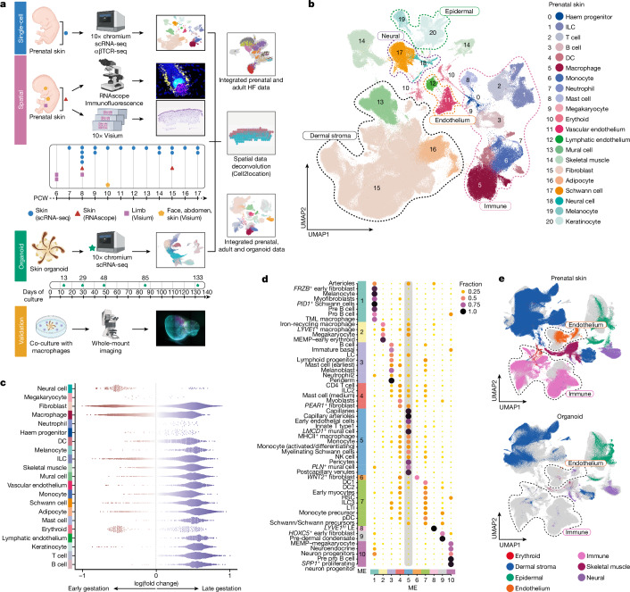
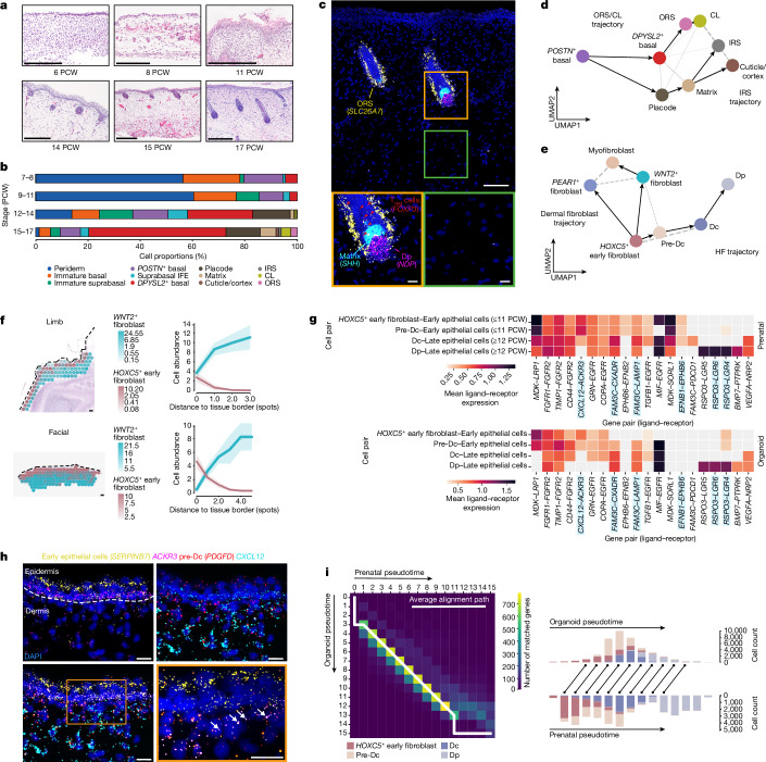
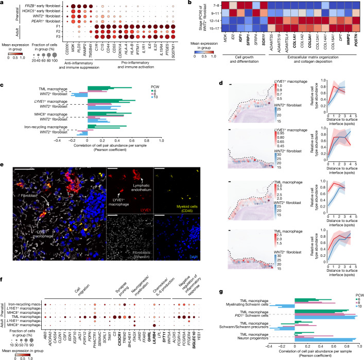
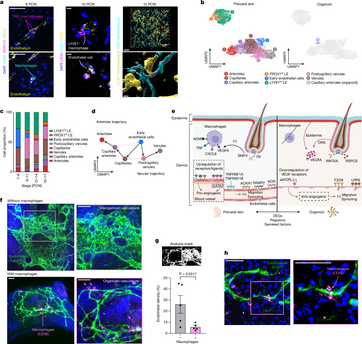
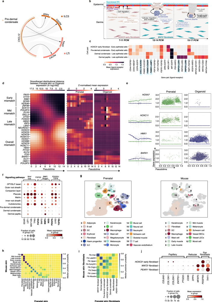
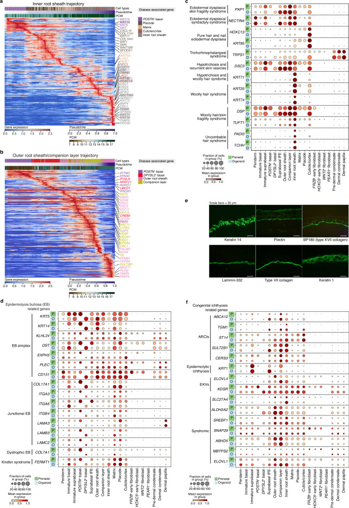
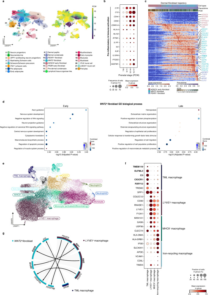
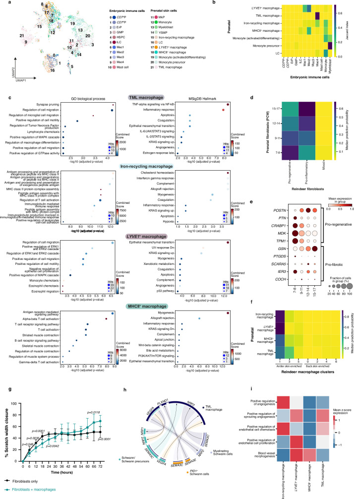
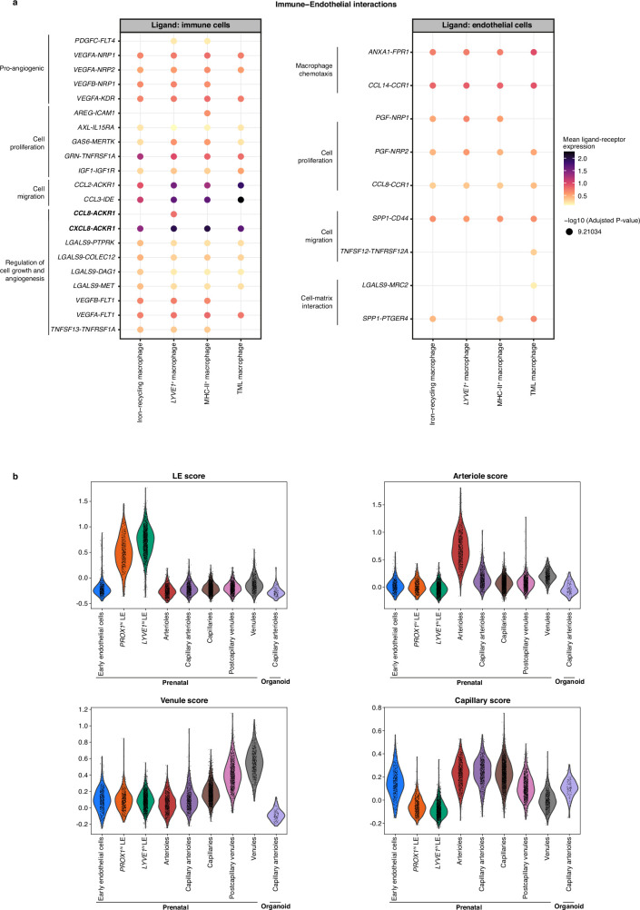
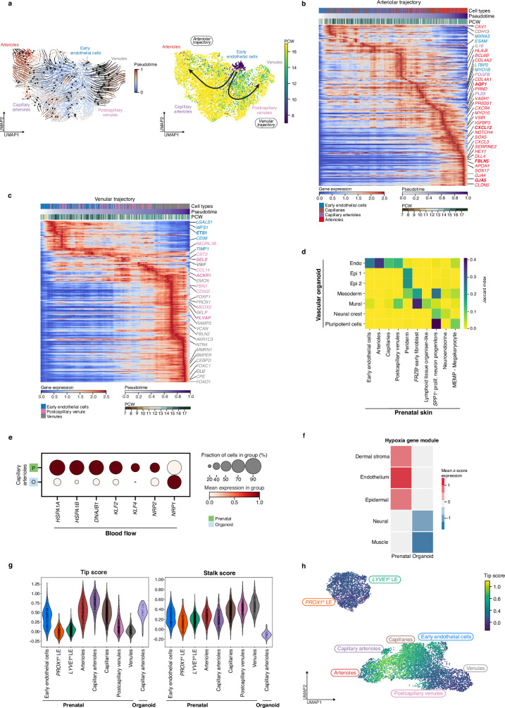
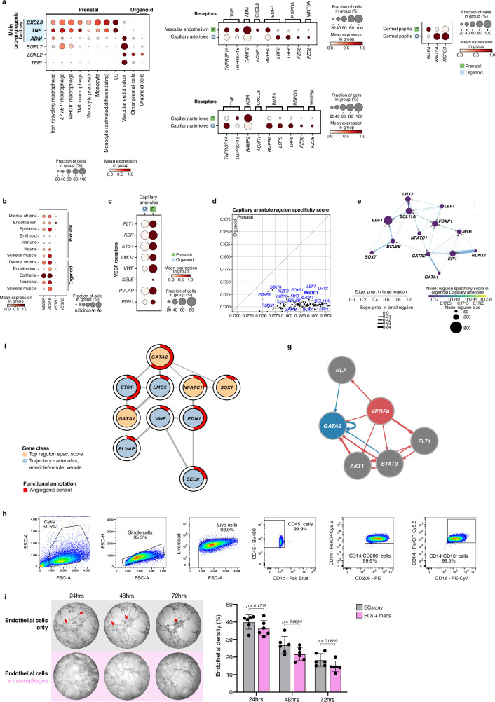
. The SkO model closely recapitulated in vivo skin epidermal and dermal cell types during hair follicle development and expression of genes implicated in the pathogenesis of genetic hair and skin disorders. However, the SkO model lacked immune cells and had markedly reduced endothelial cell heterogeneity and quantity. Our in vivo prenatal skin cell atlas indicated that macrophages and macrophage-derived growth factors have a role in driving endothelial development. Indeed, vascular network remodelling was enhanced following transfer of autologous macrophages derived from induced pluripotent stem cells into SkO cultures. Innate immune cells are therefore key players in skin morphogenesis beyond their conventional role in immunity, a function they achieve through crosstalk with non-immune cells.
Overview
The claims and statistics of this paper haven’t been checked yet.
Run the check — free, no account needed. It reads the open-access text and checks the sources it cites.
Integrity signals
Registry
Cites
Statistics
Figures
Verification
Claims the paper makes
The claims and statistics of this paper haven’t been checked yet.
Run the check — free, no account needed. It reads the open-access text and checks the sources it cites.
Claims other papers make about this one
Each is checked against this paper’s own text: does it say what the citing paper says it does?
The cited source doesn’t support the claim as written.
In the citing paper
“The dataset, collected by Jin et al.,8 consists of scratch assays performed on PC-3 prostate cancer cells with varying initial confluence prior to the scratch.”
From Inference of weak-form partial differential equations describing migration and proliferation mechanisms in wound healing experiments on cancer cells (2025) · cites this paper as [8]
Checked by a reasoning model — may be wrong.
Experimental — accuracy not yet measuredThe source backs part of the claim, not all of it — or backs it for a different population, condition or comparison.
In the citing paper
“By estimating parameters for these models, past work has helped identify quantitative effects of biological perturbations in scratch assays8,10,11.”
From Inference of weak-form partial differential equations describing migration and proliferation mechanisms in wound healing experiments on cancer cells (2025) · cites this paper as [8]
Experimental — accuracy not yet measuredThe source backs part of the claim, not all of it — or backs it for a different population, condition or comparison.
In the citing paper
“Moreover, vascular-immune interactions were primarily inferred from histological proximity, flow cytometry and gene-expression patterns; spatial transcriptomics and single-cell multiomics approaches will be valuable for elucidating cell-type-specific crosstalk at single-cell resolution 54,55 .”
From Vascularized human skin assembloids model compartmental skin organization and immune responsiveness in vitro (2026) · cites this paper as [55]
Experimental — accuracy not yet measuredThe source backs part of the claim, not all of it — or backs it for a different population, condition or comparison.
In the citing paper
“Recent advances in human induced pluripotent stem cell (hiPSC) technology have enabled the generation of skin organoids (SKO) with hair follicles, pigmentation, dermal stratification, appendage patterning, and innervation.[ 25 , 26 , 28 , 29 , 30 , 31 , 32 , 33 , 34 , 35 ]”
From Coordinated Development of Immune Cell Populations in Vascularized Skin Organoids from Human Induced Pluripotent Stem Cells (2025) · cites this paper as [35]
Experimental — accuracy not yet measuredThe source backs part of the claim, not all of it — or backs it for a different population, condition or comparison.
In the citing paper
“Previous human SKO models were devoid of vascularization and immune cells.[ 25 , 26 , 28 , 29 , 30 , 31 , 32 , 33 , 34 , 35 , 36 , 37 , 38 , 59 ]”
From Coordinated Development of Immune Cell Populations in Vascularized Skin Organoids from Human Induced Pluripotent Stem Cells (2025) · cites this paper as [35]
Experimental — accuracy not yet measured
The cited source says what the claim attributes to it.
In the citing paper
“Although macrophage co‐culture with SKOs has been reported,[ 35 ] a physiologically relevant vascularized hiPSC‐derived SKO has yet to be achieved.[ 36 , 37 , 38 ]”
From Coordinated Development of Immune Cell Populations in Vascularized Skin Organoids from Human Induced Pluripotent Stem Cells (2025) · cites this paper as [35]
The cited source says what the claim attributes to it.
In the citing paper
“Currently, SKOs lack vasculature and immune cells in a physiologically relevant context.[ 25 , 35 , 36 , 37 , 38 ]”
From Coordinated Development of Immune Cell Populations in Vascularized Skin Organoids from Human Induced Pluripotent Stem Cells (2025) · cites this paper as [35]
The check wasn’t confident either way, so it gives no verdict — never disputed.
In the citing paper
“First-principles modeling has established a variety of partial differential equation (PDE) models that accurately represent the behavior of scratch assays with a high density of cells and under a variety of conditions8,9.”
From Inference of weak-form partial differential equations describing migration and proliferation mechanisms in wound healing experiments on cancer cells (2025) · cites this paper as [8]
Experimental — accuracy not yet measuredThe check wasn’t confident either way, so it gives no verdict — never disputed.
In the citing paper
“There exist models that have considered density-dependent effects on migration, for instance representing cells interacting with each other, and on cell proliferation, for instance, modeling its saturation at high cell density8,27.”
From Inference of weak-form partial differential equations describing migration and proliferation mechanisms in wound healing experiments on cancer cells (2025) · cites this paper as [8]
Experimental — accuracy not yet measuredThe check wasn’t confident either way, so it gives no verdict — never disputed.
In the citing paper
“With it, we were able to rapidly identify an accurate model describing the evolution of cell concentration over time for a variety of initial seeding densities with accuracy that matched or improved upon results in the literature8.”
From Inference of weak-form partial differential equations describing migration and proliferation mechanisms in wound healing experiments on cancer cells (2025) · cites this paper as [8]
Experimental — accuracy not yet measuredThe check wasn’t confident either way, so it gives no verdict — never disputed.
In the citing paper
“As seen in Fig. 6A, the cell densities achieved are comparable to those observed by Jin et al.,8 and admit treatment of the problem using continuum advection-diffusion-reaction PDEs.”
From Inference of weak-form partial differential equations describing migration and proliferation mechanisms in wound healing experiments on cancer cells (2025) · cites this paper as [8]
Experimental — accuracy not yet measuredThe check wasn’t confident either way, so it gives no verdict — never disputed.
In the citing paper
“Lagergren et al. demonstrated the power of this approach on the Jin et al. dataset8 (also analyzed by Jin et al.), when they inferred density-dependent reaction-diffusion equations for cell migration10. They identified non-linear functions of concentration for diffusivity and reaction terms, and found that these functions vary with initial seeding concentration, consistent with our findings.”
From Inference of weak-form partial differential equations describing migration and proliferation mechanisms in wound healing experiments on cancer cells (2025) · cites this paper as [8]
Experimental — accuracy not yet measured
Self-check
Percentages, sums and reported differences re-computed from the paper’s own numbers.
The self-check needs the paper’s full text.
Citations
Cites
Fast, sensitive and accurate integration of single-cell data with Harmony
Korsunsky, I., Millard, N., Fan, J., Slowikowski, K., Zhang, F., Wei, K., Baglaenko, Y., Brenner, M., Loh, P.-R., & Raychaudhuri, S. (2019) · Nature Methods · doi:10.1038/s41592-019-0619-0 · Cited by 10,488
QuPath: Open source software for digital pathology image analysis
Bankhead, P., Loughrey, M. B., Fernández, J. A., Dombrowski, Y., McArt, D. G., Dunne, P. D., McQuaid, S., Gray, R. T., Murray, L. J., Coleman, H. G., et al. (2017) · Scientific Reports · doi:10.1038/s41598-017-17204-5 · Cited by 8,908
SCANPY: large-scale single-cell gene expression data analysis
Wolf, F. A., Angerer, P., & Theis, F. J. (2018) · Genome Biology · doi:10.1186/s13059-017-1382-0 · Cited by 8,881
Enrichr: interactive and collaborative HTML5 gene list enrichment analysis tool
Chen, E. Y., Tan, C. M., Kou, Y., Duan, Q., Wang, Z., Meirelles, G. V., Clark, N. R., & Ma’ayan, A. (2013) · BMC Bioinformatics · doi:10.1186/1471-2105-14-128 · Cited by 7,814
ImageJ2: ImageJ for the next generation of scientific image data
Rueden, C., Schindelin, J., Hiner, M., DeZonia, B. E., Walter, A., Arena, E. T., & Eliceiri, K. W. (2017) · BMC Bioinformatics · doi:10.1186/s12859-017-1934-z · Cited by 6,087
Macrophages in Tissue Repair, Regeneration, and Fibrosis
Wynn, T. A., & Vannella, K. M. (2016) · Immunity · doi:10.1016/j.immuni.2016.02.015 · Cited by 4,481
circlize implements and enhances circular visualization in R
Gu, Z., Gu, L., Eils, R., Schlesner, M., & Brors, B. (2014) · Bioinformatics · doi:10.1093/bioinformatics/btu393 · Cited by 4,290
Efremova, M., Vento-Tormo, M., Teichmann, S. A., & Vento-Tormo, R. (2020) · Nature Protocols · doi:10.1038/s41596-020-0292-x · Cited by 3,449
Self-citationThe Human Transcription Factors
Lambert, S. A., Jolma, A., Campitelli, L. F., Das, P. K., Yin, Y., Albu, M., Chen, X., Taipale, J., Hughes, T. R., & Weirauch, M. T. (2018) · Cell · doi:10.1016/j.cell.2018.01.029 · Cited by 3,399
Scrublet: computational identification of cell doublets in single-cell transcriptomic data
Wolock, S. L., Lopez, R., & Klein, A. M. (2018) · doi:10.1101/357368 · Cited by 2,758
Scrublet: computational identification of cell doublets in single-cell transcriptomic data
Wolock, S. L., Lopez, R., & Klein, A. M. (2019) · Cell Systems · doi:10.1016/j.cels.2018.11.005 · Cited by 2,758
Deep generative modeling for single-cell transcriptomics
Lopez, R., Regier, J., Cole, M. B., Jordan, M. I., & Yosef, N. (2018) · Nature Methods · doi:10.1038/s41592-018-0229-2 · Cited by 2,530
NicheNet: Modeling intercellular communication by linking ligands to target genes
Browaeys, R., Saelens, W., & Saeys, Y. (2020) · Nature Methods · doi:10.1038/s41592-019-0667-5 · Cited by 2,192
Metagenes and molecular pattern discovery using matrix factorization
Brunet, J.-P., Tamayo, P., Golub, T. R., & Mesirov, J. P. (2004) · Proceedings of the National Academy of Sciences · doi:10.1073/pnas.0308531101 · Cited by 2,183
A flexible R package for nonnegative matrix factorization
Gaujoux, R., & Seoighe, C. (2010) · BMC Bioinformatics · doi:10.1186/1471-2105-11-367 · Cited by 1,516
A scalable SCENIC workflow for single-cell gene regulatory network analysis
Van de Sande, B., Flerin, C., Davie, K., De Waegeneer, M., Hulselmans, G., Aibar, S., Seurinck, R., Saelens, W., Cannoodt, R., Rouchon, Q., et al. (2020) · Nature Protocols · doi:10.1038/s41596-020-0336-2 · Cited by 1,494
Distinct fibroblast lineages determine dermal architecture in skin development and repair
Driskell, R. R., Lichtenberger, B. M., Hoste, E., Kretzschmar, K., Simons, B. D., Charalambous, M., Ferron, S. R., Herault, Y., Pavlovic, G., Ferguson-Smith, A. C., et al. (2013) · Nature · doi:10.1038/nature12783 · Cited by 1,291
Cell2location maps fine-grained cell types in spatial transcriptomics
Kleshchevnikov, V., Shmatko, A., Dann, E., Aivazidis, A., King, H. W., Li, T., Elmentaite, R., Lomakin, A., Kedlian, V., Gayoso, A., et al. (2022) · Nature Biotechnology · doi:10.1038/s41587-021-01139-4 · Cited by 1,279
Self-citationCross-tissue immune cell analysis reveals tissue-specific features in humans
Conde, C. D., Xu, C., Jarvis, L. B., Rainbow, D. B., Wells, S. B., Gomes, T., Howlett, S. K., Suchánek, O., Polański, K., King, H. W., et al. (2022) · Science · doi:10.1126/science.abl5197 · Cited by 1,246
Self-citationFantin, A., Vieira, J. M., Gestri, G., Denti, L., Schwarz, Q., Prykhozhij, S., Peri, F., Wilson, S. W., & Ruhrberg, C. (2010) · Blood · doi:10.1182/blood-2009-12-257832 · Cited by 1,153
A single-cell molecular map of mouse gastrulation and early organogenesis
Pijuan-Sala, B., Griffiths, J. A., Guibentif, C., Hiscock, T. W., Jawaid, W., Calero-Nieto, F. J., Mulas, C., Ibarra-Soria, X., Tyser, R. C. V., Ho, D. L. L., et al. (2019) · Nature · doi:10.1038/s41586-019-0933-9 · Cited by 1,033
Differential abundance testing on single-cell data using k-nearest neighbor graphs
Dann, E., Henderson, N. C., Teichmann, S. A., Morgan, M. D., & Marioni, J. C. (2022) · Nature Biotechnology · doi:10.1038/s41587-021-01033-z · Cited by 938
Self-citationBBKNN: fast batch alignment of single cell transcriptomes
Polański, K., Young, M. D., Miao, Z., Meyer, K. B., Teichmann, S. A., & Park, J.-E. (2020) · Bioinformatics · doi:10.1093/bioinformatics/btz625 · Cited by 932
Self-citationHas, C., Bauer, J. W., Bodemer, C., Bolling, M. C., Bruckner‐Tuderman, L., Diem, A., Fine, J., Heagerty, A., Hovnanian, A., Marinkovich, M. P., et al. (2020) · British Journal of Dermatology · doi:10.1111/bjd.18921 · Cited by 800
Self-citationCellRank for directed single-cell fate mapping
Lange, M., Bergen, V., Klein, M., Setty, M. N., Reuter, B., Bakhti, M., Lickert, H., Ansari, M., Gote-Schniering, J., Schiller, H. B., et al. (2022) · Nature Methods · doi:10.1038/s41592-021-01346-6 · Cited by 783
Alivernini, S., MacDonald, L., Elmesmari, A., Finlay, S., Tolusso, B., Gigante, M. R., Petricca, L., Di Mario, C., Bui, L., Perniola, S., et al. (2020) · Nature Medicine · doi:10.1038/s41591-020-0939-8 · Cited by 740
Regulatory T Cells in Skin Facilitate Epithelial Stem Cell Differentiation
Ali, N., Zirak, B., Rodriguez, R. S., Pauli, M. L., Truong, H.-A., Lai, K., Ahn, R., Corbin, K., Lowe, M. M., Scharschmidt, T. C., et al. (2017) · Cell · doi:10.1016/j.cell.2017.05.002 · Cited by 680
Generation of vascular endothelial and smooth muscle cells from human pluripotent stem cells
Patsch, C., Meylan, L. C., Thoma, E., Urich, E., Heckel, T., O’Sullivan, J., Grainger, S. J., Kapp, F., Sun, L., Christensen, K., et al. (2015) · Nature Cell Biology · doi:10.1038/ncb3205 · Cited by 643
Tissue-specific macrophages: how they develop and choreograph tissue biology
Mass, E., Nimmerjahn, F., Kierdorf, K., & Schlitzer, A. (2023) · Nature Reviews Immunology · doi:10.1038/s41577-023-00848-y · Cited by 621
Single-cell transcriptomes of the human skin reveal age-related loss of fibroblast priming
Solé-Boldo, L., Raddatz, G., Schütz, S., Mallm, J.-P., Rippe, K., Lonsdorf, A. S., Rodríguez-Paredes, M., & Lyko, F. (2020) · Communications Biology · doi:10.1038/s42003-020-0922-4 · Cited by 559
Developmental cell programs are co-opted in inflammatory skin disease
Reynolds, G., Vegh, P., Fletcher, J., Poyner, E. F. M., Stephenson, E., Goh, I., Botting, R. A., Huang, N., Olabi, B., Dubois, A., et al. (2021) · Science · doi:10.1126/science.aba6500 · Cited by 529
Self-citationSouporcell: robust clustering of single-cell RNA-seq data by genotype without reference genotypes
Heaton, H., Talman, A. M., Knights, A., Imaz, M., Gaffney, D. J., Durbin, R., Hemberg, M., & Lawniczak, M. K. N. (2020) · Nature Methods · doi:10.1038/s41592-020-0820-1 · Cited by 506 · cited 1×
“Possible maternal contamination (total of 118 cells) was identified using the souporcell pipeline (v.2.4.0) 88 as previously described 5,58 .”
InfluentialFibroblast-macrophage reciprocal interactions in health, fibrosis, and cancer
Buechler, M. B., Fu, W., & Turley, S. J. (2021) · Immunity · doi:10.1016/j.immuni.2021.04.021 · Cited by 494
Deciphering human macrophage development at single-cell resolution
Bian, Z., Gong, Y., Huang, T., Lee, C., Bian, L., Bai, Z., Shi, H., Zeng, Y., Liu, C., He, J., et al. (2020) · Nature · doi:10.1038/s41586-020-2316-7 · Cited by 494
Hair-bearing human skin generated entirely from pluripotent stem cells
Lee, J., Rabbani, C. C., Gao, H., Steinhart, M. R., Woodruff, B. M., Pflum, Z. E., Kim, A., Heller, S., Liu, Y., Shipchandler, T. Z., et al. (2020) · Nature · doi:10.1038/s41586-020-2352-3 · Cited by 487
Self-citationScarless Fetal Wound Healing: A Basic Science Review
Larson, B. J., Longaker, M. T., & Lorenz, H. P. (2010) · Plastic & Reconstructive Surgery · doi:10.1097/prs.0b013e3181eae781 · Cited by 467
Integrated Single-Cell Atlas of Endothelial Cells of the Human Lung
Schupp, J. C., Adams, T. S., Cosme, C., Raredon, M. S. B., Yuan, Y., Omote, N., Poli, S., Chioccioli, M., Rose, K.-A., Manning, E. P., et al. (2021) · Circulation · doi:10.1161/circulationaha.120.052318 · Cited by 453
Self-citationQuestioning the fetal microbiome illustrates pitfalls of low-biomass microbial studies
Kennedy, K. M., de Goffau, M. C., Pérez-Muñoz, M. E., Arrieta, M., Bäckhed, F., Bork, P., Braun, T., Bushman, F. D., Dore, J. M., de Vos, W. M., et al. (2023) · Nature · doi:10.1038/s41586-022-05546-8 · Cited by 415
BigStitcher: reconstructing high-resolution image datasets of cleared and expanded samples
Hörl, D., Rusak, F. R., Preußer, F., Tillberg, P. W., Randel, N., Chhetri, R. K., Cardona, A., Keller, P., Harz, H., Leonhardt, H., et al. (2019) · Nature Methods · doi:10.1038/s41592-019-0501-0 · Cited by 366
The Molecular Anatomy of Mouse Skin during Hair Growth and Rest
Joost, S., Annusver, K., Jacob, T., Sun, X., Dalessandri, T., Sivan, U., Sequeira, I., Sandberg, R., & Kasper, M. (2020) · Cell stem cell · doi:10.1016/j.stem.2020.01.012 · Cited by 333
Self-citationMapping the developing human immune system across organs
Suo, C., Dann, E., Goh, I., Jardine, L., Kleshchevnikov, V., Park, J.-E., Botting, R. A., Stephenson, E., Engelbert, J., Tuong, Z. K., et al. (2022) · Science · doi:10.1126/science.abo0510 · Cited by 323
Self-citationDIMINISHED INTERLEUKIN 6 (IL-6) PRODUCTION DURING SCARLESS HUMAN FETAL WOUND REPAIR
Liechty, K. W., Adzick, N. S., & Crombleholme, T. M. (2000) · Cytokine · doi:10.1006/cyto.1999.0598 · Cited by 259
Abbasi, S., Sinha, S., Labit, E., Rosin, N. L., Yoon, G., Rahmani, W. M., Jaffer, A., Sharma, N. C., Hagner, A., Shah, P. T., et al. (2020) · Cell stem cell · doi:10.1016/j.stem.2020.07.008 · Cited by 203
Microglia-containing human brain organoids for the study of brain development and pathology
Zhang, W., Jiang, J., Xu, Z., Yan, H., Tang, B., Liu, C., Chen, C., & Meng, Q. (2022) · Molecular Psychiatry · doi:10.1038/s41380-022-01892-1 · Cited by 195
Fibroblast inflammatory priming determines regenerative versus fibrotic skin repair in reindeer
Sinha, S., Sparks, H. D., Labit, E., Robbins, H. N., Gowing, K., Jaffer, A., Kutluberk, E., Arora, R., Raredon, M. S. B., Cao, L., et al. (2022) · Cell · doi:10.1016/j.cell.2022.11.004 · Cited by 188
The Biology of Fetal Wound Healing
Longaker, M. T., & Adzick, N. S. (1991) · Plastic & Reconstructive Surgery · doi:10.1097/00006534-199104000-00032 · Cited by 182
Quinonez, S. C., & Innis, J. W. (2013) · Molecular Genetics and Metabolism · doi:10.1016/j.ymgme.2013.10.012 · Cited by 181
Ghersi, G., Zhao, Q., Salamone, M., Yeh, Y., Zucker, S., & Chen, W. (2006) · Cancer Research · doi:10.1158/0008-5472.can-05-1245 · Cited by 169
Blood and immune development in human fetal bone marrow and Down syndrome
Jardine, L., Webb, S., Goh, I., Londoño, M. Q., Reynolds, G., Mather, M. W., Olabi, B., Stephenson, E., Botting, R. A., Horsfall, D., et al. (2021) · Nature · doi:10.1038/s41586-021-03929-x · Cited by 164
Self-citationLGR5 expressing skin fibroblasts define a major cellular hub perturbed in scleroderma
Gur, C., Wang, S.-Y., Sheban, F., Zada, M., Li, B., Kharouf, F., Peleg, H., Aamar, S., Yalin, A., Kirschenbaum, D., et al. (2022) · Cell · doi:10.1016/j.cell.2022.03.011 · Cited by 159
Wang, Z., Wu, Z., Wang, H., Feng, R., Wang, G., Li, M., Wang, S.-Y., Chen, X., Su, Y., Wang, J., et al. (2023) · Cell · doi:10.1016/j.cell.2023.08.019 · Cited by 158
Congenital Ichthyosis: An Overview of Current and Emerging Therapies
Vahlquist, A., Gånemo, A., & Virtanen, M. (2008) · Acta Dermato Venereologica · doi:10.2340/00015555-0415 · Cited by 151
VCAM-1+ macrophages guide the homing of HSPCs to a vascular niche
Li, D., Xue, W., Li, M., Dong, M., Wang, J., Wang, X., Li, X., Chen, K., Zhang, W., Wu, S., et al. (2018) · Nature · doi:10.1038/s41586-018-0709-7 · Cited by 150
An updated classification of hair follicle morphogenesis
Saxena, N., Mok, K., & Rendl, M. (2019) · Experimental Dermatology · doi:10.1111/exd.13913 · Cited by 146
Snail-induced claudin-11 prompts collective migration for tumour progression
Li, C.-F., Chen, J.-Y., Ho, Y.-H., Hsu, W., Wu, L.-C., Lan, H., Hsu, D. S., Tai, S., Chang, Y., & Yang, M. (2019) · Nature Cell Biology · doi:10.1038/s41556-018-0268-z · Cited by 144
A human embryonic limb cell atlas resolved in space and time
Zhang, B., He, P., Lawrence, J. E. G., Wang, S., Tuck, E., Williams, B. A., Roberts, K., Kleshchevnikov, V., Mamanova, L., Bolt, L., et al. (2024) · Nature · doi:10.1038/s41586-023-06806-x · Cited by 143
Self-citationThe Duffy antigen/receptor for chemokines (DARC) regulates prostate tumor growth
Shen, H., Schuster, R. M., Stringer, K. F., Waltz, S. E., & Lentsch, A. B. (2006) · The FASEB Journal · doi:10.1096/fj.05-4764com · Cited by 139
Human Dermal Fibroblast Subpopulations Are Conserved across Single-Cell RNA Sequencing Studies
Ascensión, A. M., Fuertes-Álvarez, S., Ibáñez-Solé, O., Izeta, A., & Araúzo-Bravo, M. J. (2020) · Journal of Investigative Dermatology · doi:10.1016/j.jid.2020.11.028 · Cited by 131
The role of macrophages in skin homeostasis
Yanez, D. A., Lacher, R. K., Vidyarthi, A., & Colegio, O. R. (2017) · Pflügers Archiv - European Journal of Physiology · doi:10.1007/s00424-017-1953-7 · Cited by 117
Yolk sac cell atlas reveals multiorgan functions during human early development
Goh, I., Botting, R. A., Rose, A., Webb, S., Engelbert, J., Gitton, Y., Stephenson, E., Londoño, M. Q., Mather, M., Mende, N., et al. (2023) · Science · doi:10.1126/science.add7564 · Cited by 111
Self-citationMok, K., Saxena, N., Heitman, N., Grisanti, L., Srivastava, D., Muraro, M. J., Jacob, T., Sennett, R., Wang, Z., Su, Y., et al. (2018) · Developmental Cell · doi:10.1016/j.devcel.2018.11.034 · Cited by 110
Self-citationHu, B., Lefort, K., Qiu, W., Nguyen, B.-C., Rajaram, R. D., Castillo, E., He, F., Chen, Y., Angel, P. E., Brisken, C., et al. (2010) · Genes & Development · doi:10.1101/gad.1886910 · Cited by 106
Single-cell transcriptome profiling reveals vascular endothelial cell heterogeneity in human skin
Li, Q., Zhu, Z., Wang, L., Lin, Y., Fang, H., Lei, J., Cao, T., Wang, G., & Dang, E. (2021) · Theranostics · doi:10.7150/thno.54917 · Cited by 103
Biggs, L. C., Mäkelä, O. J., Myllymäki, S.-M., Roy, R. D., Närhi, K. M., Pispa, J., Mustonen, T., & Mikkola, M. L. (2018) · eLife · doi:10.7554/elife.36468 · Cited by 92
Cariboni, A. M., Hickok, J. R., Rakić, S., Andrews, W. D., Maggi, R., Tischkau, S. A., & Parnavelas, J. G. (2007) · Journal of Neuroscience · doi:10.1523/jneurosci.5075-06.2007 · Cited by 92
Origin and Differentiation of Nerve-Associated Macrophages
Kolter, J., Kierdorf, K., & Henneke, P. (2020) · The Journal of Immunology · doi:10.4049/jimmunol.1901077 · Cited by 91
Nunan, R. D., Campbell, J., Mori, R., Pitulescu, M. E., Jiang, W. G., Harding, K. G., Adams, R. H., Nobes, C. D., & Martin, P. B. (2015) · Cell Reports · doi:10.1016/j.celrep.2015.09.085 · Cited by 85
Dillenburg-Pilla, P., Patel, V., Mikelis, C. M., Zárate-Bladés, C. R., Doçi, C. L., Amornphimoltham, P., Wang, Z., Martı́n, D., Leelahavanichkul, K., Dorsam, R. T., et al. (2014) · The FASEB Journal · doi:10.1096/fj.14-260083 · Cited by 83
Defining Transcriptional Signatures of Human Hair Follicle Cell States
Takahashi, R., Grzenda, A. L., Allison, T. F., Rawnsley, J. D., Balin, S. J., Sabri, S., Plath, K., & Lowry, W. E. (2019) · Journal of Investigative Dermatology · doi:10.1016/j.jid.2019.07.726 · Cited by 81
Molecular and spatial landmarks of early mouse skin development
Jacob, T., Annusver, K., Czarnewski, P., Dalessandri, T., Kalk, C., Levron, C. L., Sanz, N. C., Kastriti, M. E., Mikkola, M. L., Rendl, M., et al. (2023) · Developmental Cell · doi:10.1016/j.devcel.2023.07.015 · Cited by 80
Self-citationGoto, M., Sawamura, D., Ito, K., Abe, M., Nishie, W., Sakai, K., Shibaki, A., Akiyama, M., & Shimizu, H. (2006) · Journal of Investigative Dermatology · doi:10.1038/sj.jid.5700117 · Cited by 79
Fate and state transitions during human blood vessel organoid development
Nikolova, M. T., He, Z., Seimiya, M., Jonsson, G., Cao, W., Okuda, R., Wimmer, R., Okamoto, R., Nikoloff, J. M., Dittrich, P. S., et al. (2025) · Cell · doi:10.1016/j.cell.2025.03.037 · Cited by 79
Self-citationHair Follicle Terminal Differentiation Is Orchestrated by Distinct Early and Late Matrix Progenitors
Mesler, A. L., Veniaminova, N. A., Lull, M. V., & Wong, S. Y. (2017) · Cell Reports · doi:10.1016/j.celrep.2017.03.077 · Cited by 69
Shamloul, G., & Khachemoune, A. (2020) · Dermatologic Therapy · doi:10.1111/dth.14695 · Cited by 67
The chemokine SDF-1/CXCL12 regulates the migration of melanocyte progenitors in mouse hair follicles
Belmadani, A. A., Jung, H., Ren, D., & Miller, R. J. (2009) · Differentiation · doi:10.1016/j.diff.2008.10.015 · Cited by 66
Coma, S., Allard‐Ratick, M., Akino, T., van Meeteren, L. A., Mammoto, A., & Klagsbrun, M. (2013) · Angiogenesis · doi:10.1007/s10456-013-9370-9 · Cited by 60
A Crucial Role of CXCL14 for Promoting Regulatory T Cells Activation in Stroke
Lee, H., Liu, S., Lin, C.-H., Lee, S. W., Hsu, C. Y., Sytwu, H., Hsieh, C., & Shyu, W. (2017) · Theranostics · doi:10.7150/thno.17558 · Cited by 60
Henn, D., Chen, K., Fehlmann, T., Trotsyuk, A. A., Sivaraj, D., Maan, Z. N., Bonham, C. A., Barrera, J. A., Mays, C. J., Greco, A. H., et al. (2021) · Science Advances · doi:10.1126/sciadv.abi4528 · Cited by 53
Hagner, A., Shin, W., Sinha, S., Alpaugh, W., Workentine, M. L., Abbasi, S., Rahmani, W. M., Agabalyan, N. A., Sharma, N. C., Sparks, H. D., et al. (2020) · iScience · doi:10.1016/j.isci.2020.101019 · Cited by 51
Strobel, H. A., Moss, S. M., & Hoying, J. B. (2023) · Bioengineering · doi:10.3390/bioengineering10020124 · Cited by 49
Zheng, C., Yin, S., Yang, Y., Yu, Y., & Xie, X. (2017) · Cellular and Molecular Immunology · doi:10.1038/cmi.2016.57 · Cited by 45
Immune and vascular contributions to organogenesis of the testis and ovary
Gu, X., Li, S., & DeFalco, T. (2021) · FEBS Journal · doi:10.1111/febs.15848 · Cited by 45
Robust temporal map of human in vitro myelopoiesis using single-cell genomics
Alsinet, C., Primo, M. N., Lorenzi, V., Bello, E., Kelava, I., Jones, C. P., Vilarrasa‐Blasi, R., Sancho‐Serra, C., Knights, A. J., Park, J.-E., et al. (2022) · Nature Communications · doi:10.1038/s41467-022-30557-4 · Cited by 45
Self-citationSynaptotagmin‐11 inhibits cytokine secretion and phagocytosis in microglia
Du, C., Wang, Y., Zhang, F., Yan, S., Guan, Y., Gong, X., Zhang, T., Cui, X., Wang, X., & Zhang, C. X. (2017) · Glia · doi:10.1002/glia.23186 · Cited by 44
Randall, V. A., & Botchkareva, N. V. (2009) · Elsevier eBooks · doi:10.1016/b978-0-8155-1572-2.50006-3 · Cited by 41
Huitema, L. F. A., Phillips, T., Alexeev, V. Y., & Igoucheva, O. (2021) · Experimental Dermatology · doi:10.1111/exd.14411 · Cited by 41
Gene-level alignment of single-cell trajectories
Sumanaweera, D., Suo, C., Cujba, A.-M., Muraro, D., Dann, E., Polański, K., Steemers, A. S., Lee, W., Oliver, A. J., Park, J.-E., et al. (2024) · Nature Methods · doi:10.1038/s41592-024-02378-4 · Cited by 41
Self-citationEmbryology of the hair follicle
Muller‐Bolla, M., Jasmin, J. R., Monteil, R. A., & Loubière, R. (1991) · Early Human Development · doi:10.1016/0378-3782(91)90155-v · Cited by 37
Abbasi, S., Sinha, S., Labit, E., Rosin, N. L., Yoon, G., Rahmani, W. M., Jaffer, A., Sharma, N. C., Hagner, A., Shah, P. T., et al. (2021) · Cell stem cell · doi:10.1016/j.stem.2021.02.004 · Cited by 35
WebAtlas pipeline for integrated single-cell and spatial transcriptomic data
Li, T., Horsfall, D., Basurto-Lozada, D., Roberts, K., Prete, M., Lawrence, J. E. G., He, P., Tuck, E., Moore, J., Yoldaş, A., et al. (2024) · Nature Methods · doi:10.1038/s41592-024-02371-x · Cited by 33
Self-citationBack to the Future: From Appendage Development Toward Future Human Hair Follicle Neogenesis
de Groot, S. C., Ulrich, M. M. W., Gho, C. G., & Huisman, M. A. (2021) · Frontiers in Cell and Developmental Biology · doi:10.3389/fcell.2021.661787 · Cited by 33
Hair-bearing human skin generated entirely from pluripotent stem cellsPreprint
Lee, J., Rabbani, C. C., Gao, H., Steinhart, M. R., Woodruff, B. M., Pflum, Z. E., Kim, A., Heller, S., Liu, Y., Shipchandler, T. Z., et al. (2019) · bioRxiv (Cold Spring Harbor Laboratory) · doi:10.1101/684282 · Cited by 31
Self-citationHe, Y., Tacconi, C., Dieterich, L. C., Kim, J., Restivo, G., Gousopoulos, E., Lindenblatt, N., Levesque, M. P., Claassen, M., & Detmar, M. J. (2022) · Cells · doi:10.3390/cells11071111 · Cited by 30
Self-citationXu, Y., Zhang, J., Hu, Y., Li, X., Sun, L., Yu, P., Sun, Y., Liu, B., Bian, Z., & Rong, Z. (2021) · Cell Reports · doi:10.1016/j.celrep.2021.109524 · Cited by 27
Developing Human Skin Contains Lymphocytes Demonstrating a Memory Signature
Dhariwala, M. O., Karthikeyan, D., Vasquez, K. S., Farhat, S., Weckel, A., Taravati, K., Leitner, E. G., Clancy, S., Pauli, M. L., Piper, M. L., et al. (2020) · Cell Reports Medicine · doi:10.1016/j.xcrm.2020.100132 · Cited by 26
Huang, M., Smith, A., Watson, M. C., Bhandari, R., Baugh, L. M., Ivanovska, I. L., Watkins, T., Lang, I. M., Trojanowska, M. E., Black, L. D., et al. (2022) · Arthritis & Rheumatology · doi:10.1002/art.42097 · Cited by 23
Optimal binning: mathematical programming formulationPreprint
Navas-Palencia, G. (2020) · arXiv (Cornell University) · doi:10.48550/arxiv.2001.08025 · Cited by 22
The developing immune network in human prenatal skin
Botting, R. A., & Haniffa, M. A. (2020) · Immunology · doi:10.1111/imm.13192 · Cited by 19
Self-citationThe Tempo of Mammalian Embryogenesis: Variation in the Pace of Brain and Body Development
Halley, A. C. (2022) · Brain Behavior and Evolution · doi:10.1159/000523715 · Cited by 19
Robins, E. J., & Breathnach, A. S. (1970) · Europe PMC (PubMed Central) · Cited by 18
Understanding immune profiles in ichthyosis may lead to novel therapeutic targets
Akiyama, M. (2022) · Journal of Allergy and Clinical Immunology · doi:10.1016/j.jaci.2022.02.010 · Cited by 15
Martišová, A., Sommerová, L., Krejčí, A., Selingerová, I., Kolářová, T., Kokas, F. Z., Holánek, M., Podhorec, J., Kazda, T., & Hrstka, R. (2022) · International Journal of Molecular Sciences · doi:10.3390/ijms231810845 · Cited by 12
Immunological Aspects of AXL/GAS‐6 in the Context of Human Liver Regeneration
Ortmayr, G., Brunnthaler, L., Pereyra, D., Huber, H., Santol, J., Rumpf, B., Najarnia, S., Smoot, R. L., Ammon, D., Sorz, T., et al. (2021) · Hepatology Communications · doi:10.1002/hep4.1832 · Cited by 10
Yin, S., Chen, F., Ye, P., & Yang, G. (2018) · Europe PMC (PubMed Central) · Cited by 10
Li, Q., Zhu, Z., Wang, L., Lin, Y., Fang, H., Lei, J., Cao, T., Gang, W., & Dang, E. (2021) · Journal of Investigative Dermatology · doi:10.1016/j.jid.2021.02.627 · Cited by 8
haniffalab/FCA_fetal_skin: 0.1.0
Nhgopee, Horsfall, D., Dannda, E., & Goh, I. (2023) · Zenodo (CERN European Organization for Nuclear Research) · doi:10.5281/zenodo.8164271 · Cited by 1
Self-citation
Research works printed but not linked: 4
- Sadler, T. W. & Langman, J. Langman’s Medical Embryology (Lippincott Raven, 2000).
- Grubbs, H., Nassereddin, A. & Morrison, M. Embryology, Hair. StatPearls [Internet] https://www.ncbi.nlm.nih.gov/books/NBK534794/ (2022).
- Darby, I. A. & Desmoulière, A. in Textbook on Scar Management: State of the Art Management and Emerging Technologies (eds Téot, L. et al.) (Springer, 2020).
- Wallace, C. S. Statistical and Inductive Inference by Minimum Message Length (Springer Science & Business Media, 2005).
Cited by
Sorted by citations
The Human Cell Atlas from a cell census to a unified foundation model
Rood, J., Wynne, S., Robson, L., Hupalowska, A., Randell, J., Teichmann, S. A., & Regev, A. (2024) · Nature · doi:10.1038/s41586-024-08338-4 · Cited by 173
Self-citationHuman Wound and Its Burden: Updated 2025 Compendium of Estimates
Sen, C. K. (2025) · Advances in Wound Care · doi:10.1177/21621918251359554 · Cited by 115
Steele, L., Olabi, B., Roberts, K., Mazin, P., Koplev, S., Tudor, C. L., Rumney, B. M., Admane, C., Jiang, T., Correa‐Gallegos, D., et al. (2025) · Nature Immunology · doi:10.1038/s41590-025-02267-8 · Cited by 95
Self-citationThe spatial and temporal activation of macrophages during fibrosis
Behmoaras, J. V., Mulder, K., Ginhoux, F., & Petretto, E. G. (2025) · Nature reviews. Immunology · doi:10.1038/s41577-025-01186-x · Cited by 75
Sui, J., Hou, Y., Ding, C., Zheng, Z., Chen, M., Yu, H., Liu, L., Liu, R., Zhang, X., Xu, S., et al. (2025) · Advanced Materials · doi:10.1002/adma.202505061 · Cited by 38
Metadata
- Authors
- Gopee, N. H., Winheim, E., Olabi, B., Admane, C., Foster, A. R., Huang, N., Botting, R. A., Torabi, F., Sumanaweera, D., Le, A. P., Kim, J., Verger, L., Stephenson, E., Adão, D., Ganier, C., Gim, K. Y., Serdy, S. A., Deakin, C., Goh, I., Steele, L., Annusver, K., Miah, M.-U., Tun, W. M., Moghimi, P., Kwakwa, K. A., Li, T., Basurto Lozada, D., Rumney, B., Tudor, C. L., Roberts, K., Chipampe, N.-J., Sidhpura, K., Englebert, J., Jardine, L., Reynolds, G., Rose, A., Rowe, V., Pritchard, S., Mulas, I., Fletcher, J., Popescu, D.-M., Poyner, E., Dubois, A., Guy, A., Filby, A., Lisgo, S., Barker, R. A., Glass, I. A., Park, J.-E., Vento-Tormo, R., Nikolova, M. T., He, P., Lawrence, J. E. G., Moore, J., Ballereau, S., Hale, C. B., Shanmugiah, V., Horsfall, D., Rajan, N., McGrath, J. A., O’Toole, E. A., Treutlein, B., Bayraktar, O., Kasper, M., Progatzky, F., Mazin, P., Lee, J., Gambardella, L., Koehler, K. R., Teichmann, S. A., & Haniffa, M.
- Affiliations
- Newcastle upon Tyne Hospitals NHS Foundation Trust (GB)
- Wellcome Sanger Institute (GB)
- Newcastle University (GB)
- Boston Children's Hospital (US)
- University of Oxford (GB)
- Karolinska Institutet (SE)
- Guy's Hospital (GB)
- Wellcome/MRC Cambridge Stem Cell Institute (GB)
- University of Washington (US)
- ETH Zurich (CH)
- University of California, San Francisco (US)
- German BioImaging – Gesellschaft für Mikroskopie und Bildanalyse (DE)
- King's College London (GB)
- Queen Mary University of London (GB)
- Corresponding author
- Koehler, Karl R.
- Teichmann, Sarah A.
- Haniffa, Muzlifah
- ORCID
- Gopee, Nusayhah Hudaa
- Winheim, Elena
- Olabi, Bayanne
- Admane, Chloe
- Foster, April Rose
- Huang, Ni
- Botting, Rachel A.
- Torabi, Fereshteh
- Sumanaweera, Dinithi
- Le, Anh Phuong
- Kim, Jin
- Verger, Luca
- Stephenson, Emily
- Adão, Diana
- Gim, Kelly Y.
- Serdy, Sara A.
- Goh, Issac
- Steele, Lloyd
- Annusver, Karl
- Miah, Mohi-Uddin
- Tun, Win Min
- Li, Tong
- Tudor, Catherine L.
- Roberts, Kenny
- Chipampe, Nana-Jane
- Sidhpura, Keval
- Jardine, Laura
- Reynolds, Gary
- Rose, Antony
- Pritchard, Sophie
- Mulas, Ilaria
- Poyner, Elizabeth
- Guy, Alyson
- Filby, Andrew
- Lisgo, Steven
- Barker, Roger A.
- Glass, Ian A.
- Park, Jong-Eun
- Vento-Tormo, Roser
- Nikolova, Marina Tsvetomilova
- He, Peng
- Lawrence, John E. G.
- Moore, Josh
- Hale, Christine B.
- Horsfall, David
- Rajan, Neil
- McGrath, John A.
- O’Toole, Edel A.
- Treutlein, Barbara
- Bayraktar, Omer
- Kasper, Maria
- Progatzky, Fränze
- Mazin, Pavel
- Lee, Jiyoon
- Gambardella, Laure
- Koehler, Karl R.
- Teichmann, Sarah A.
- Haniffa, Muzlifah
- Venue
- Nature
- Published
- 2024-10-16
- Volume
- 635
- Issue
- 8039
- Pages
- 679-689
- ISSN
- 0028-0836 (ISSN-L)
- 1476-4687
- PMC
- PMC11578897
- OpenAlex
- W4403450831
- Cited by
- 108
- References
- 108
- Citations by year
- 2026: 44 · 2025: 52 · 2024: 11
- FWCI
- 50.45
- Type
- journal article
- Language
- en
- Open access
- hybrid
- doi.org
- Licence
- Locations
- Nature (publishedVersion, cc-by-nc-nd)
- PubMed Central (publishedVersion, cc-by-nc-nd)
- PubMed (publishedVersion)
- RePEc: Research Papers in Economics (submittedVersion)
- eScholarship (California Digital Library) (submittedVersion, cc-by-nc-nd)
- Queen Mary Research Online (Queen Mary University of London) (submittedVersion, cc-by-nc-nd)
- Apollo (University of Cambridge) (submittedVersion, cc-by-nc-nd)
- Repository for Publications and Research Data (ETH Zurich) (submittedVersion, cc-by-nc-nd)
- ETH Zürich Research Collection
- Funders
- Wellcome Trust
- National Institute for Health and Care Research ((NIHR203312)
- Medical Research Council (MR/W015625/1)
- National Institute of Arthritis and Musculoskeletal and Skin Diseases (R01AR075018, R01AR083139)
- Eunice Kennedy Shriver National Institute of Child Health and Human Development (R24HD000836)
- Topics
- Health Sciences › Medicine › Dermatology › Hair Growth and Disorders
- Life Sciences › Biochemistry, Genetics and Molecular Biology › Cell Biology › Skin and Cellular Biology Research
- Life Sciences › Biochemistry, Genetics and Molecular Biology › Molecular Biology › Developmental Biology and Gene Regulation
- Keywords
- skin morphogenesis, immune regulation, macrophages, endothelial development, hair follicle development, induced pluripotent stem cells, innate immune cells, skin diseases
- MeSH terms
- Human Embryonic Stem Cells, Multiomics, Adult, Atlases as Topic, Female, Fetus, Humans, Immunity, Innate, Macrophages, Morphogenesis, Organoids, Skin, Skin Diseases, Wound Healing, Hair Follicle, Neovascularization, Physiologic, Endothelial Cells, Induced Pluripotent Stem Cells, Single-Cell Analysis, Transcriptome
About these checks
Registry, retraction, Cites and statistics results are computed, not judged, with one exception: a printed reference that no index lists is read once by a model before it counts as a citation error. Claims are extracted by a model and kept only when their quote appears verbatim in the text; each cited source is then checked, with a model’s help, for whether it says what the claim relies on. Those verdicts can be wrong — read the claim’s quote and the cited source before relying on one.
Statistics and arithmetic are recomputed, not judged.
Confidence bands are shown only for fields calibrated on our annotated test set. None is calibrated yet, so no band is shown.
What counts as a claim ›How we measure confidence ›How we check this ›
Are you an author, or do you see a problem with this page? Request a review
