

The DOI matches its Crossref record; 35 of the 35 references it cites were found in a registry or an index; 13 of the 19 sources its claims cite were checked for support.
Detecting structural heart disease from electrocardiograms using AI
Open accessAbstract
Early detection of structural heart disease is critical to improving outcomes, but widespread screening remains limited by the cost and accessibility of imaging tools such as echocardiography1,2. Recent advances in machine learning applied to heart rhythm recordings have shown promise in identifying disease3,4, although previous work has been limited by development in narrow populations or targeting only select heart conditions5. Here we introduce a deep learning model, EchoNext, trained on more than 1 million heart rhythm and imaging records across a large and diverse health system to detect many forms of structural heart disease. The model demonstrated high diagnostic accuracy in internal and external validation, outperforming cardiologists in a controlled evaluation and showing consistent performance across different care settings and racial and/or ethnic groups. The models were prospectively evaluated in a clinical trial of patients without previous cardiac imaging, successfully identifying previously undiagnosed heart disease. These findings support the potential of artificial intelligence to expand access to heart disease screening at scale. To enable further development and transparency, we have publicly released model weights and a large, annotated dataset linking heart rhythm data to imaging-based diagnoses. EchoNext, a deep learning model for electrocardiograms trained and validated in diverse health systems, successfully detects many forms of structural heart disease, supporting the potential of artificial intelligence to expand access to heart disease screening at scale.
Overview
AI snapshot
Written from the paper’s full text.
TL;DR
EchoNext, a deep learning model trained on more than 1 million ECG–echocardiogram pairs, detects a broad composite of structural heart disease (SHD) from 12-lead ECGs with high internal accuracy and moderate external generalization. It outperformed cardiologists in a controlled ECG review and identified previously undiagnosed SHD in a 100-patient pilot trial, with model weights and an annotated ECG dataset publicly released.
Key findings
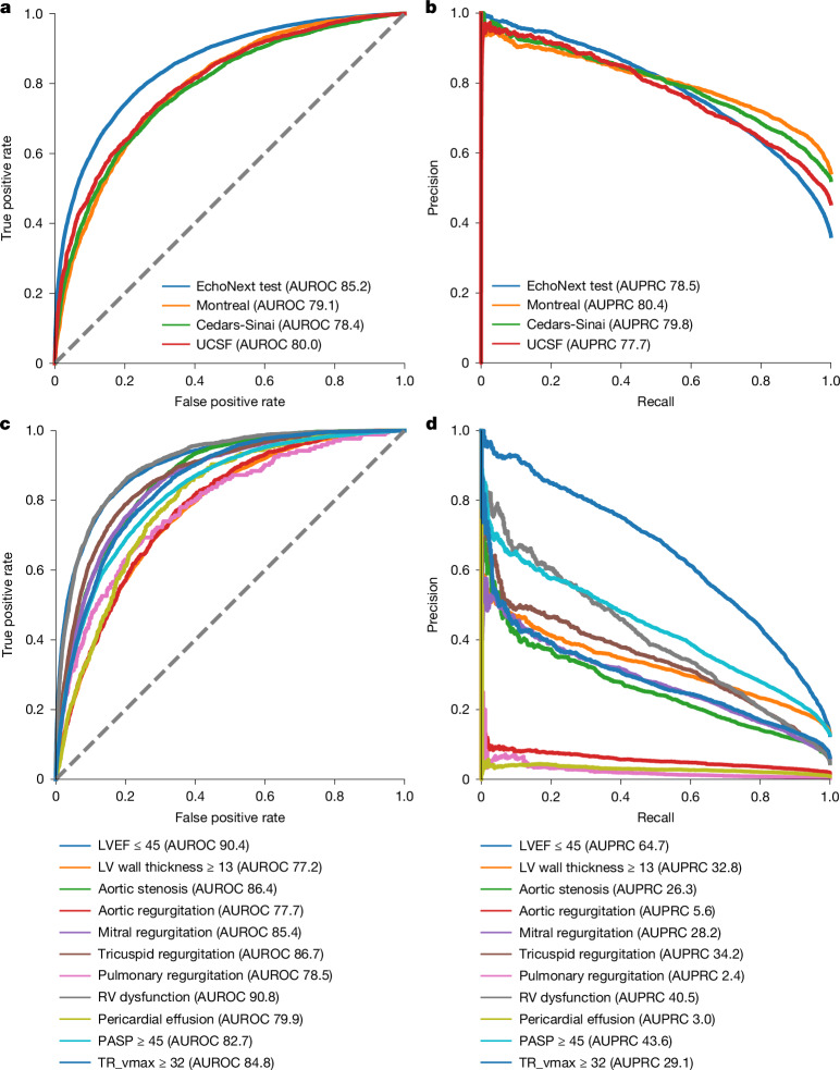
- In the NYP multicentre test set, EchoNext achieved an AUROC of 85.2% (95% CI 84.5–85.9%), AUPRC of 78.5% (95% CI 77.2–79.6%) and diagnostic odds ratio of 12.8 (95% CI 11.6–14.1) for the SHD composite, where test-set SHD prevalence was 36%.
- External validation at Cedars-Sinai (n = 10,177), Montreal Heart Institute (n = 10,862) and UCSF (n = 6,106) showed AUROC 78–80%, a 5–7% drop versus internal NYP hospitals; at 70% sensitivity, external cohorts had comparable positive predictive value but a 10% drop in specificity.
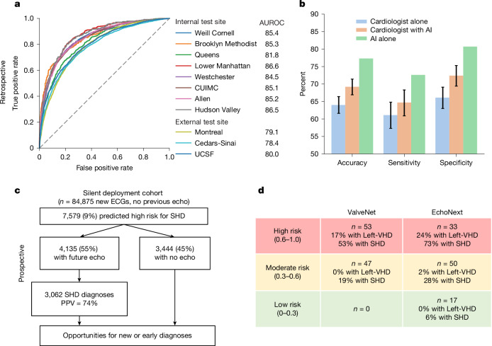
- In a cardiologist survey using 150 ECGs and 3,200 reviews from 13 cardiologists, EchoNext alone had accuracy 77.3%, sensitivity 72.6% and specificity 80.7%; cardiologists without AI had accuracy 64.0% (95% CI 61.6–66.4%), improving to 69.2% (95% CI 66.9–71.4%) with AI assistance, still below AI alone.
- In the 100-patient DISCOVERY pilot trial, post hoc EchoNext risk groups showed SHD in 73% of high-risk, 28% of moderate-risk and 6% of low-risk patients (P < 0.001); silent deployment on 124,027 ECGs found AUROC 83% and AUPRC 81%, with 38% of patients who underwent first echocardiography newly diagnosed with SHD.
Methods
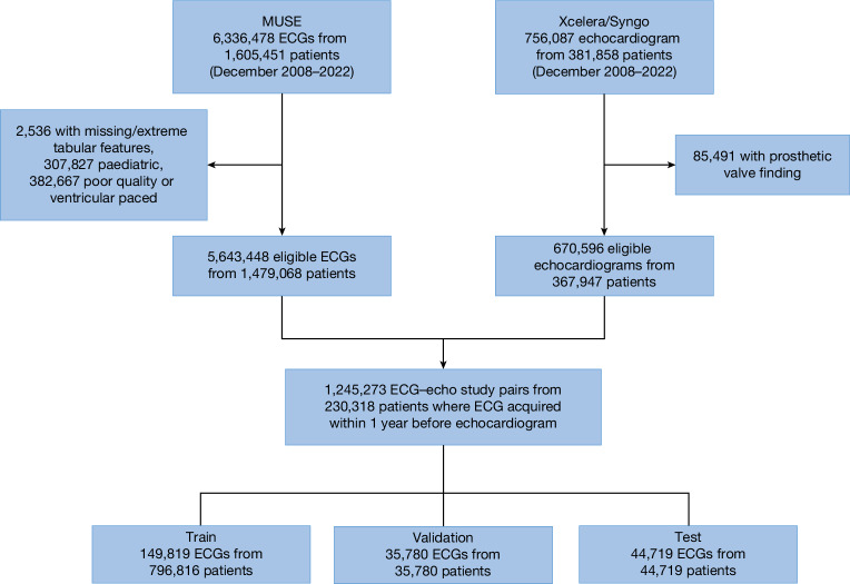
- Retrospective NYP multicentre cohort of 1,245,273 ECG–echocardiogram pairs from 230,318 unique patients aged 18 years or older across eight NYP-affiliated hospitals (December 2008–2022), split at patient level into train (149,819 patients), validation (35,780) and test (44,719) sets.
- EchoNext is a convolutional neural network using the 12-lead ECG waveform plus demographics and seven standard ECG values; it was trained as a multitask classifier for the SHD composite and individual component labels, with SHD defined by echocardiographic cutoffs including LVEF ≤45%, LV wall thickness ≥1.3 cm, moderate/severe right ventricular dysfunction, pulmonary hypertension, moderate/severe valvular disease and moderate/large pericardial effusion.
- Validation included three external cohorts, a temporally distinct silent deployment (January–September 2023), a cardiologist comparison survey, and the single-arm DISCOVERY pilot trial; performance was assessed mainly by AUROC, AUPRC, sensitivity, specificity and positive predictive value, with 95% CIs from 1,000 bootstrapped estimates.
Limitations
- Stated by the paper: binary cutoffs were arbitrarily chosen for continuous measures such as LVEF and LV wall thickness; AUPRCs come from retrospective ECG-plus-echocardiogram populations with higher SHD prevalence than a screening population; some component labels performed suboptimally, including LV wall thickness (label noise) and pulmonary regurgitation (rare and correlated with other conditions); multilabel optimization may compromise certain labels; and DISCOVERY was small and recruited using the earlier ValveNet model, so larger pragmatic trials are needed.
- Evident in the text: external AUROC dropped to 78–80% and external SHD prevalence was higher (54%, 52%, 46%) than in the NYP cohort (36%), indicating performance variation across settings and case mix.
- Evident in the text: for patients with ECGs but no follow-up echocardiograms, true SHD prevalence is unknown, so positive predictive value was modeled across assumed prevalences; the cardiologist comparison also excluded clinical history, physical exam and other testing data typically available in practice.
Model-written summary. Check it against the paper before relying on it.
Integrity signals
Registry
Cites
Statistics
Figures
Verification
Claims the paper makes
42 Own result · 14 Own method · 13 Own interpretation · 12 No basis given · 6 Partly supported · 3 Supported · 1 Supported so far · accuracy not yet measured
The cited sources support only part of this claim — read each source’s verdict below.
In the paper
“although previous work has been limited by development in narrow populations or targeting only select heart conditions5.”
§ Detecting structural heart disease from electrocardiograms using AI > Abstract
What the sources say
- Partly supportedcites [5] Siontis, K. C., Noseworthy, P. A., Attia, Z. I. & Friedman, P. A. Artificial intelligence-enhanced electrocardiography in cardiovascular disease management. Nat. Rev. Cardiol. 18, 465–478 (2021). sourceExperimental — accuracy not yet measured
In “Potential challenges and solutions” (page unavailable), the review warns that models may not generalize across populations and says validation data for most AI–ECG algorithms are pending. Its Introduction (Table 1; page unavailable) lists applications targeting particular conditions, but it does not establish that previous work as a whole was limited to only select heart conditions. The claim overstates that part of the review. (LLM, conf=0.82)
plain- Asserted: passes — The authors put it forward as true.
- Checkable: passes — It can be checked against evidence.
- Load-bearing: passes — The paper’s conclusions depend on it.
- Based: passes — The paper gives a reason why it holds.
- Partly supported
The cited sources support only part of this claim — read each source’s verdict below.
In the paper
“The application of deep learning, a subset of artificial intelligence (AI), has been shown to be beneficial in the detection of specific heart diseases from the 12-lead electrocardiogram (ECG). This includes conditions such as aortic stenosis, low left ventricular ejection fraction (LVEF) and low left ventricular hypertrophy, as well as composites of many valvular diseases3,17–21.”
§ Detecting structural heart disease from electrocardiograms using AI > Main
What the sources say
- Partly supportedcites [3] Elias, P. et al. Deep learning electrocardiographic analysis for detection of left-sided valvular heart disease. J. Am. Coll. Cardiol. 80, 613–626 (2022). sourceExperimental — accuracy not yet measured
The Discussion reports that the ECG model detected aortic stenosis and a composite of three valvular diseases. The Conclusions also describe prior ECG-based detection of left ventricular systolic dysfunction, consistent with low LVEF. But the paper does not report detection of “low left ventricular hypertrophy”; it mentions hypertrophy as a possible consequence of aortic stenosis, not as a detected condition. Page numbers are unavailable in our copy. (LLM, conf=0.88)
- Not checked — no full text of the sourcecites [17] Cohen-Shelly, M. et al. Electrocardiogram screening for aortic valve stenosis using artificial intelligence. Eur. Heart J. 42, 2885–2896 (2021). source
- Supportedcites [18] Kwon, J. M. et al. Deep learning-based algorithm for detecting aortic stenosis using electrocardiography. J. Am. Heart Assoc. 9, e014717 (2020). source
The Abstract Conclusions report high accuracy for detecting significant aortic stenosis using a deep-learning algorithm with 12-lead ECGs. In this multi-citation sentence, aortic stenosis is the condition this source directly supports; the other listed conditions are not needed to establish that attribution. (LLM, conf=0.92)
- Not checked — no full text of the sourcecites [19] Adedinsewo, D. et al. Artificial intelligence-enabled ECG algorithm to identify patients with left ventricular systolic dysfunction presenting to the emergency department with dyspnea. Circ. Arrhythm. Electrophysiol. 13, e008437 (2020). source
- Partly supportedcites [20] Ko, W. Y. et al. Detection of hypertrophic cardiomyopathy using a convolutional neural network-enabled electrocardiogram. J. Am. Coll. Cardiol. 75, 722–733 (2020). sourceExperimental — accuracy not yet measured
The Abstract reports that a deep-learning model detected hypertrophic cardiomyopathy from 12-lead ECGs and mentions earlier success detecting left ventricular dysfunction. The Subgroup Analyses treat left ventricular hypertrophy and aortic stenosis as patient subgroups or comparators, not conditions the model was trained to detect. The paper does not establish detection of the listed valvular diseases. Page numbers were not supplied. (LLM, conf=0.86)
- Partly supportedcites [21] Tison, G. H. et al. Assessment of disease status and treatment response with artificial intelligence−enhanced electrocardiography in obstructive hypertrophic cardiomyopathy. J. Am. Coll. Cardiol. 79, 1032–1034 (2022). sourceExperimental — accuracy not yet measured
The Abstract reports accurate automated detection of hypertrophic cardiomyopathy using AI analysis of standard 12-lead ECGs, supporting the general point that AI can detect a heart disease from an ECG. It does not report detection of aortic stenosis, low LVEF, low left ventricular hypertrophy, or composites of valvular diseases. (LLM, conf=0.86)
plaindirect- Asserted: passes — The authors put it forward as true.
- Checkable: passes — It can be checked against evidence.
- Load-bearing: passes — The paper’s conclusions depend on it.
- Based: passes — The paper gives a reason why it holds.
- Partly supported
The cited sources support only part of this claim — read each source’s verdict below.
In the paper
“More general AI-ECG models have also been developed that can accurately detect a composite of low left ventricular systolic dysfunction, low left ventricular hypertrophy and moderate or greater VHD4.”
§ Detecting structural heart disease from electrocardiograms using AI > Main
What the sources say
- Partly supportedcites [4] Ulloa-Cerna, A. E. et al. rECHOmmend: an ECG-based machine learning approach for identifying patients at increased risk of undiagnosed structural heart disease detectable by echocardiography. Circulation 146, 36–47 (2022). sourceExperimental — accuracy not yet measured
The Abstract (Methods) defines a composite of reduced ejection fraction (<50%), increased interventricular septal thickness (>15 mm), and moderate or severe valvular disease; the Abstract (Results) reports an AUROC of 0.91 for the composite model. The claim's “low left ventricular hypertrophy” does not match the paper's increased septal thickness criterion. (LLM, conf=0.88)
plaincorrelational- Asserted: passes — The authors put it forward as true.
- Checkable: passes — It can be checked against evidence.
- Load-bearing: passes — The paper’s conclusions depend on it.
- Based: passes — The paper gives a reason why it holds.
- Partly supported
Every source the paper cites for this claim was checked and supports it.
In the paper
“Before development of EchoNext, study investigators had created ValveNet, a similarly architected AI-ECG model trained to detect moderate or greater left-sided VHD (specifically aortic stenosis, aortic regurgitation and mitral regurgitation), a subset of SHD3.”
§ Detecting structural heart disease from electrocardiograms using AI > Prospective validation
What the sources say
- Supportedcites [3] Elias, P. et al. Deep learning electrocardiographic analysis for detection of left-sided valvular heart disease. J. Am. Coll. Cardiol. 80, 613–626 (2022). source
The Methods (page not specified) describe ValveNet as a convolutional neural network using ECG data and define its targets as moderate or severe aortic stenosis, aortic regurgitation, and mitral regurgitation. The comparison with EchoNext is the citing authors’ framing, not a finding attributed to this earlier paper. (LLM, conf=0.92)
plain- Asserted: passes — The authors put it forward as true.
- Checkable: passes — It can be checked against evidence.
- Load-bearing: unclear — It is unclear whether the paper’s conclusions depend on it.
- Based: passes — The paper gives a reason why it holds.
- Supported
Every source the paper cites for this claim was checked and supports it.
In the paper
“the performance of rECHOmmend was observed to drop (AUROC 0.88) when restricting to patients with echocardiography-confirmed disease, thereby increasing disease prevalence, as was done for the present study.”
§ Detecting structural heart disease from electrocardiograms using AI > Relationship to previous work
What the sources say
- Supportedcites [4] Ulloa-Cerna, A. E. et al. rECHOmmend: an ECG-based machine learning approach for identifying patients at increased risk of undiagnosed structural heart disease detectable by echocardiography. Circulation 146, 36–47 (2022). source
The Results (Sensitivity Analyses) report that using echocardiography-confirmed labels lowered rECHOmmend’s AUROC to 0.88 relative to the primary results. The paper also reports a higher disease prevalence of 53% under this restriction, which required echocardiographic confirmation of absence of disease for negative patients. (LLM, conf=0.93)
plaindirect- Asserted: passes — The authors put it forward as true.
- Checkable: passes — It can be checked against evidence.
- Load-bearing: unclear — It is unclear whether the paper’s conclusions depend on it.
- Based: passes — The paper gives a reason why it holds.
- Supported
A result the paper reports from its own data — it cites no other source for it, so there is none to check. The quote shows it is in the paper.
In the paper
“Performance remained robust when tested on different combinations of component disease labels (left or right ventricular dysfunction, all valvular disease, left-sided heart disease and right-sided heart disease).”
§ Detecting structural heart disease from electrocardiograms using AI > Model development and validation
plain- Asserted: passes — The authors put it forward as true.
- Checkable: passes — It can be checked against evidence.
- Load-bearing: unclear — It is unclear whether the paper’s conclusions depend on it.
- Based: passes — The paper gives a reason why it holds.
A result the paper reports from its own data — it cites no other source for it, so there is none to check. The quote shows it is in the paper.
In the paper
“Furthermore, performance was stable for both academic and community hospitals.”
§ Detecting structural heart disease from electrocardiograms using AI > Model development and validation
plain- Asserted: passes — The authors put it forward as true.
- Checkable: passes — It can be checked against evidence.
- Load-bearing: passes — The paper’s conclusions depend on it.
- Based: passes — The paper gives a reason why it holds.
A result the paper reports from its own data — it cites no other source for it, so there is none to check. The quote shows it is in the paper.
In the paper
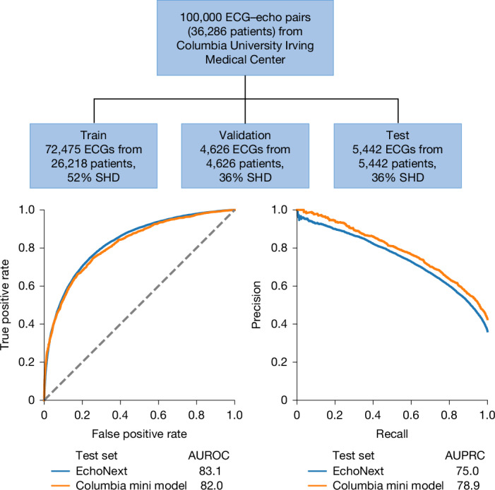
“The Columbia mini-model was highly performant for SHD detection across all eight hospitals, with AUROC of 82.0% (95% CI 80.9–83.0%) (Fig. 4).”
§ Detecting structural heart disease from electrocardiograms using AI > AI benchmark in SHD
plain- Asserted: passes — The authors put it forward as true.
- Checkable: passes — It can be checked against evidence.
- Load-bearing: passes — The paper’s conclusions depend on it.
- Based: passes — The paper gives a reason why it holds.
The paper states this without a source or data behind it here, so there is nothing to check it against. The quote shows it is in the paper.
In the paper
“performance was robust with respect to patient demographics (age, sex, race and/or ethnicity) across clinical contexts in a highly diverse patient population”
§ Detecting structural heart disease from electrocardiograms using AI > Discussion
plain- Asserted: passes — The authors put it forward as true.
- Checkable: passes — It can be checked against evidence.
- Load-bearing: passes — The paper’s conclusions depend on it.
- Based: passes — The paper gives a reason why it holds.
A result the paper reports from its own data — it cites no other source for it, so there is none to check. The quote shows it is in the paper.
In the paper
“For example, if SHD prevalence is 10% in this population, the projected positive predictive value would be 46.5% at 50% sensitivity.”
§ Detecting structural heart disease from electrocardiograms using AI > Silent deployment validation
hedgedcorrelational- Asserted: passes — The authors put it forward as true.
- Checkable: passes — It can be checked against evidence.
- Load-bearing: unclear — It is unclear whether the paper’s conclusions depend on it.
- Based: passes — The paper gives a reason why it holds.
- The authors state this as an assumption: The prevalence of SHD among the 69,781 patients with an ECG but no follow-up echocardiogram is 10% (or can be represented by the scenarios in Table 3) for the estimate of 1,998 newly diagnosed cases.
- It may state two separate claims as one. 39
- Probably not a substantial claim. 19
The cited sources support only part of this claim — read each source’s verdict below.
In the paper
“Early detection of structural heart disease is critical to improving outcomes, but widespread screening remains limited by the cost and accessibility of imaging tools such as echocardiography1,2.”
§ Detecting structural heart disease from electrocardiograms using AI > Abstract
What the sources say
- Not checked — no full text of the sourcecites [1] Otto, C. M. et al. 2020 ACC/AHA guideline for the management of patients with valvular heart disease: a report of the American College of Cardiology/American Heart Association Joint Committee on Clinical Practice Guidelines. Circulation 143, e72–e227 (2021). source
- Partly supportedcites [2] Heidenreich, P. A. et al. 2022 AHA/ACC/HFSA guideline for the management of heart failure: executive summary: a report of the American College of Cardiology/American Heart Association Joint Committee on Clinical Practice Guidelines. J. Am. Coll. Cardiol. 79, 1757–1780 (2022). sourceExperimental — accuracy not yet measured
Take-Home Message No. 9 (Methods; page unavailable) supports the prevention aspect: it says screening followed by team-based care can help prevent left ventricular dysfunction or new-onset heart failure. It does not say that the cost or accessibility of echocardiography limits widespread screening; searches for those terms found no supporting passage in our copy. (LLM, conf=0.83)
plaindirect- Asserted: passes — The authors put it forward as true.
- Checkable: passes — It can be checked against evidence.
- Load-bearing: passes — The paper’s conclusions depend on it.
- Based: passes — The paper gives a reason why it holds.
It may state two separate claims as one.
The cited sources support only part of this claim — read each source’s verdict below.
In the paper
“A study of 2,500 people 65 years of age or older found that 4.9% were previously diagnosed with clinically significant (graded moderate or severe) VHD and found another 6.4% with undiagnosed VHD more than doubling the overall prevalence14.”
§ Detecting structural heart disease from electrocardiograms using AI > Main
What the sources say
- Partly supportedcites [14] d’Arcy, J. L. et al. Large-scale community echocardiographic screening reveals a major burden of undiagnosed valvular heart disease in older people: the OxVALVE Population Cohort Study. Eur. Heart J. 37, 3515–3522 (2016). sourceExperimental — accuracy not yet measured
The Abstract reports 2,500 participants aged ≥65, 6.4% with undiagnosed moderate or severe VHD, and 4.9% with pre-existing VHD, yielding 11.3% overall—more than twice 4.9%. However, the Discussion says the previously diagnosed group was excluded from screening and its clinical significance was assumed, not confirmed by moderate-or-severe grading. Page numbers are not available in the supplied copy. (LLM, conf=0.87)
plainn = 2,500- Asserted: passes — The authors put it forward as true.
- Checkable: passes — It can be checked against evidence.
- Load-bearing: passes — The paper’s conclusions depend on it.
- Based: passes — The paper gives a reason why it holds.
It may state two separate claims as one.
- Partly supported
The cited sources support only part of this claim — read each source’s verdict below.
In the paper
“Finding patients with SHD earlier in the disease process has been shown to reduce mortality, decrease costs and improve quality of life, but getting to diagnosis remains challenging1,15,16.”
§ Detecting structural heart disease from electrocardiograms using AI > Main
What the sources say
- Not checked — no full text of the sourcecites [1] Otto, C. M. et al. 2020 ACC/AHA guideline for the management of patients with valvular heart disease: a report of the American College of Cardiology/American Heart Association Joint Committee on Clinical Practice Guidelines. Circulation 143, e72–e227 (2021). source
- Not checked — no full text of the sourcecites [15] Kang, D. H. et al. Early surgery or conservative care for asymptomatic aortic stenosis. N. Engl. J. Med. 382, 111–119 (2020). source
- Partly supportedcites [16] Suri, R. M. et al. Association between early surgical intervention vs watchful waiting and outcomes for mitral regurgitation due to flail mitral valve leaflets. JAMA 310, 609–616 (2013). sourceExperimental — accuracy not yet measured
The Conclusion (page unavailable) reports that early surgery after detection of mitral regurgitation due to flail mitral leaflets was associated with greater long-term survival. That supports the mortality component only for this condition and intervention; the paper does not show that finding patients with structural heart disease earlier reduces mortality, nor does it report decreased costs or improved quality of life. (LLM, conf=0.88)
boosteddirect- Asserted: passes — The authors put it forward as true.
- Checkable: passes — It can be checked against evidence.
- Load-bearing: passes — The paper’s conclusions depend on it.
- Based: passes — The paper gives a reason why it holds.
It may state two separate claims as one.
The sources with a verdict support this claim; the rest weren’t checked yet, have no full text we could read, or their verdicts aren’t rated yet.
In the paper
“This method of human–machine comparison is similar to typical methods and one we have used previously34,35.”
§ Detecting structural heart disease from electrocardiograms using AI > Methods > Cardiologist survey
What the sources say
- Not checked — no full text of the sourcecites [34] Oakden-Rayner, L. & Palmer, L. J. in Artificial Intelligence in Medical Imaging: Opportunities, Applications and Risks (eds Ranschaert, E. R. et al.) 83–104 (Springer, 2019). source
- Supportedcites [35] Bhave, S. et al. Deep learning to detect left ventricular structural abnormalities in chest X-rays. Eur. Heart J. 45, 2002–2012 (2024). source
The Methods section (‘Comparison with radiologists’; page not provided) describes a human–machine comparison using the same chest X-rays read by radiologists and evaluated by the model. The Results section compares their performance at matched specificity, confirming this paper as an example of the previously used method. The claim does not require this source to establish how typical the method is. (LLM, conf=0.88)
plain- Asserted: passes — The authors put it forward as true.
- Checkable: passes — It can be checked against evidence.
- Load-bearing: fails — The paper’s conclusions may not depend on it.
- Based: passes — The paper gives a reason why it holds.
Probably not a substantial claim. The paper’s conclusions may not depend on it. It may state two separate claims as one.
Every source the paper cites for this claim was checked and supports it.
In the paper
“Other ECG-based SHD models, such as rECHOmmend, have demonstrated excellent performance with an AUROC of 91% in its retrospective dataset4.”
§ Detecting structural heart disease from electrocardiograms using AI > Relationship to previous work
What the sources say
- Supportedcites [4] Ulloa-Cerna, A. E. et al. rECHOmmend: an ECG-based machine learning approach for identifying patients at increased risk of undiagnosed structural heart disease detectable by echocardiography. Circulation 146, 36–47 (2022). source
The Results section (Cross-Validation Performance of rECHOmmend Model; page unavailable) reports an AUROC of 0.91 for the composite model. This is the reported cross-validation result; the separate simulated retrospective deployment had an AUROC of 0.86. (LLM, conf=0.90)
boosted- Asserted: passes — The authors put it forward as true.
- Checkable: passes — It can be checked against evidence.
- Load-bearing: fails — The paper’s conclusions may not depend on it.
- Based: passes — The paper gives a reason why it holds.
Probably not a substantial claim. The paper’s conclusions may not depend on it.
- Supported
A result the paper reports from its own data — it cites no other source for it, so there is none to check. The quote shows it is in the paper.
In the paper
“When the model was trained on data from four of the eight NYP hospitals and tested on independent data from the four unseen hospitals, the performance change was minimal. The same was true when switching the hospitals used for training and testing.”
§ Detecting structural heart disease from electrocardiograms using AI > Model development and validation
plain- Asserted: passes — The authors put it forward as true.
- Checkable: passes — It can be checked against evidence.
- Load-bearing: passes — The paper’s conclusions depend on it.
- Based: passes — The paper gives a reason why it holds.
It may state two separate claims as one.
A result the paper reports from its own data — it cites no other source for it, so there is none to check. The quote shows it is in the paper.
In the paper
“In this cohort, 18% (15,094 patients) underwent their first echocardiogram as part of routine clinical care after the ECG; of which, 38% (5,744 patients) were newly diagnosed with SHD.”
§ Detecting structural heart disease from electrocardiograms using AI > Silent deployment validation
plainn = 5,744- Asserted: passes — The authors put it forward as true.
- Checkable: passes — It can be checked against evidence.
- Load-bearing: passes — The paper’s conclusions depend on it.
- Based: passes — The paper gives a reason why it holds.
It may state two separate claims as one.
A result the paper reports from its own data — it cites no other source for it, so there is none to check. The quote shows it is in the paper.
In the paper
“Model performance varied across each of these individual components with right ventricular (AUROC 91%) and low left ventricular systolic dysfunction (90%) as the best performing (Fig. 2 and Supplementary Table 2).”
§ Detecting structural heart disease from electrocardiograms using AI > Model development and validation
plain- Asserted: passes — The authors put it forward as true.
- Checkable: passes — It can be checked against evidence.
- Load-bearing: unclear — It is unclear whether the paper’s conclusions depend on it.
- Based: passes — The paper gives a reason why it holds.
It may state two separate claims as one.
A result the paper reports from its own data — it cites no other source for it, so there is none to check. The quote shows it is in the paper.
In the paper
“The rates of disease were strongly correlated within these groups as follows: high risk (n = 33, 24% with left-sided VHD and 73% with SHD), moderate risk (n = 50, 2% with left-sided VHD, 28% with SHD) and low risk (n = 17, 0% with left-sided VHD and 6% with SHD).”
§ Detecting structural heart disease from electrocardiograms using AI > Prospective validation
boostedcorrelationaln = 100 ·- Asserted: passes — The authors put it forward as true.
- Checkable: passes — It can be checked against evidence.
- Load-bearing: passes — The paper’s conclusions depend on it.
- Based: passes — The paper gives a reason why it holds.
It may state two separate claims as one.
A result the paper reports from its own data — it cites no other source for it, so there is none to check. The quote shows it is in the paper.
In the paper
“All differences between risk groups for EchoNext were significant (P = 0.002 for left-sided VHD and P < 0.001 for SHD).”
§ Detecting structural heart disease from electrocardiograms using AI > Prospective validation
plaincorrelationaln = 100 ·- Asserted: passes — The authors put it forward as true.
- Checkable: passes — It can be checked against evidence.
- Load-bearing: passes — The paper’s conclusions depend on it.
- Based: passes — The paper gives a reason why it holds.
It may state two separate claims as one.
A result the paper reports from its own data — it cites no other source for it, so there is none to check. The quote shows it is in the paper.
In the paper
“EchoNext had superior accuracy, sensitivity and specificity in detecting SHD from ECGs compared to cardiologists both when given the AI prediction to consult and when not”
- Also stated in: § Expanding ECG use through AI · boosted
“the performance of the EchoNext model was significantly superior, improving on both diagnostic sensitivity and specificity compared with the human experts”
§ Detecting structural heart disease from electrocardiograms using AI > Discussion
boostedcorrelational- Asserted: passes — The authors put it forward as true.
- Checkable: passes — It can be checked against evidence.
- Load-bearing: passes — The paper’s conclusions depend on it.
- Based: passes — The paper gives a reason why it holds.
It may state two separate claims as one.
- Also stated in: § Expanding ECG use through AI · boosted
A result the paper reports from its own data — it cites no other source for it, so there is none to check. The quote shows it is in the paper.
In the paper
“For the 1,600 non-AI-assisted reviews, the accuracy of the cardiologists was 64.0% (95% CI 61.6–66.4%) with sensitivity 61.1% (95% CI 57.3–64.8%) and specificity 66.1% (95% CI 63.0–69.1%).”
§ Detecting structural heart disease from electrocardiograms using AI > Model performance versus cardiologists
plain- Asserted: passes — The authors put it forward as true.
- Checkable: passes — It can be checked against evidence.
- Load-bearing: passes — The paper’s conclusions depend on it.
- Based: passes — The paper gives a reason why it holds.
It may state two separate claims as one.
A result the paper reports from its own data — it cites no other source for it, so there is none to check. The quote shows it is in the paper.
In the paper
“In the set of 150 ECGs, the EchoNext model had an accuracy of 77.3%, sensitivity 72.6% and specificity of 80.7%.”
§ Detecting structural heart disease from electrocardiograms using AI > Model performance versus cardiologists
plain- Asserted: passes — The authors put it forward as true.
- Checkable: passes — It can be checked against evidence.
- Load-bearing: passes — The paper’s conclusions depend on it.
- Based: passes — The paper gives a reason why it holds.
It may state two separate claims as one.
A result the paper reports from its own data — it cites no other source for it, so there is none to check. The quote shows it is in the paper.
In the paper
“Across hospitals (AUROC range 82–87%) (Fig. 3) and clinical contexts (AUROC range 79–84%), the model showed stable, generalizable performance.”
§ Detecting structural heart disease from electrocardiograms using AI > Model development and validation
plain- Asserted: passes — The authors put it forward as true.
- Checkable: passes — It can be checked against evidence.
- Load-bearing: passes — The paper’s conclusions depend on it.
- Based: passes — The paper gives a reason why it holds.
It may state two separate claims as one.
A result the paper reports from its own data — it cites no other source for it, so there is none to check. The quote shows it is in the paper.
In the paper
“There was a significant difference in the number of patients positive for left-sided VHD (P = 0.005) and SHD (P = 0.003) when comparing high- to moderate-risk ValveNet scores.”
§ Detecting structural heart disease from electrocardiograms using AI > Prospective validation
plaincorrelationaln = 100 ·- Asserted: passes — The authors put it forward as true.
- Checkable: passes — It can be checked against evidence.
- Load-bearing: passes — The paper’s conclusions depend on it.
- Based: passes — The paper gives a reason why it holds.
It may state two separate claims as one.
A result the paper reports from its own data — it cites no other source for it, so there is none to check. The quote shows it is in the paper.
In the paper
“The lowest performance was seen for low left ventricular wall thickness (AUROC 77%), AR (78%), pulmonary regurgitation (79%) and pericardial effusion (80%).”
§ Detecting structural heart disease from electrocardiograms using AI > Model development and validation
plain- Asserted: passes — The authors put it forward as true.
- Checkable: passes — It can be checked against evidence.
- Load-bearing: unclear — It is unclear whether the paper’s conclusions depend on it.
- Based: passes — The paper gives a reason why it holds.
It may state two separate claims as one.
A result the paper reports from its own data — it cites no other source for it, so there is none to check. The quote shows it is in the paper.
In the paper
“The models were prospectively evaluated in a clinical trial of patients without previous cardiac imaging, successfully identifying previously undiagnosed heart disease.”
§ Detecting structural heart disease from electrocardiograms using AI > Abstract
plaindirect- Asserted: passes — The authors put it forward as true.
- Checkable: passes — It can be checked against evidence.
- Load-bearing: passes — The paper’s conclusions depend on it.
- Based: passes — The paper gives a reason why it holds.
It may state two separate claims as one.
A result the paper reports from its own data — it cites no other source for it, so there is none to check. The quote shows it is in the paper.
In the paper
“A total of 47 patients were recruited with moderate-risk ValveNet scores, with 0% positive for left-sided VHD and 19% positive for SHD.”
§ Detecting structural heart disease from electrocardiograms using AI > Prospective validation
plainn = 47- Asserted: passes — The authors put it forward as true.
- Checkable: passes — It can be checked against evidence.
- Load-bearing: passes — The paper’s conclusions depend on it.
- Based: passes — The paper gives a reason why it holds.
It may state two separate claims as one.
A result the paper reports from its own data — it cites no other source for it, so there is none to check. The quote shows it is in the paper.
In the paper
“Notably, the accuracy of the cardiologists differed between clinically normal and abnormal ECGs (69% versus 62%, respectively), whereas EchoNext performed equally well (77% for both) despite the substantial difference in SHD prevalence (25% versus 46.5% in normal versus abnormal ECGs) (Supplementary Table 12).”
§ Detecting structural heart disease from electrocardiograms using AI > Model performance versus cardiologists
plaincorrelational- Asserted: passes — The authors put it forward as true.
- Checkable: passes — It can be checked against evidence.
- Load-bearing: passes — The paper’s conclusions depend on it.
- Based: passes — The paper gives a reason why it holds.
It may state two separate claims as one.
A result the paper reports from its own data — it cites no other source for it, so there is none to check. The quote shows it is in the paper.
In the paper
“EchoNext performance on the NYP multicentre test set was high and well calibrated (Figs. 2 and 3 and Supplementary Fig. 1), with an area under the receiver operating characteristic curve (AUROC) of 85.2% (95% confidence interval (CI) 84.5–85.9%), area under the precision–recall curve (AUPRC) of 78.5% (95% CI 77.2–79.6%) and diagnostic odds ratio of 12.8 (95% CI 11.6–14.1) (Fig. 2).”
§ Detecting structural heart disease from electrocardiograms using AI > Model development and validation
plain- Asserted: passes — The authors put it forward as true.
- Checkable: passes — It can be checked against evidence.
- Load-bearing: passes — The paper’s conclusions depend on it.
- Based: passes — The paper gives a reason why it holds.
It may state two separate claims as one.
A result the paper reports from its own data — it cites no other source for it, so there is none to check. The quote shows it is in the paper.
In the paper
“With AI assistance, the accuracy of the cardiologists significantly improved to 69.2% (95% CI 66.9–71.4%) with sensitivity 64.7% (95% CI 60.9–68.3%) and specificity 72.4% (95% CI 69.4–75.3%).”
§ Detecting structural heart disease from electrocardiograms using AI > Model performance versus cardiologists
plaindirect- Asserted: passes — The authors put it forward as true.
- Checkable: passes — It can be checked against evidence.
- Load-bearing: passes — The paper’s conclusions depend on it.
- Based: passes — The paper gives a reason why it holds.
It may state two separate claims as one.
A result the paper reports from its own data — it cites no other source for it, so there is none to check. The quote shows it is in the paper.
In the paper
“Of note, despite the smaller and single-centre training set, this model’s performance was only modestly lower than the full multicentre-trained EchoNext model, with AUROC in the same multicentre test set of 83.1%.”
§ Detecting structural heart disease from electrocardiograms using AI > AI benchmark in SHD
plain- Asserted: passes — The authors put it forward as true.
- Checkable: passes — It can be checked against evidence.
- Load-bearing: passes — The paper’s conclusions depend on it.
- Based: passes — The paper gives a reason why it holds.
It may state two separate claims as one.
A result the paper reports from its own data — it cites no other source for it, so there is none to check. The quote shows it is in the paper.
In the paper
“At a fixed sensitivity of 70%, the external cohorts showed comparable positive predictive value, but a 10% drop in specificity (Supplementary Table 10).”
§ Detecting structural heart disease from electrocardiograms using AI > External validation
plain- Asserted: passes — The authors put it forward as true.
- Checkable: passes — It can be checked against evidence.
- Load-bearing: passes — The paper’s conclusions depend on it.
- Based: passes — The paper gives a reason why it holds.
It may state two separate claims as one.
A result the paper reports from its own data — it cites no other source for it, so there is none to check. The quote shows it is in the paper.
In the paper
“Using the predefined model score cutoff of 0.6, 27% (4,135 patients) of the ECG and/or echocardiogram population were predicted as high risk, corresponding to a precision (positive predictive value) and recall (sensitivity) of 74% and 53%, respectively.”
§ Detecting structural heart disease from electrocardiograms using AI > Silent deployment validation
plainn = 4,135- Asserted: passes — The authors put it forward as true.
- Checkable: passes — It can be checked against evidence.
- Load-bearing: passes — The paper’s conclusions depend on it.
- Based: passes — The paper gives a reason why it holds.
It may state two separate claims as one.
A result the paper reports from its own data — it cites no other source for it, so there is none to check. The quote shows it is in the paper.
In the paper
“Across hospitals (AUROC range 82–87%) (Fig. 3) and clinical contexts (AUROC range 79–84%), the model showed stable, generalizable performance.”
§ Detecting structural heart disease from electrocardiograms using AI > Model development and validation
plain- Asserted: passes — The authors put it forward as true.
- Checkable: passes — It can be checked against evidence.
- Load-bearing: passes — The paper’s conclusions depend on it.
- Based: passes — The paper gives a reason why it holds.
It may state two separate claims as one.
A result the paper reports from its own data — it cites no other source for it, so there is none to check. The quote shows it is in the paper.
In the paper
“A total of 53 patients with high-risk ValveNet scores were recruited, with 17% positive for moderate or greater left-sided VHD and 53% positive for SHD.”
§ Detecting structural heart disease from electrocardiograms using AI > Prospective validation
plainn = 53- Asserted: passes — The authors put it forward as true.
- Checkable: passes — It can be checked against evidence.
- Load-bearing: passes — The paper’s conclusions depend on it.
- Based: passes — The paper gives a reason why it holds.
It may state two separate claims as one.
The paper states this without a source or data behind it here, so there is nothing to check it against. The quote shows it is in the paper.
In the paper
“Compared to other models that have been developed for similar pathologies, the current study had significantly increased racial and/or ethnic diversity across many internal or external institutes.”
§ Detecting structural heart disease from electrocardiograms using AI > Relationship to previous work
boosted- Asserted: passes — The authors put it forward as true.
- Checkable: passes — It can be checked against evidence.
- Load-bearing: fails — The paper’s conclusions may not depend on it.
- Based: fails — The paper may give no reason why it holds.
Probably not a substantial claim. The paper’s conclusions may not depend on it. The paper may give no reason why it holds.
The paper states this without a source or data behind it here, so there is nothing to check it against. The quote shows it is in the paper.
In the paper
“the SHD label in the current study captures 98.9% of all echo-based diagnoses as compared to 65.6% by rECHOmmend”
§ Detecting structural heart disease from electrocardiograms using AI > Relationship to previous work
plaincorrelational- Asserted: passes — The authors put it forward as true.
- Checkable: passes — It can be checked against evidence.
- Load-bearing: passes — The paper’s conclusions depend on it.
- Based: passes — The paper gives a reason why it holds.
It may state two separate claims as one.
The paper states this without a source or data behind it here, so there is nothing to check it against. The quote shows it is in the paper.
In the paper
“low left ventricular wall thickness is a highly prevalent label but subjective to high interobserver variation, thus introducing inherent label noise.”
§ Detecting structural heart disease from electrocardiograms using AI > Limitations
plaindirect- Asserted: passes — The authors put it forward as true.
- Checkable: passes — It can be checked against evidence.
- Load-bearing: fails — The paper’s conclusions may not depend on it.
- Based: passes — The paper gives a reason why it holds.
Probably not a substantial claim. The paper’s conclusions may not depend on it. It may state two separate claims as one.
A result the paper reports from its own data — it cites no other source for it, so there is none to check. The quote shows it is in the paper.
In the paper
“Restricting the model to a different arbitrary threshold showed a similar AUROC of 87.7% providing evidence that performance was not dependent on any specific threshold for defining disease.”
§ Detecting structural heart disease from electrocardiograms using AI > Limitations
plain- Asserted: passes — The authors put it forward as true.
- Checkable: passes — It can be checked against evidence.
- Load-bearing: unclear — It is unclear whether the paper’s conclusions depend on it.
- Based: passes — The paper gives a reason why it holds.
It may state two separate claims as one.
A result the paper reports from its own data — it cites no other source for it, so there is none to check. The quote shows it is in the paper.
In the paper
“Rare conditions such as pulmonary regurgitation are under-represented and are highly correlated with other more prevalent conditions, such as tricuspid regurgitation max velocity (Supplementary Fig. 4), which makes it hard to learn patterns that are exclusive to pulmonary regurgitation.”
§ Detecting structural heart disease from electrocardiograms using AI > Limitations
plaincorrelational- Asserted: passes — The authors put it forward as true.
- Checkable: passes — It can be checked against evidence.
- Load-bearing: passes — The paper’s conclusions depend on it.
- Based: passes — The paper gives a reason why it holds.
It may state two separate claims as one.
The paper states this without a source or data behind it here, so there is nothing to check it against. The quote shows it is in the paper.
In the paper
“Most importantly, these models and the underlying training data are typically proprietary, limiting comparisons and broader evaluation of generalizability.”
§ Detecting structural heart disease from electrocardiograms using AI > Main
plaindirect- Asserted: passes — The authors put it forward as true.
- Checkable: passes — It can be checked against evidence.
- Load-bearing: fails — The paper’s conclusions may not depend on it.
- Based: passes — The paper gives a reason why it holds.
Probably not a substantial claim. The paper’s conclusions may not depend on it.
A conclusion the paper draws from its own results — it cites no other source for it, so there is none to check. The quote shows it is in the paper.
In the paper
“which was strengthened by the multilabel approach to best capture collinearity and correlation across component disease labels”
§ Detecting structural heart disease from electrocardiograms using AI > Discussion
plaindirect- Asserted: passes — The authors put it forward as true.
- Checkable: passes — It can be checked against evidence.
- Load-bearing: passes — The paper’s conclusions depend on it.
- Based: fails — The paper may give no reason why it holds.
Probably not a substantial claim. The paper may give no reason why it holds.
A conclusion the paper draws from its own results — it cites no other source for it, so there is none to check. The quote shows it is in the paper.
In the paper
“Thus, whereas AI assistance for cardiologists in this task improved predictive accuracy, the combined performance still lagged that of the AI alone.”
§ Detecting structural heart disease from electrocardiograms using AI > Model performance versus cardiologists
plaindirect- Asserted: passes — The authors put it forward as true.
- Checkable: passes — It can be checked against evidence.
- Load-bearing: passes — The paper’s conclusions depend on it.
- Based: passes — The paper gives a reason why it holds.
It may state two separate claims as one.
What the paper did — it cites no source for this, so there is none to check. The quote shows it is in the paper.
In the paper
“This task focused on using the ECG for SHD detection without taking advantage of other information (clinical history, physical exam, other testing data) that would typically be available to a physician in clinical practice.”
§ Detecting structural heart disease from electrocardiograms using AI > Model performance versus cardiologists
plain- Asserted: passes — The authors put it forward as true.
- Checkable: passes — It can be checked against evidence.
- Load-bearing: passes — The paper’s conclusions depend on it.
- Based: passes — The paper gives a reason why it holds.
Probably not a substantial claim.
A conclusion the paper draws from its own results — it cites no other source for it, so there is none to check. The quote shows it is in the paper.
In the paper
“The AUPRCs presented in this study are based on retrospective data in which patients have both ECGs and echocardiograms, which is a population with a much higher prevalence of SHD, and thus not representative of AUPRCs we would expect to see if this model was used as a screening study among patients with ECGs and no echocardiograms.”
§ Detecting structural heart disease from electrocardiograms using AI > Limitations
plain- Asserted: passes — The authors put it forward as true.
- Checkable: passes — It can be checked against evidence.
- Load-bearing: passes — The paper’s conclusions depend on it.
- Based: passes — The paper gives a reason why it holds.
It may state two separate claims as one.
- Rests on an assumption: The underlying prevalence of undiagnosed SHD among patients with ECGs and no echocardiograms is not well known.
A conclusion the paper draws from its own results — it cites no other source for it, so there is none to check. The quote shows it is in the paper.
In the paper
“deep learning analysis can accurately detect SHDs individually and as a composite, with high accuracy across diverse populations in several clinical contexts and geographically distinct external test sets.”
§ Detecting structural heart disease from electrocardiograms using AI > Relationship to previous work
plaincorrelational- Asserted: passes — The authors put it forward as true.
- Checkable: passes — It can be checked against evidence.
- Load-bearing: passes — The paper’s conclusions depend on it.
- Based: passes — The paper gives a reason why it holds.
It may state two separate claims as one.
What the paper did — it cites no source for this, so there is none to check. The quote shows it is in the paper.
In the paper
“The digital ECG was accessed as a pdf and the name, date and clinical interpretation was cropped out of the image leaving only the waveform and the ECG measurements (ventricular rate, pulmonary regurgitation interval, QRS interval, Q wave-to-T wave interval and axis). The age (truncated to greater than 90) and sex were added to each ECG.”
§ Detecting structural heart disease from electrocardiograms using AI > Methods > Cardiologist survey
plain- Asserted: passes — The authors put it forward as true.
- Checkable: passes — It can be checked against evidence.
- Load-bearing: passes — The paper’s conclusions depend on it.
- Based: passes — The paper gives a reason why it holds.
Probably not a substantial claim. It may state two separate claims as one.
What the paper did — it cites no source for this, so there is none to check. The quote shows it is in the paper.
In the paper
“For each statistical test, 95% CIs were generated using 1,000 bootstrapped estimates.”
§ Detecting structural heart disease from electrocardiograms using AI > Methods > Statistical analysis
plain- Asserted: passes — The authors put it forward as true.
- Checkable: passes — It can be checked against evidence.
- Load-bearing: fails — The paper’s conclusions may not depend on it.
- Based: passes — The paper gives a reason why it holds.
Probably not a substantial claim. The paper’s conclusions may not depend on it.
A conclusion the paper draws from its own results — it cites no other source for it, so there is none to check. The quote shows it is in the paper.
In the paper
“This small trial showed promising results, but larger, more pragmatic interventions will be necessary to determine the potential benefit in excess of usual clinical care for EchoNext.”
§ Detecting structural heart disease from electrocardiograms using AI > Limitations
hedgedconditionaln = 100- Asserted: passes — The authors put it forward as true.
- Checkable: passes — It can be checked against evidence.
- Load-bearing: fails — The paper’s conclusions may not depend on it.
- Based: passes — The paper gives a reason why it holds.
Probably not a substantial claim. The paper’s conclusions may not depend on it. It may state two separate claims as one.
What the paper did — it cites no source for this, so there is none to check. The quote shows it is in the paper.
In the paper
“The threshold chosen for this study was 45% and results may have differed if a different cutoff was chosen.”
§ Detecting structural heart disease from electrocardiograms using AI > Limitations
hedgedconditional- Asserted: passes — The authors put it forward as true.
- Checkable: passes — It can be checked against evidence.
- Load-bearing: fails — The paper’s conclusions may not depend on it.
- Based: passes — The paper gives a reason why it holds.
Probably not a substantial claim. The paper’s conclusions may not depend on it. It may state two separate claims as one.
What the paper did — it cites no source for this, so there is none to check. The quote shows it is in the paper.
In the paper
“Patients included in the development cohort were excluded from this analysis.”
§ Detecting structural heart disease from electrocardiograms using AI > Methods > Silent deployment validation
plain- Asserted: passes — The authors put it forward as true.
- Checkable: passes — It can be checked against evidence.
- Load-bearing: passes — The paper’s conclusions depend on it.
- Based: passes — The paper gives a reason why it holds.
Probably not a substantial claim.
What the paper did — it cites no source for this, so there is none to check. The quote shows it is in the paper.
In the paper
“data from all eight hospital campuses were blended and split by patient into training, validation and test sets (64%, 16% and 20%)”
§ Detecting structural heart disease from electrocardiograms using AI > Methods > Patient identification and data sources
plain- Asserted: passes — The authors put it forward as true.
- Checkable: passes — It can be checked against evidence.
- Load-bearing: passes — The paper’s conclusions depend on it.
- Based: passes — The paper gives a reason why it holds.
Probably not a substantial claim.
What the paper did — it cites no source for this, so there is none to check. The quote shows it is in the paper.
In the paper
“These data were harmonized across each echocardiographic reading system and hospital with a minimum of 100 cases audited per label to confirm the accuracy of each analysis.”
§ Detecting structural heart disease from electrocardiograms using AI > Methods > Patient identification and data sources
plainn = 100- Asserted: passes — The authors put it forward as true.
- Checkable: passes — It can be checked against evidence.
- Load-bearing: passes — The paper’s conclusions depend on it.
- Based: passes — The paper gives a reason why it holds.
Probably not a substantial claim.
A conclusion the paper draws from its own results — it cites no other source for it, so there is none to check. The quote shows it is in the paper.
In the paper
“The multilabel approach helps to best capture the collinearity and correlation among component disease labels and provides insight on the specific disease(s) that leads to a high-risk prediction result, although not conclusive.”
§ Detecting structural heart disease from electrocardiograms using AI > Limitations
plaindirect- Asserted: passes — The authors put it forward as true.
- Checkable: passes — It can be checked against evidence.
- Load-bearing: passes — The paper’s conclusions depend on it.
- Based: passes — The paper gives a reason why it holds.
It may state two separate claims as one.
What the paper did — it cites no source for this, so there is none to check. The quote shows it is in the paper.
In the paper
“we subsequently collected ECG–echocardiogram pairs acquired at NYP from January to 16 September 2023, as a temporally distinct validation set.”
§ Detecting structural heart disease from electrocardiograms using AI > Methods > Silent deployment validation
plain- Asserted: passes — The authors put it forward as true.
- Checkable: passes — It can be checked against evidence.
- Load-bearing: passes — The paper’s conclusions depend on it.
- Based: passes — The paper gives a reason why it holds.
Probably not a substantial claim.
What the paper did — it cites no source for this, so there is none to check. The quote shows it is in the paper.
In the paper
“After the trial completion, EchoNext model performance was assessed on the 100 patients, none of whom were included in model training, validation or testing.”
§ Detecting structural heart disease from electrocardiograms using AI > Limitations
plainn = 100- Asserted: passes — The authors put it forward as true.
- Checkable: passes — It can be checked against evidence.
- Load-bearing: passes — The paper’s conclusions depend on it.
- Based: passes — The paper gives a reason why it holds.
Probably not a substantial claim.
What the paper did — it cites no source for this, so there is none to check. The quote shows it is in the paper.
In the paper
“This retrospective study was conducted with approval of the Columbia University and Weill Cornell Institutional Research Boards with waiver of patient consent.”
§ Detecting structural heart disease from electrocardiograms using AI > Methods > Patient identification and data sources
plain- Asserted: passes — The authors put it forward as true.
- Checkable: passes — It can be checked against evidence.
- Load-bearing: fails — The paper’s conclusions may not depend on it.
- Based: passes — The paper gives a reason why it holds.
Probably not a substantial claim. The paper’s conclusions may not depend on it. It may state two separate claims as one.
What the paper did — it cites no source for this, so there is none to check. The quote shows it is in the paper.
In the paper
“Subgroup analyses were performed using subsets of age, sex, race and ethnicity.”
§ Detecting structural heart disease from electrocardiograms using AI > Methods > Statistical analysis
plain- Asserted: passes — The authors put it forward as true.
- Checkable: passes — It can be checked against evidence.
- Load-bearing: fails — The paper’s conclusions may not depend on it.
- Based: passes — The paper gives a reason why it holds.
Probably not a substantial claim. The paper’s conclusions may not depend on it.
A conclusion the paper draws from its own results — it cites no other source for it, so there is none to check. The quote shows it is in the paper.
In the paper
“because we took a multilabel approach to train all labels in one model to best capture collinearity, the model optimization was regularized to minimize overall loss across all labels, performance on certain labels might be compromised to ensure better over performance.”
§ Detecting structural heart disease from electrocardiograms using AI > Limitations
hedgedconditional- Asserted: passes — The authors put it forward as true.
- Checkable: passes — It can be checked against evidence.
- Load-bearing: unclear — It is unclear whether the paper’s conclusions depend on it.
- Based: passes — The paper gives a reason why it holds.
It may state two separate claims as one.
What the paper did — it cites no source for this, so there is none to check. The quote shows it is in the paper.
In the paper
“several ECG–echocardiogram pairs were used in training, but the most recent ECG–echocardiogram pair was selected for each unique patient in the validation and test sets.”
§ Detecting structural heart disease from electrocardiograms using AI > Methods > Patient identification and data sources
plain- Asserted: passes — The authors put it forward as true.
- Checkable: passes — It can be checked against evidence.
- Load-bearing: passes — The paper’s conclusions depend on it.
- Based: passes — The paper gives a reason why it holds.
Probably not a substantial claim. It may state two separate claims as one.
The authors state this as an assumption.
The prevalence of SHD among the 69,781 patients with an ECG but no follow-up echocardiogram is 10% (or can be represented by the scenarios in Table 3) for the estimate of 1,998 newly diagnosed cases.
“Given that the actual prevalence of disease in this population is unknown, Table 3 provides estimates of model precision as a function of varying prevalence and chosen sensitivity. For example, if SHD prevalence is 10% in this population, the projected positive predictive value would be 46.5% at 50% sensitivity.”
The paper relies on this without support.
For heart failure and VHD, symptoms can be attributed to many potential diagnoses and are often only present late in the disease course.
“For at least two forms of SHD, heart failure and VHD, symptoms can be attributed to many potential diagnoses and are often only present late in the disease course.”
The paper relies on this without support.
All forms of SHD can be definitively diagnosed with echocardiography, but cost, required expertise and appropriate patient selection limit its total use.
“All forms of SHD can be definitively diagnosed with echocardiography, but cost, required expertise and appropriate patient selection limit its total use.”
The paper relies on this without support.
SHD is a growing epidemic that remains substantially underdiagnosed.
“Structural heart disease (SHD) is a growing epidemic that remains substantially underdiagnosed.”
The paper relies on this without support.
Explicit detection of most SHD from ECG, particularly without other aspects of clinical history and physical exam, is not standard clinical practice.
“explicit detection of most SHD from ECG—particularly without other aspects of clinical history and physical exam—is not a standard clinical practice”
The paper relies on this without support.
There have been few prospective studies of AI-ECG models thus far.
“There have been few prospective studies of AI-ECG models thus far.”
The paper relies on this without support.
The DISCOVERY trial is the first focused specifically on the detection of VHD and the broader composite of all SHD.
“The DISCOVERY trial is the first focused specifically on the detection of VHD and the broader composite of all SHD.”
The paper relies on this without support.
The underlying prevalence of undiagnosed SHD among patients with ECGs and no echocardiograms is not well known.
“Because it is not well known what the underlying prevalence of undiagnosed SHD among patients with ECGs and no echocardiograms, we modelled a range of potential prevalences”
Claims other papers make about this one
Each is checked against this paper’s own text: does it say what the citing paper says it does?
The source backs part of the claim, not all of it — or backs it for a different population, condition or comparison.
In the citing paper
“Equitable AI-ECG deployment depends not only on broadly similar performance across demographic subgroups, but also on transportability across institutions, acquisition workflows, and care environments.”
From AI-ECG for Echocardiography Triage in Structural Heart Disease: Evidence, Implementation, and Future Directions (2026) · cites this paper as [3]
Experimental — accuracy not yet measuredThe source backs part of the claim, not all of it — or backs it for a different population, condition or comparison.
In the citing paper
“By contrast, composite SHD screening and rarer-disease applications such as cardiac amyloidosis remain at an earlier stage, with the literature still dominated by model development, retrospective validation, and limited prospective implementation evidence.”
From AI-ECG for Echocardiography Triage in Structural Heart Disease: Evidence, Implementation, and Future Directions (2026) · cites this paper as [3]
Experimental — accuracy not yet measuredThe source backs part of the claim, not all of it — or backs it for a different population, condition or comparison.
In the citing paper
“Published AI-ECG studies have so far focused predominantly on opportunistic, clinic-based, or referral-enrichment strategies rather than organized population screening programmes with predefined invitation, recall, quality assurance, and long-term outcome monitoring structures.3,14”
From AI-ECG for Echocardiography Triage in Structural Heart Disease: Evidence, Implementation, and Future Directions (2026) · cites this paper as [3]
Experimental — accuracy not yet measuredThe cited source says what the claim attributes to it.
In the citing paper
“Valvular and composite SHD models are promising for referral enrichment”
From AI-ECG for Echocardiography Triage in Structural Heart Disease: Evidence, Implementation, and Future Directions (2026) · cites this paper as [3]
The cited source says what the claim attributes to it.
In the citing paper
“Echocardiography is the principal confirmatory test, but its broad use as a screening tool is constrained by imaging capacity, cost, and referral efficiency.”
From AI-ECG for Echocardiography Triage in Structural Heart Disease: Evidence, Implementation, and Future Directions (2026) · cites this paper as [3]
The cited source says what the claim attributes to it.
In the citing paper
“In a temporally distinct silent-deployment cohort of EchoNext, 45% of patients labeled high risk did not undergo follow-up echocardiography as part of routine clinical care, highlighting a substantial action gap between model-based risk identification and downstream imaging.3”
From AI-ECG for Echocardiography Triage in Structural Heart Disease: Evidence, Implementation, and Future Directions (2026) · cites this paper as [3]
The cited source says what the claim attributes to it.
In the citing paper
“EchoNext explicitly framed AI-ECG deployment along a spectrum ranging from a “safety-net” strategy that triggers additional echocardiography to a later-stage “gatekeeper” strategy intended to reduce unnecessary imaging.3”
From AI-ECG for Echocardiography Triage in Structural Heart Disease: Evidence, Implementation, and Future Directions (2026) · cites this paper as [3]
The cited source says what the claim attributes to it.
In the citing paper
“What remains unproven is not detection capability, but whether a negative or low-risk AI-ECG output can safely support echocardiography deferral.3,36,39,40”
From AI-ECG for Echocardiography Triage in Structural Heart Disease: Evidence, Implementation, and Future Directions (2026) · cites this paper as [3]
The cited source says what the claim attributes to it.
In the citing paper
“EchoNext extended composite SHD screening to a multicenter, demographically diverse eight-hospital cohort and showed that broad referral-oriented detection can generalize across academic and community settings.3”
From AI-ECG for Echocardiography Triage in Structural Heart Disease: Evidence, Implementation, and Future Directions (2026) · cites this paper as [3]
The cited source says what the claim attributes to it.
In the citing paper
“EchoNext, a deep learning model trained on 1.2 million ECG–echocardiogram pairs from 230,018 unique patients across a diverse eight-hospital system.”
From AI-ECG for Echocardiography Triage in Structural Heart Disease: Evidence, Implementation, and Future Directions (2026) · cites this paper as [3]
The cited source says what the claim attributes to it.
In the citing paper
“EchoNext reported broadly consistent performance across racial and care-setting strata, and federated-learning work in HCM supports improved cross-site generalizability.”
From AI-ECG for Echocardiography Triage in Structural Heart Disease: Evidence, Implementation, and Future Directions (2026) · cites this paper as [3]
The cited source says what the claim attributes to it.
In the citing paper
“EchoNext broadened the same idea to structural abnormalities, again with echocardiography as the confirmatory pathway [42]”
From Deep Learning Applied to 12-Lead ECGs for Detection of Structural, Metabolic and Systemic Disease (2026) · cites this paper as [42]
The check wasn’t confident either way, so it gives no verdict — never disputed.
In the citing paper
“The regulatory maturity of AI-ECG for SHD screening remains uneven.”
From AI-ECG for Echocardiography Triage in Structural Heart Disease: Evidence, Implementation, and Future Directions (2026) · cites this paper as [3]
Experimental — accuracy not yet measured
Self-check
Percentages, sums and reported differences re-computed from the paper’s own numbers.
Each contradiction between two passages listed here was confirmed by an independent check.
Rated major: it would touch a result — a value a result rests on, reported and recomputed p-values on opposite sides of the significance level, two passages that conflict with no sign which is right, or a cited work retracted before this paper was published.
In the paper
§ Detecting structural heart disease from electrocardiograms using AI > Prospective validation
“A total of 53 patients with high-risk ValveNet scores were recruited, with 17% positive for moderate or greater left-sided VHD and 53% positive for SHD. A total of 47 patients were recruited with moderate-risk ValveNet scores, with 0% positive for left-sided VHD and 19% positive for SHD.”
§ Detecting structural heart disease from electrocardiograms using AI > Prospective validation
“high risk (n = 33, 24% with left-sided VHD and 73% with SHD), moderate risk (n = 50, 2% with left-sided VHD, 28% with SHD) and low risk (n = 17, 0% with left-sided VHD and 6% with SHD)”
Rated minor: it would not touch a result — a value no result rests on, a p-value that stays on the same side of the significance level, or a conflict that is clerical, resolved elsewhere in the paper, or outside its own findings.
In the paper
§ Detecting structural heart disease from electrocardiograms using AI > Model development and validation
“Tricuspid regurgitation Vmax ≥ 3.2 cm s−1”
§ Detecting structural heart disease from electrocardiograms using AI > Model development and validation
“tricuspid regurgitation jet velocity greater than or equal to 3.2 m s−1”
Rated minor: it would not touch a result — a value no result rests on, a p-value that stays on the same side of the significance level, or a conflict that is clerical, resolved elsewhere in the paper, or outside its own findings.
In the paper
§ Detecting structural heart disease from electrocardiograms using AI > Model development and validation
“We curated a dataset comprising 1,245,273 ECG–echocardiogram pairs from 230,318 unique patients”
§ Detecting structural heart disease from electrocardiograms using AI > Model development and validation
“This yielded 1.2 million ECG–echocardiogram pairs in 230,018 unique patients with data split into train, validation and test sets.”
Citations
Cites
James, S. L., Abate, D., Abate, K. H., Abay, S. M., Abbafati, C., Abbasi, N., Abbastabar, H., Abd-Allah, F., Abdela, J., Abdelalim, A., et al. (2018) · The Lancet · doi:10.1016/s0140-6736(18)32279-7 · Cited by 13,867
Lang, R. M., Badano, L. P., Mor-Avi, V., Afilalo, J., Armstrong, A., Ernande, L., Flachskampf, F. A., Foster, E., Goldstein, S. A., Kuznetsova, T., et al. (2015) · Journal of the American Society of Echocardiography · doi:10.1016/j.echo.2014.10.003 · Cited by 12,362
Heart Disease and Stroke Statistics—2023 Update: A Report From the American Heart Association
Tsao, C. W., Aday, A. W., Almarzooq, Z. I., Anderson, C. A. M., Arora, P., Avery, C. L., Baker-Smith, C. M., Beaton, A. Z., Boehme, A. K., Buxton, A. E., et al. (2023) · Circulation · doi:10.1161/cir.0000000000001123 · Cited by 8,300 · cited 1×
“SHD represents more than US $100 billion in annual direct and indirect costs in the USA, a number that will continue to rise as the disease burden increases 7–9 .”
Heart Disease and Stroke Statistics—2022 Update: A Report From the American Heart Association
Tsao, C. W., Aday, A. W., Almarzooq, Z. I., Alonso, A., Beaton, A. Z., Bittencourt, M. S., Boehme, A. K., Buxton, A. E., Carson, A. P., Commodore-Mensah, Y., et al. (2022) · Circulation · doi:10.1161/cir.0000000000001052 · Cited by 5,987
Otto, C. M., Nishimura, R. A., Bonow, R. O., Carabello, B. A., Erwin, J. P., Gentile, F., Jneid, H., Krieger, E. V., Mack, M., McLeod, C., et al. (2021) · Circulation · doi:10.1161/cir.0000000000000923 · Cited by 3,686
Recommendations for Noninvasive Evaluation of Native Valvular Regurgitation
Zoghbi, W. A., Adams, D., Bonow, R. O., Enriquez-Sarano, M. L., Foster, E., Grayburn, P. A., Hahn, R. T., Han, Y., Hung, J., Lang, R. M., et al. (2017) · Journal of the American Society of Echocardiography · doi:10.1016/j.echo.2017.01.007 · Cited by 3,675
Self-citationMitchell, C., Rahko, P. S., Blauwet, L. A., Canaday, B., Finstuen, J. A., Foster, M., Horton, K. D., Ogunyankin, K. O., Palma, R. A., & Velazquez, E. J. (2018) · Journal of the American Society of Echocardiography · doi:10.1016/j.echo.2018.06.004 · Cited by 2,628
Angiotensin–Neprilysin Inhibition in Heart Failure with Preserved Ejection Fraction
Solomon, S. D., McMurray, J. J. V., Anand, I. S., Ge, J., Lam, C. S. P., Maggioni, A. P., Martinez, F., Packer, M., Pfeffer, M. A., Pieske, B., et al. (2019) · New England Journal of Medicine · doi:10.1056/nejmoa1908655 · Cited by 2,430
Global Burden of Cardiovascular Diseases and Risks, 1990-2022
Mensah, G. A., Fuster, V., Murray, C. J. L., Roth, G. A., Mensah, G. A., Abate, Y. H., Abbasian, M., Abd-Allah, F., Abdollahi, A., Abdollahi, M. R., et al. (2023) · Journal of the American College of Cardiology · doi:10.1016/j.jacc.2023.11.007 · Cited by 1,808
Self-citationAortic Stenosis in the Elderly
Osnabrugge, R. L. J., Mylotte, D., Head, S. J., Van Mieghem, N. M., Nkomo, V. T., LeReun, C. M., Bogers, A. J. J. C., Piazza, N., & Kappetein, A. P. (2013) · Journal of the American College of Cardiology · doi:10.1016/j.jacc.2013.05.015 · Cited by 1,378
Cited by
Sorted by citations
Tasmurzayev, N., Amangeldy, B., Иманбек, Б., Baigarayeva, Z., Imankulov, T., Dikhanbayeva, G., Amangeldi, I., & Sharipova, S. (2025) · Sensors · doi:10.3390/s25175272 · Cited by 62
Population Prevalence of Valvular Heart Disease in the United States
Brener, M. I., Chuang, M. L., Nishimura, R., et al. (2026) · Journal of the American College of Cardiology · doi:10.1016/j.jacc.2026.02.5137 · Cited by 17
Self-citationOikonomou, E. K., Batinica, B., Dhingra, L. S., Aminorroaya, A., Coppi, A. C., & Khera, R. (2026) · NEJM AI · doi:10.1056/aioa2500588 · Cited by 17
Artificial Intelligence for Cardiovascular Care in Action
Biswas, D., Thangaraj, P., Pedroso, A. F., Ogunniyi, M. O., Maddox, T. M., Ahmad, F. S., & Khera, R. (2025) · JACC Advances · doi:10.1016/j.jacadv.2025.102307 · Cited by 17
Wu, S., Han, Y., Kang, M., Zhou, Y., & Zhang, Y. (2025) · Nano Energy · doi:10.1016/j.nanoen.2025.111615 · Cited by 14
Shinge, S. A. U., Zhang, B., Zheng, B., Qiang, Y., Ali, H. M., Melchiade, Y. T. V., Zhang, L., Gao, M., Feng, G., Zeng, K., et al. (2025) · Journal of Epidemiology and Global Health · doi:10.1007/s44197-025-00455-1 · Cited by 14
Leveraging Single-Cell Technologies to Advance Understanding of Myocardial Disease
Gardner, R. S., Tucker, N. R., & Amancherla, K. V. (2026) · Circulation Research · doi:10.1161/circresaha.125.326002 · Cited by 9
Precision cardiovascular medicine with big data and AI
Xu, Q., Li, Y., Zhu, M., Cai, Y., Cheng, X., Wan, M., Ju, J., Xu, Y., Li, Y., & Liu, Y. (2026) · npj Digital Medicine · doi:10.1038/s41746-026-02538-0 · Cited by 8
Alexandrino, F. B., Schlesinger, R., Bird, J., Lee, E., Deshmukh, A. J., Nkomo, V. T., Oh, J. K., Noseworthy, P. A., Pellikka, P. A., Attia, Z., et al. (2026) · American Heart Journal · doi:10.1016/j.ahj.2025.107337 · Cited by 5
Perlman, G., Leroux, J., Mendoza, C. O. P., & Sharma, A. (2026) · Heart Failure Reviews · doi:10.1007/s10741-026-10596-5 · Cited by 5
Current State of Artificial Intelligence in Assessing Cardiac Function
Yuan, V., Lee, K. K., Ambrosy, A. P., Ouyang, D., & Ieki, H. (2025) · Current Cardiology Reports · doi:10.1007/s11886-025-02314-8 · Cited by 4
Self-citationArtificial Intelligence-Enabled Electrocardiography in Practice: A State-of-the-Art Review
Lee, H. S., Croon, P. M., Lee, M. S., Poterucha, T. J., Lin, C. H., Son, J. M., Jeon, K. H., Tarabanis, C., Lee, S., Kim, K.-H., et al. (2026) · Korean Circulation Journal · doi:10.4070/kcj.2025.0486 · Cited by 4
Self-citationPedroso, A. F., Nascimento, B. R., Dhingra, L. S., Shankar, S. V., Vinhal, W., Reges, R. B. E., Cardoso, C. S., Sable, C., Ribeiro, A. L. P., & Khera, R. (2026) · American Journal of Preventive Cardiology · doi:10.1016/j.ajpc.2026.101539 · Cited by 4
Hughes, J. W., Jing, L., Finer, J., Hartzel, D. N., Kelsey, C., Long, A., Rocha, D., Ruhl, J. A., Poterucha, T. J., & Elias, P. A. (2026) · NEJM AI · doi:10.1056/aidbp2500516 · Cited by 5
Self-citationBig Data and Trustworthy AI for Heart Failure: A Review
Llussà, J. P., Skorupko, G., Rao, S., Pujadas, E. R., Jouide, S., Stepin, I., Mamouei, M., Boonstra, M., Triantafyllidis, A., Asselbergs, F. W., et al. (2026) · Circulation Heart Failure · doi:10.1161/circheartfailure.125.013823 · Cited by 8
Current and future use of artificial intelligence in valvular heart disease imaging
Sengupta, P. P., Poterucha, T. J., Pezel, T., Tsang, T. S. M., & Cosyns, B. (2025) · European Heart Journal - Cardiovascular Imaging · doi:10.1093/ehjci/jeaf348 · Cited by 3
Self-citationThe global roadmap for cardiovascular imaging: bridging the diagnostic divide
Sengupta, P. P., Chandrashekhar, Y., & Narula, J. (2026) · European Heart Journal - Imaging Methods and Practice · doi:10.1093/ehjimp/qyag031 · Cited by 3
Pediatric reflex syncope: updated insights and future directions
Russo, V., Comune, A., Nardo, G. D., Marco, G. M. D., Gaudieri, G., Parente, E., Caturano, A., Papa, A. A., Rago, A., Russo, M. G., et al. (2026) · European Journal of Pediatrics · doi:10.1007/s00431-026-06786-w · Cited by 3
Tat, E., Oliver, F. R., Malta, P. P., et al. (2026) · JACC Case Reports · doi:10.1016/j.jaccas.2026.107184 · Cited by 3
Self-citationECGomics: An Open Platform for AI-ECG Digital Biomarker Discovery
Zhang, D., Li, J., Geng, S., Wang, Y., Chen, S., Fan, S., Zhao, Q., & Hong, S. (2026) · Health Data Science · doi:10.34133/hds.0427 · Cited by 4
Soubh, N., Rasenack, E. C. L., Haarmann, H., Wiedmann, F., Zabel, M., Schmidt, C., Suliman, R., & Bergau, L. (2026) · Journal of Medical Internet Research · doi:10.2196/86692 · Cited by 3
Joseph, J., & Kartheeban, K. (2025) · Frontiers in Cardiovascular Medicine · doi:10.3389/fcvm.2025.1651324 · Cited by 6
Poterucha, T. J., Elias, P. A., Lukyanenko, P., & Mayourian, J. (2025) · Journal of the American College of Cardiology · doi:10.1016/j.jacc.2025.08.029 · Cited by 2
Self-citationElantary, R., & Othman, S. (2025) · Cureus · doi:10.7759/cureus.94065 · Cited by 3
Vela, V., Sonay, A. Y., Limani, P., Graf, L., Sabani, B., Gjermeni, D., Rroku, A., Zela, A., Gorica, E., Biefer, H. R. C., et al. (2025) · Journal of Clinical Medicine · doi:10.3390/jcm14217555 · Cited by 4
Yang, Z.-Y., Hsing, S.-C., Tsai, D., Lin, C., Lin, C.-S., Wang, C., & Fang, W. (2026) · BioData Mining · doi:10.1186/s13040-026-00536-2 · Cited by 2
Stabilizing Extreme Few‐Shot ECG Classification via Self‐Supervised Contrastive Pretraining
Zeng, L., Pan, J., Lu, Y., & Pan, X. (2026) · Annals of Noninvasive Electrocardiology · doi:10.1111/anec.70188 · Cited by 2
Deep Learning and Cardiovascular Diseases: An Updated Narrative Review
Myśliwiec, A., Bartusik‐Aebisher, D., Xavierselvan, M., Paul, A., & Aebisher, D. (2026) · Journal of Clinical Medicine · doi:10.3390/jcm15083053 · Cited by 5
Lucki, M., Lucka, E., Żak, J., Mitkowski, P. P., & Lesiak, M. (2026) · Journal of Clinical Medicine · doi:10.3390/jcm15134885 · Cited by 2
Advances in the Interpretation of the Electrocardiogram by Artificial Intelligence
Lobodzinski, S. S., & Piotrowicz, R. (2026) · Diagnostics · doi:10.3390/diagnostics16142167 · Cited by 2
Palermi, S., Zeidaabadi, B., Vecchiato, M., Anselmino, M., Adorisio, R., Biffi, A., Borrelli, F., Brugin, E., Cantarutti, N., Cavarretta, E., et al. (2026) · International Journal of Cardiology · doi:10.1016/j.ijcard.2026.134677 · Cited by 2
Kranz, D. D., Kahriman, O., Dischl, D., Treskatsch, S., Sander, A., Brachmann, J., Park, J.-W., & Wessel, N. (2025) · doi:10.64898/2025.11.30.25341301 · Cited by 1
Factors associated with physician modifications to automated ECG interpretations
Chiu, I.-M., Sahashi, Y., Torbati, S. S., Chugh, S. S., & Ouyang, D. (2025) · European Heart Journal - Digital Health · doi:10.1093/ehjdh/ztaf119 · Cited by 2
Self-citationForouzannia, S. M., Faggioni, M., Zier, L. S., & Elmariah, S. (2025) · Journal of the Heart Valve Society · doi:10.1177/30494826251399512 · Cited by 1
Nie, M., Yao, Y., Kim, J., & Liu, C. (2025) · BMC Medical Informatics and Decision Making · doi:10.1186/s12911-025-03277-0 · Cited by 2
The Emerging Role of ECG-AI as a Digital Risk Biomarker for Incident Heart Failure
Oikonomou, E. (2026) · Journal of the American College of Cardiology · doi:10.1016/j.jacc.2025.11.039 · Cited by 1
Feasibility of detecting aortic stenosis with mobile phone auscultation data: a pilot study
Close, R. M., Judson, G. L., Zhang, J., Kowalski, K., Martinez, D., Diaz, M., & Huecker, M. R. (2026) · Frontiers in Cardiovascular Medicine · doi:10.3389/fcvm.2026.1768473 · Cited by 1
The role of artificial intelligence in early detection and risk prediction of ischemic heart disease
Azami, P., Kojuri, J., & Razeghian‐Jahromi, I. (2026) · Annals of Medicine and Surgery · doi:10.1097/ms9.0000000000004812 · Cited by 2
Gene Therapy for Cardiomyopathy: Tools, Targets, and Trials
Agarwal, R. (2026) · Circulation Heart Failure · doi:10.1161/circheartfailure.125.013361 · Cited by 1
Abbawy, M., Holden, F., Fisher, T., et al. (2026) · Open Heart · doi:10.1136/openhrt-2026-004057 · Cited by 1
Kranz, D. D., Kahriman, O., Dischl, D., Treskatsch, S., Sander, A., Brachmann, J., Park, J. W., & Wessel, N. (2026) · npj Digital Medicine · doi:10.1038/s41746-026-02819-8 · Cited by 2
Choi, R. B., & Khera, R. (2026) · European Heart Journal - Digital Health · doi:10.1093/ehjdh/ztag091 · Cited by 1
A case of artificial intelligence-enhanced diagnostics leading to heart transplantation
Hartman, H. S., Finer, J., Hartzel, D. N., et al. (2026) · Nature Medicine · doi:10.1038/s41591-026-04454-y · Cited by 1
Self-citationOng, Q. C., Ang, C., Bojić, I., Johnson, C. L., Aggour, H., Ng, F. S., Car, J., Leeson, P., Gonçalves, J., & Lai, N. M. (2026) · EClinicalMedicine · doi:10.1016/j.eclinm.2026.104123 · Cited by 4
Xia, X., Khan, W. U., Khan, Q., Zhong, C., & Khan, M. S. (2026) · Trends in Cardiovascular Medicine · doi:10.1016/j.tcm.2026.08.005 · Cited by 1
AI-ECG Detection of Structural Heart Disease in the Community Setting
Poterucha, T. J., Hughes, J. W., Brener, M. I., Chuang, M. L., Hamid, N. B., Snyder, C., Thompson, J. B., Ungarten, J., Takahashi, K., Lindman, B. R., et al. (2026) · Journal of the American College of Cardiology · doi:10.1016/j.jacc.2026.06.013 · Cited by 1
Self-citationEDJIMBI, J. A. C., Shah, N. B., Donisi, L., Gajjar, A., See, C., Grimshaw, A. A., Diop, H. B., Ribeiro, A. L. P., Marijon, É., Clifford, G. D., et al. (2026) · European Heart Journal - Digital Health · doi:10.1093/ehjdh/ztag130
Sequential Gating Is a Restricted Form of Multimodal Fusion in TARGET-AI
Lin, C. S., & Chen, K. (2026) · NEJM AI · doi:10.1056/ailtr2600179 · Cited by 1
Zhang, A. A., Sia, C., & Poh, K. K. (2025) · Singapore Medical Journal · doi:10.4103/singaporemedj.smj-2025-189 · Cited by 1
An ECG Foundation Model for Generalizable Cardiac Function Prediction across the LifespanPreprint
Yang, Y., Peracchio, L., Mayourian, J., Miller, T., & Cava, W. L. (2026) · medRxiv · doi:10.2139/ssrn.6786041 · Cited by 1
Tang, Q., Deng, K., Cui, Y., Liu, H., & Qian, B. (2026) · International Journal of General Medicine · doi:10.2147/ijgm.s610298
“3 A central economic concern in broader SHD screening is downstream testing burden: referral-oriented AI-ECG models can improve enrichment, yet moderate positive predictive value may still translate into substantial confirmatory echocardiography use.”
InfluentialDeep Learning Applied to 12-Lead ECGs for Detection of Structural, Metabolic and Systemic Disease
Shaulian, S., Zeltser, R., & Makaryus, A. N. (2026) · Diagnostics · doi:10.3390/diagnostics16172834
Tovar, C. E. C., Castellares, J. R. R., Gómez, M., Araujo, J. E. G., López, E. T., López, A. M. C., Ramirez, S. C., & Carreño, C. A. D. (2025) · Educational Regent Multidisciplinary Journal · doi:10.63969/2k2psg72
The Moment AI Arrived in the Clinic: Insights from the SAIL 2025 Year in Review
Elias, P. A., Black, K. C., Chandak, P., Healey, E., Kumar, R., Li, M. M., Morales, H. M. P., Beaulieu‐Jones, B. K., & Alsentzer, E. (2025) · NEJM AI · doi:10.1056/aip2500788
Self-citationA survey on clinical practice assisted by artificial intelligence in cardiovascular medicine
Liang, C., Zhang, Y., & Shi, J. (2025) · Asian Heart Journal · doi:10.1097/ah9.0000000000000019
Zheng, N., Zhang, X., Li, C., et al. (2025) · The Innovation · doi:10.1016/j.xinn.2025.101222
Estimating Ascending Aortic Diameter From the Electrocardiogram
Demarais, Z. S., Olgin, J. E., & Pirruccello, J. P. (2025) · Journal of the American College of Cardiology · doi:10.1016/j.jacc.2025.10.045
Opening Up the Future of Valvular Heart Disease
Messika–Zeitoun, D., Labinaz, M. X., LAU, L. S. W., Tavoosi, A., Grinberg, T., Alfreidi, H., Rashid, M. K., Mulloy, A. J., Thériault-Lauzier, P., Abdel‐Razek, O., et al. (2025) · Canadian Journal of Cardiology · doi:10.1016/j.cjca.2025.12.015 · Cited by 1
Artificial intelligence for aortic valve calcium score quantification by echocardiographyPreprint
López-Gutiérrez, P., Morales-Galán, A., Galián-Gay, L., Garrido-Oliver, J., Dux‐Santoy, L., Craig, N. J., Ye, Z., Alegret, J. M., Bermejo, J., Calvo, F., et al. (2025) · medRxiv · doi:10.64898/2025.12.26.25343075
Beemari, S., Yajjala, A., Muntha, R., et al. (2025) · International Conference on Emerging Applications of Material Science and Technology (ICEAMST) · doi:10.1109/iceamst67459.2025.11335675
Troponins and echocardiography: role in detecting myocardial injury in burn patients
AbuBaha, M., Aldwaik, S. A., Awashra, A., et al. (2026) · Therapeutic Advances in Cardiovascular Disease · doi:10.1177/17539447251414564
Young community perspectives for 2026—ESC acute cardiovascular care
Dippenaar, A. P., Oleksiak, A., Țica, O., et al. (2026) · European Heart Journal Acute Cardiovascular Care · doi:10.1093/ehjacc/zuag013
Zhang, X., Wang, C., Cheng, C., Xiang, Z., & Huang, Y. (2026) · The International Journal of Cardiovascular Imaging · doi:10.1007/s10554-026-03797-4
Al-Zubaidi, A. S., Muhammad, R. S., Radeaf, H. S., Mahmmod, B. M., Abdulhussain, S. H., Alsabah, M. Q., & Hussain, A. J. (2025) · International Conference on Developments in eSystems Engineering (DeSE) · doi:10.1109/dese68208.2025.11367909
Loewe, A. (2026) · Die Kardiologie · doi:10.1007/s12181-026-00797-5
Primäre und sekundäre Prävention des plötzlichen Herztodes
Shamloo, A. S., & Hindricks, G. (2026) · Die Kardiologie · doi:10.1007/s12181-026-00805-8
Beyond Labels: Generating Clinical Text for ECGs with Domain-Adapted VLMs
Donoso, J., Vallejo, G., Pedemonte, O., et al. (2025) · Proceedings/Proceedings - International Conference of the Chilean Computer Science Society · doi:10.1109/sccc67219.2025.11420761
Ainiwaer, A., Konings, T. J. A. J., Kadier, K., et al. (2026) · npj Digital Medicine · doi:10.1038/s41746-026-02530-8
CARDIAC-FM: A Generalizable Multimodal Foundation Model Integrating ECG and Cardiac MRIPreprint
Liu, S., Li, S., Chen, B., Qian, Y., Brody, J. A., Yogeswaran, V., Wiggins, K. L., Sitlani, C. M., Bis, J. C., Shojaie, A., et al. (2026) · medRxiv · doi:10.64898/2026.03.16.26348526 · Cited by 1
Self-citationDong, X., Pan, G., Tang, J., Zhang, X. M., Hou, Y., Zhang, P., Mao, X., Ke, Z., Zhao, Z., & Zho, X. (2026) · Science of Traditional Chinese Medicine · doi:10.1097/st9.0000000000000110 · Cited by 1
Response to ‘promising advances and remaining challenges in AI-driven QTc estimation’ by Zekai Yu
Plagwitz, L., Doldi, F., Magerfleisch, J., et al. (2026) · EP Europace · doi:10.1093/europace/euag043
Korobchenko, L. E., Rykov, S. V., Gorev, M. V., et al. (2026) · Russian Journal of Cardiology · doi:10.15829/1560-4071-2026-6898
Multicellular Cross Talk and Therapeutic Vaccines in Heart Failure
Katoh, M., Nomura, S., Ko, T., Komuro, J., Hayashi, H., Nakagami, H., & Komuro, I. (2026) · Physiology · doi:10.1152/physiol.00007.2026
Lyu, H., Liao, H., Ruan, Q., et al. (2026) · Colloids and Surfaces B Biointerfaces · doi:10.1016/j.colsurfb.2026.115794
Emerging Artificial Intelligence Tools for the Screening of Structural and Valvular Heart Disease
Abbaoui, Y., Nolin-Lapalme, A., Morisset, J., Adib, I. E., Généreux, P., Poterucha, T. J., Elias, P. A., Yao, X., & Avram, R. (2026) · Current Heart Failure Reports · doi:10.1007/s11897-026-00757-w
Self-citationNolin-Lapalme, A., Tastet, O., Sowa, A., Delfrate, J., Adib, I. E., Morisset, J., Ahuja, R., Banerjee, R., Lessard, M.-G., Vadeboncœur, A., et al. (2026) · Journal of Electrocardiology · doi:10.1016/j.jelectrocard.2026.154359
Self-citationCroon, P. M., Dhingra, L. S., Pedroso, A. F., & Khera, R. (2026) · Current Heart Failure Reports · doi:10.1007/s11897-026-00764-x · Cited by 1
Advances in the diagnosis and management of aortic stenosis
Gupta, A., Pawar, S., Leon, M. B., Chikwe, J., Bax, J. J., Borger, M. A., & Makkar, R. (2026) · BMJ · doi:10.1136/bmj-2023-078952
Self-citationStenhede, E., Orstad, E. B., Omland, T., Schirmer, H., & Ranjbar, A. (2026) · European Heart Journal - Digital Health · doi:10.1093/ehjdh/ztag141
AI-ECG for structural heart disease screening: from retrospective validation to clinical impact
Croon, P. M., Khan, S. U., Khera, R., et al. (2026) · American Journal of Preventive Cardiology · doi:10.1016/j.ajpc.2026.101697
AI-augmented ECG for pre-echocardiography triage: a tool to optimize cardiac imaging utilization
Swamy, A. K., Krishnan, D., Umredkar, P. N., et al. (2026) · European Heart Journal - Digital Health · doi:10.1093/ehjdh/ztag082
A Hybrid Multi-Level Computational Framework for Latent Risk Modeling from Tabular Data
MUKHAMETZHANOVA, B., Naizagarayeva, A. A., Аңсабекова, Г., Turmaganbetova, S., Sarsikeyev, Y. Z., Kassymova, А., Dnekeshev, A. A., Dunayev, P., & Manbetova, Z. (2026) · Computers · doi:10.3390/computers15070402
Agentic Autodiscovery of Diastolic Dysfunction Phenotypes from Surface ElectrocardiogramPreprint
Jamthikar, A. D., Shanmugham, A., Singh, S., Radhakrishnan, A., Dong, J., Maganti, K., Yanamala, N. V. K., & Sengupta, P. P. (2026) · medRxiv · doi:10.64898/2026.06.17.26355897
Tejashree, V., & Kumar, S. S. (2026) · International Journal of Systems Assurance Engineering and Management · doi:10.1007/s13198-026-03372-z
Idakwo, V. O., Strote, C., Goelz, C., Shafei, Q., Stocker, T. J., Hausleiter, J., & Vieluf, S. (2026) · Frontiers in Cardiovascular Medicine · doi:10.3389/fcvm.2026.1855775
Multi-view Masked ECG Representation Learning with Hierarchical Fuzzy Memory
Zhou, T., Chen, Y., Zhang, W., Fu, F., & Ding, W. (2026) · Lecture notes in computer science · doi:10.1007/978-981-92-2369-5_17
Alavi, R., Li, J., Matthews, R. V., Pahlevan, N. M., Kloner, R. A., & Gharib, M. (2026) · medRxiv · doi:10.64898/2026.07.08.26357521
Scaling ECG Foundation Models and Identifying a Threshold for Effective Representation LearningPreprint
Sriram, R., Nenadic, I., Shahrabani, E., Goonewardena, S. N., Yao, S., Farrell, B., Loring, Z., & Murthy, V. L. (2026) · medRxiv · doi:10.64898/2026.07.15.26358182
Lee, H. S., Kang, S., Lee, M. S., Pandey, A., Kim, M. U., Jang, J.-H., Jo, Y., Lim, J., Son, J. M., Kim, K. S., et al. (2026) · medRxiv · doi:10.64898/2026.07.21.26358539
Peck, S. J., Tabuena, J., Jakher, Y., Abdelati, A., Shemesh, A. J., Poterucha, T. J., Probst, M. A., Elias, P. A., Sharma, R., & Daniels, B. (2026) · doi:10.2196/preprints.107550
Self-citationWhen physicians and AI work together, who is accountable? How to lay out medical liability
Lam, K., Duffourc, M., Sun, J., Topol, E. J., & Qiu, J. (2026) · Nature · doi:10.1038/d41586-026-02315-9
Ortiz, P. M. B., & Qamar, M. N. (2026) · Lecture notes in computer science · doi:10.1007/978-3-032-35387-0_40
Lequeux, B., Gardent, C., & Girerd, N. (2026) · Archives of cardiovascular diseases · doi:10.1016/j.acvd.2026.07.002
Bollmann, A., Pradler, V., Husser, D., Kim, I., Hohenstein, S., Nitsche, A., Kwast, S., Kannt, O., Pawlu, C., Moscho, A., et al. (2026) · Frontiers in Cardiovascular Medicine · doi:10.3389/fcvm.2026.1899224
Sorour, S. E., & Elbaz, M. (2026) · Research Square · doi:10.21203/rs.3.rs-10344352/v1
Allgårdh, G., Helgesson, G., & Kihlbom, U. (2026) · BMC Medical Ethics · doi:10.1186/s12910-026-01572-7
James, J. K., Dasari, S., Amadio, J. M., Scott, C. G., Muchtar, E., Gertz, M. A., Kumar, S. K., Buadi, F. K., Dingli, D., Kourelis, T., et al. (2026) · European Heart Journal - Digital Health · doi:10.1093/ehjdh/ztag121
Yıldırım, A., & Özdemır, Ö. (2026) · Artificial Intelligence in Medical Imaging · doi:10.35711/aimi.v7.i1.117331
ECG-based longitudinal risk prediction across diseases and organ systemsPreprint
ye, Y., Zeng, Z., Tian, X., Yuan, Z., Wang, J., & Zhu, Y. (2026) · medRxiv · doi:10.64898/2026.08.29.26361697
Metadata
- Authors
- Poterucha, T. J., Jing, L., Ricart, R. P., Adjei-Mosi, M., Finer, J., Hartzel, D., Kelsey, C., Long, A., Rocha, D., Ruhl, J. A., vanMaanen, D., Probst, M. A., Daniels, B., Joshi, S. D., Tastet, O., Corbin, D., Avram, R., Barrios, J. P., Tison, G. H., Chiu, I.-M., Ouyang, D., Volodarskiy, A., Castillo, M., Roedan Oliver, F. A., Malta, P. P., Ye, S., Rosner, G. F., Dizon, J. M., Ali, S. R., Liu, Q., Bradley, C. K., Vaishnava, P., Waksmonski, C. A., DeFilippis, E. M., Agarwal, V., Lebehn, M., Kampaktsis, P. N., Shames, S., Beecy, A. N., Kumaraiah, D., Homma, S., Schwartz, A., Hahn, R. T., Leon, M., Einstein, A. J., Maurer, M. S., Hartman, H. S., Hughes, J. W., Haggerty, C. M., & Elias, P.
- Affiliations
- Columbia University Irving Medical Center (US)
- NewYork–Presbyterian Hospital (US)
- Columbia University (US)
- Weill Cornell Medicine (US)
- Montreal Heart Institute (CA)
- University of California, San Francisco (US)
- Cedars-Sinai Medical Center (US)
- Corresponding author
- Elias, Pierre
- ORCID
- Poterucha, Timothy J.
- Jing, Linyuan
- Ricart, Ramon Pimentel
- Finer, Joshua
- Hartzel, Dustin
- Rocha, Daniel
- Ruhl, Jeffrey A.
- Probst, Marc A.
- Daniels, Brock
- Joshi, Shalmali D.
- Tastet, Olivier
- Corbin, Denis
- Avram, Robert
- Barrios, Joshua P.
- Tison, Geoffrey H.
- Chiu, I-Min
- Ouyang, David
- Volodarskiy, Alexander
- Castillo, Michelle
- Malta, Paloma P.
- Ye, Siqin
- Dizon, Jose M.
- Ali, Shah R.
- Liu, Qi
- Bradley, Corey K.
- DeFilippis, Ersilia M.
- Agarwal, Vratika
- Lebehn, Mark
- Shames, Sofia
- Beecy, Ashley N.
- Schwartz, Allan
- Hahn, Rebecca T.
- Leon, Martin
- Einstein, Andrew J.
- Maurer, Mathew S.
- Hartman, Heidi S.
- Hughes, John Weston
- Haggerty, Christopher M.
- Elias, Pierre
- Venue
- Nature
- Published
- 2025-07-16
- Volume
- 644
- Issue
- 8075
- Pages
- 221-230
- ISSN
- 0028-0836 (ISSN-L)
- 1476-4687
- PMC
- PMC12328201
- OpenAlex
- W4412479499
- Cited by
- 116
- References
- 35
- Citations by year
- 2026: 89 · 2025: 27
- FWCI
- 71.13
- Type
- journal article
- Language
- en
- Open access
- hybrid
- doi.org
- Licence
- Locations
- Nature (publishedVersion, cc-by-nc-nd)
- PubMed (publishedVersion)
- PubMed Central (submittedVersion, cc-by-nc-nd)
- Funders
- American Heart Association (58275/AHA, 23SCISA1077494, 933452)
- Patient-Centered Outcomes Research Institute
- Amyloidosis Foundation
- Fondation Institut de Cardiologie de Montréal
- Institut de Cardiologie de Montréal
- National Institutes of Health (R01‐grant)
- National Heart, Lung, and Blood Institute (R01HL149680, R01HL177055)
- Topics
- Health Sciences › Medicine › Cardiology and Cardiovascular Medicine › ECG Monitoring and Analysis
- Health Sciences › Medicine › Health Informatics › Artificial Intelligence in Healthcare and Education
- Physical Sciences › Computer Science › Artificial Intelligence › Machine Learning in Healthcare
- Keywords
- structural heart disease, electrocardiogram, deep learning, cardiac screening, cardiac imaging, cardiac rhythm, racial and ethnic diversity, model validation
- MeSH terms
- Deep Learning, Adult, Aged, Artificial Intelligence, Electrocardiography, Female, Heart Diseases, Heart Rate, Humans, Male, Middle Aged, Prospective Studies, Reproducibility of Results, Early Diagnosis
About these checks
Registry, retraction, Cites and statistics results are computed, not judged, with one exception: a printed reference that no index lists is read once by a model before it counts as a citation error. Claims are extracted by a model and kept only when their quote appears verbatim in the text; each cited source is then checked, with a model’s help, for whether it says what the claim relies on. Those verdicts can be wrong — read the claim’s quote and the cited source before relying on one.
A language model drew the claims from the paper on 2026-10-27, under version 3 of the claim definition (model deepseek-v4.1-flash, prompt extract-v3).
A second model labelled each claim on 2026-10-27 (model jev-1.13-20260917, definition revision gold-guideline-v0.2).
Cited sources are checked with a language model’s help (gpt-6-sol).
Statistics and arithmetic are recomputed, not judged.
Confidence bands are shown only for fields calibrated on our annotated test set. None is calibrated yet, so no band is shown.
What counts as a claim ›How we measure confidence ›How we check this ›
Are you an author, or do you see a problem with this page? Request a review
