

The DOI matches its Crossref record; 38 of the 38 references it cites were found in a registry or an index.
Spatial single-cell mass spectrometry defines zonation of the hepatocyte proteome
Open accessAbstract
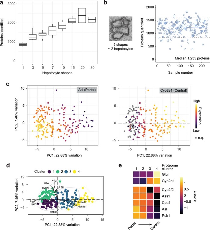
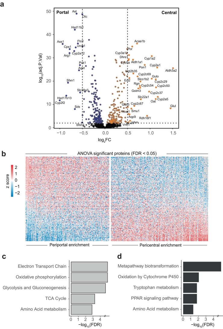
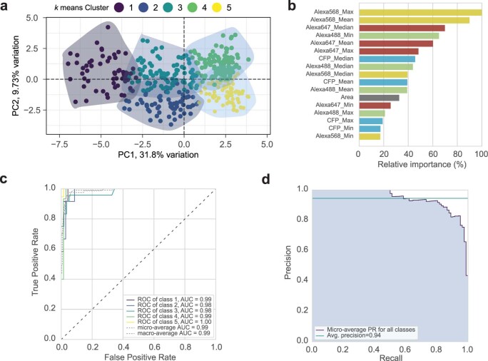
Single-cell proteomics by mass spectrometry is emerging as a powerful and unbiased method for the characterization of biological heterogeneity. So far, it has been limited to cultured cells, whereas an expansion of the method to complex tissues would greatly enhance biological insights. Here we describe single-cell Deep Visual Proteomics (scDVP), a technology that integrates high-content imaging, laser microdissection and multiplexed mass spectrometry. scDVP resolves the context-dependent, spatial proteome of murine hepatocytes at a current depth of 1,700 proteins from a cell slice. Half of the proteome was differentially regulated in a spatial manner, with protein levels changing dramatically in proximity to the central vein. We applied machine learning to proteome classes and images, which subsequently inferred the spatial proteome from imaging data alone. scDVP is applicable to healthy and diseased tissues and complements other spatial proteomics and spatial omics technologies.
Overview
The claims and statistics of this paper haven’t been checked yet.
Run the check — free, no account needed. It reads the open-access text and checks the sources it cites.
Integrity signals
Registry
Cites
Statistics
Figures
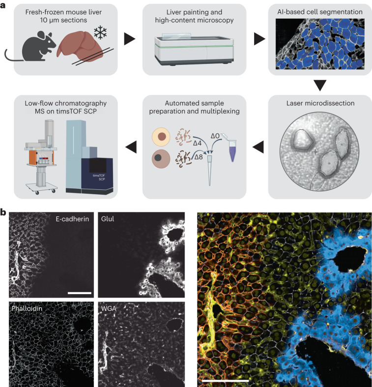
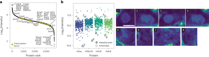
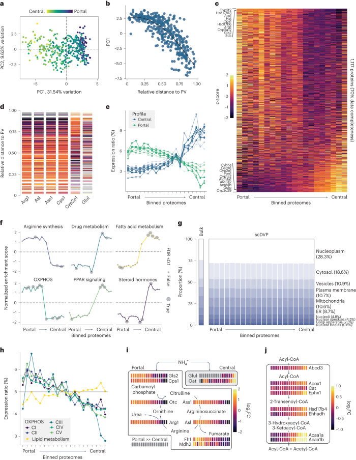
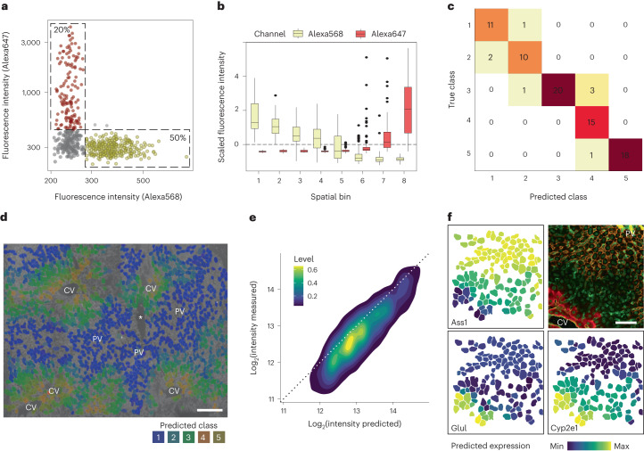
Verification
Claims the paper makes
The claims and statistics of this paper haven’t been checked yet.
Run the check — free, no account needed. It reads the open-access text and checks the sources it cites.
Self-check
Percentages, sums and reported differences re-computed from the paper’s own numbers.
The self-check needs the paper’s full text.
Citations
Cites
The PRIDE database resources in 2022: a hub for mass spectrometry-based proteomics evidences
Perez-Riverol, Y., Bai, J., Bandla, C., García-Seisdedos, D., Hewapathirana, S., Kamatchinathan, S., Kundu, D. J., Prakash, A., Frericks-Zipper, A., Eisenacher, M., et al. (2022) · Nucleic Acids Research · doi:10.1093/nar/gkab1038 · Cited by 6,125
Cox, J., Hein, M. Y., Luber, C. A., Paron, I., Nagaraj, N., & Mann, M. (2014) · Molecular & Cellular Proteomics · doi:10.1074/mcp.m113.031591 · Cited by 5,180
Self-citationDIA-NN: neural networks and interference correction enable deep proteome coverage in high throughput
Demichev, V., Messner, C. B., Vernardis, S. I., Lilley, K. S., & Ralser, M. (2019) · Nature Methods · doi:10.1038/s41592-019-0638-x · Cited by 3,452
WebGestalt 2019: gene set analysis toolkit with revamped UIs and APIs
Liao, Y., Wang, J., Jaehnig, E. J., Shi, Z., & Zhang, B. (2019) · Nucleic Acids Research · doi:10.1093/nar/gkz401 · Cited by 3,144
MSFragger: ultrafast and comprehensive peptide identification in mass spectrometry–based proteomics
Kong, A. T., Leprevost, F. D. V., Avtonomov, D. M., Mellacheruvu, D., & Nesvizhskii, A. I. (2017) · Nature Methods · doi:10.1038/nmeth.4256 · Cited by 2,406
Rath, S., Sharma, R., Gupta, R., Ast, T., Chan, C., Durham, T. J., Goodman, R. P., Grabarek, Z., Haas, M. E., Hung, W. H. W., et al. (2021) · Nucleic Acids Research · doi:10.1093/nar/gkaa1011 · Cited by 1,888
A human liver cell atlas reveals heterogeneity and epithelial progenitors
Aizarani, N., Saviano, A., Sagar, Mailly, L., Durand, S., Herman, J. S., Pessaux, P., Baumert, T. F., & Grün, D. (2019) · Nature · doi:10.1038/s41586-019-1373-2 · Cited by 1,147
Single-cell spatial reconstruction reveals global division of labour in the mammalian liver
Halpern, K. B., Shenhav, R., Matcovitch-Natan, O., Tóth, B., Lemze, D., Golan, M., Massasa, E. E., Baydatch, S., Landen, S., Moor, A. E., et al. (2017) · Nature · doi:10.1038/nature21065 · Cited by 1,059
Spatial proteogenomics reveals distinct and evolutionarily conserved hepatic macrophage niches
Guilliams, M., Bonnardel, J., Haest, B., Vanderborght, B., Wagner, C., Remmerie, A., Bujko, A., Martens, L., Thoné, T., Browaeys, R., et al. (2022) · Cell · doi:10.1016/j.cell.2021.12.018 · Cited by 985
diaPASEF: parallel accumulation–serial fragmentation combined with data-independent acquisition
Meier, F., Brunner, A., Frank, M., Ha, A., Bludau, I., Voytik, E., Kaspar‐Schoenefeld, S., Lubeck, M., Raether, O., Bache, N., et al. (2020) · Nature Methods · doi:10.1038/s41592-020-00998-0 · Cited by 930
Self-renewing diploid Axin2+ cells fuel homeostatic renewal of the liver
Wang, B., Zhao, L., Fish, M., Logan, C. Y., & Nusse, R. (2015) · Nature · doi:10.1038/nature14863 · Cited by 674
scGen predicts single-cell perturbation responses
Lotfollahi, M., Wolf, F. A., & Theis, F. J. (2019) · Nature Methods · doi:10.1038/s41592-019-0494-8 · Cited by 661
Deep Visual Proteomics defines single-cell identity and heterogeneity
Mund, A., Coscia, F., Kriston, A., Hollandi, R., Kovács, F., Brunner, A.-D., Migh, E., Schweizer, L., Santos, A., Bzorek, M., et al. (2022) · Nature Biotechnology · doi:10.1038/s41587-022-01302-5 · Cited by 562
Ultra‐high sensitivity mass spectrometry quantifies single‐cell proteome changes upon perturbation
Brunner, A., Thielert, M., Vasilopoulou, C., Ammar, C., Coscia, F., Mund, A., Hoerning, O. B., Bache, N., Apalategui, A., Lubeck, M., et al. (2022) · Molecular Systems Biology · doi:10.15252/msb.202110798 · Cited by 544
Spatial profiling of chromatin accessibility in mouse and human tissues
Deng, Y., Bartosovic, M., Ma, S., Zhang, D., Kukanja, P., Xiao, Y., Su, G., Liu, Y., Qin, X., Rosoklija, G. B., et al. (2022) · Nature · doi:10.1038/s41586-022-05094-1 · Cited by 417
A Novel LC System Embeds Analytes in Pre-formed Gradients for Rapid, Ultra-robust Proteomics
Bache, N., Geyer, P. E., Bekker‐Jensen, D. B., Hørning, O. B., Falkenby, L. G., Treit, P. V., Doll, S., Paron, I., Müller, J., Meier, F., et al. (2018) · Molecular & Cellular Proteomics · doi:10.1074/mcp.tir118.000853 · Cited by 414
Single-cell Proteomics: Progress and Prospects
Kelly, R. (2020) · Molecular & Cellular Proteomics · doi:10.1074/mcp.r120.002234 · Cited by 367
Phosphorylation Is a Central Mechanism for Circadian Control of Metabolism and Physiology
Robles, M. S., Humphrey, S. J., & Mann, M. (2016) · Cell Metabolism · doi:10.1016/j.cmet.2016.10.004 · Cited by 354
Skowronek, P., Thielert, M., Voytik, E., Tanzer, M. C., Hansen, F. M., Willems, S., Karayel, Ö., Brunner, A., Meier, F., & Mann, M. (2022) · Molecular & Cellular Proteomics · doi:10.1016/j.mcpro.2022.100279 · Cited by 305
Increasing the throughput of sensitive proteomics by plexDIA
Derks, J., Leduc, A., Wallmann, G., Huffman, R. G., Willetts, M., Khan, S., Specht, H., Ralser, M., Demichev, V., & Slavov, N. (2022) · Nature Biotechnology · doi:10.1038/s41587-022-01389-w · Cited by 234
Spatial sorting enables comprehensive characterization of liver zonation
Ben-Moshe, S., Shapira, Y., Moor, A. E., Manco, R., Veg, T., Bahar Halpern, K., & Itzkovitz, S. (2019) · Nature Metabolism · doi:10.1038/s42255-019-0109-9 · Cited by 205
Spatial sorting enables comprehensive characterization of liver zonation
Ben-Moshe, S., Shapira, Y., Moor, A. E., Halpern, K. B., & Itzkovitz, S. (2019) · doi:10.1101/529784 · Cited by 205
Liver Zonation – Revisiting Old Questions With New Technologies
Cunningham, R. P., & Porat‐Shliom, N. (2021) · Frontiers in Physiology · doi:10.3389/fphys.2021.732929 · Cited by 181
The BioImage Archive – Building a Home for Life-Sciences Microscopy Data
Hartley, M., Kleywegt, G. J., Patwardhan, A., Sarkans, U., Swedlow, J. R., & Brazma, A. (2022) · Journal of Molecular Biology · doi:10.1016/j.jmb.2022.167505 · Cited by 159
Space-time logic of liver gene expression at sub-lobular scale
Droin, C., Kholtei, J. E., Bahar Halpern, K., Hurni, C., Rozenberg, M., Muvkadi, S., Itzkovitz, S., & Naef, F. (2021) · Nature Metabolism · doi:10.1038/s42255-020-00323-1 · Cited by 148
Stitching and registering highly multiplexed whole-slide images of tissues and tumors using ASHLAR
Muhlich, J. L., Chen, Y., Yapp, C., Russell, D., Santagata, S., & Sorger, P. K. (2022) · Bioinformatics · doi:10.1093/bioinformatics/btac544 · Cited by 140
Surfactant-assisted one-pot sample preparation for label-free single-cell proteomics
Tsai, C., Zhang, P., Scholten, D., Martin, K., Wang, Y.-T., Zhao, R., Chrisler, W., Patel, D. B., Dou, M., Jia, Y., et al. (2021) · Communications Biology · doi:10.1038/s42003-021-01797-9 · Cited by 122
Pham, T. V., Henneman, A. A., & Jiménez, C. R. (2020) · Bioinformatics · doi:10.1093/bioinformatics/btz961 · Cited by 119
Segovia-Miranda, F., Morales-Navarrete, H., Kücken, M., Moser, V., Seifert, S., Repnik, U., Rost, F., Brosch, M., Hendricks, A., Hinz, S., et al. (2019) · Nature Medicine · doi:10.1038/s41591-019-0660-7 · Cited by 109
Robust dimethyl‐based multiplex‐DIA doubles single‐cell proteome depth via a reference channel
Thielert, M., Itang, E. C. M., Ammar, C., Rosenberger, F. A., Bludau, I., Schweizer, L., Nordmann, T. M., Skowronek, P., Wahle, M., Zeng, W., et al. (2023) · Molecular Systems Biology · doi:10.15252/msb.202211503 · Cited by 98
A framework for ultra-low-input spatial tissue proteomics
Makhmut, A., Qin, D., Fritzsche, S., Nimo, J., König, J., & Coscia, F. (2023) · Cell Systems · doi:10.1016/j.cels.2023.10.003 · Cited by 67
Kang, S. W. S., Cunningham, R. P., Miller, C. B., Brown, L. A., Cultraro, C. M., Harned, A., Narayan, K., Hernandez, J. M., Jenkins, L. M. M., Lobanov, A. V., et al. (2024) · Nature Communications · doi:10.1038/s41467-024-45751-9 · Cited by 66
Making single-cell proteomics biologically relevant
Rosenberger, F. A., Thielert, M., & Mann, M. (2023) · Nature Methods · doi:10.1038/s41592-023-01771-9 · Cited by 65
Harding, R., Deme, J. C., Hevler, J. F., Tamara, S., Lemak, A. S., Cantle, J. P., Szewczyk, M. M., Begeja, N., Goss, S., Zuo, X., et al. (2021) · Communications Biology · doi:10.1038/s42003-021-02895-4 · Cited by 56
Flexibility of zonation of fatty acid oxidation in rat liver
Guzmán, M., Bijleveld, C., & Geelen, M. J. H. (1995) · Biochemical Journal · doi:10.1042/bj3110853 · Cited by 46
Simplified high-throughput methods for deep proteome analysis on the timsTOF ProPreprint
Sandow, J. J., Infusini, G., Dagley, L. F., Larsen, R. H., & Webb, A. I. (2019) · bioRxiv (Cold Spring Harbor Laboratory) · doi:10.1101/657908 · Cited by 27
Kang, S., Cunningham, R. P., Miller, C. B., Brown, L. A., Cultraro, C., Harned, A., Narayan, K., Hernandez, J., Jenkins, L. M., Lobanov, A., et al. (2023) · bioRxiv · doi:10.1101/2023.04.13.536717 · Cited by 26
µPhos: a scalable and sensitive platform for functional phosphoproteomicsPreprint
Oliinyk, D., Will, A., Schneidmadel, F. R., Humphrey, S. J., & Meier, F. (2023) · bioRxiv (Cold Spring Harbor Laboratory) · doi:10.1101/2023.04.04.535617 · Cited by 4
Cited by
Sorted by citations
Spatiotemporal omics for biology and medicine
Liu, L., Chen, A., Li, Y., Mulder, J., Heyn, H., & Xu, X. (2024) · Cell · doi:10.1016/j.cell.2024.07.040 · Cited by 252
Single-cell sequencing to multi-omics: technologies and applications
Wu, X., Yang, X., Dai, Y., Zhao, Z., Zhu, J., Guo, H., & Yang, R. (2024) · Biomarker Research · doi:10.1186/s40364-024-00643-4 · Cited by 222
“Additionally, the integration of high-content imaging, LCM, and multiplexed mass spectrometry can extend single-cell proteomics to intact tissue, significantly improving biological insight [67].”
BackgroundGatto, L., Aebersold, R. H., Cox, J., Demichev, V., Derks, J., Emmott, E., Franks, A. M., Ivanov, A. R., Kelly, R., Khoury, L., et al. (2023) · Nature Methods · doi:10.1038/s41592-023-01785-3 · Cited by 187
MS-Based Proteomics of Body Fluids: The End of the Beginning
Bader, J. M., Albrecht, V., & Mann, M. (2023) · Molecular & Cellular Proteomics · doi:10.1016/j.mcpro.2023.100577 · Cited by 178
Spatial proteomics identifies JAKi as treatment for a lethal skin disease
Nordmann, T. M., Anderton, H., Hasegawa, A., Schweizer, L., Zhang, P., Stadler, P., Sinha, A., Metousis, A., Rosenberger, F. A., Zwiebel, M., et al. (2024) · Nature · doi:10.1038/s41586-024-08061-0 · Cited by 175
Metadata
- Authors
- Rosenberger, F. A., Thielert, M., Strauss, M. T., Schweizer, L., Ammar, C., Mädler, S. C., Metousis, A., Skowronek, P., Wahle, M., Madden, K., Gote-Schniering, J., Semenova, A., Schiller, H. B., Rodriguez, E., Nordmann, T. M., Mund, A., & Mann, M.
- Affiliations
- Max Planck Institute of Biochemistry (DE)
- University of Copenhagen (DK)
- Helmholtz Munich (DE)
- Corresponding author
- Mann, Matthias
- ORCID
- Venue
- Nature Methods
- Published
- 2023-10
- Volume
- 20
- Issue
- 10
- Pages
- 1530-1536
- ISSN
- 1548-7091 (ISSN-L)
- 1548-7105
- PMC
- PMC10555842
- OpenAlex
- W4387266998
- Cited by
- 176
- References
- 36
- Citations by year
- 2026: 49 · 2025: 62 · 2024: 48 · 2023: 17
- FWCI
- 16.23
- Type
- journal article
- Language
- en
- Open access
- hybrid
- doi.org
- Locations
- Nature Methods (publishedVersion, cc-by)
- bioRxiv (Cold Spring Harbor Laboratory) (acceptedVersion, cc-by-nc-nd)
- PubMed Central (publishedVersion, cc-by)
- PubMed (publishedVersion)
- Research at the University of Copenhagen (University of Copenhagen) (publishedVersion, cc-by)
- PuSH - Publication Server of Helmholtz Zentrum München (submittedVersion, cc-by)
- Funders
- Fund for Astrophysical Research
- European Respiratory Society (847462)
- Chan Zuckerberg Initiative (CZF2019-002448)
- European Commission (H2020, 825694, 874839, 847462)
- Boehringer Ingelheim Fonds
- Max-Planck-Gesellschaft
- Novo Nordisk (NNF14CC0001, NNF15CC0001)
- Novo Nordisk Fonden (NNF 14CC0001, NNF15CC0001)
- Horizon 2020 Framework Programme (874839, 825694, 847462)
- Topics
- Physical Sciences › Chemistry › Spectroscopy › Advanced Proteomics Techniques and Applications
- Physical Sciences › Chemistry › Spectroscopy › Mass Spectrometry Techniques and Applications
- Life Sciences › Biochemistry, Genetics and Molecular Biology › Molecular Biology › Single-cell and spatial transcriptomics
- Keywords
- spatial proteomics, single-cell proteomics, hepatocytes, liver zonation, central vein, laser microdissection, high-content imaging, machine learning
- MeSH terms
- Animals, Mass Spectrometry, Proteome, Proteomics, Mice, Laser Capture Microdissection
About these checks
Registry, retraction, Cites and statistics results are computed, not judged, with one exception: a printed reference that no index lists is read once by a model before it counts as a citation error. Claims are extracted by a model and kept only when their quote appears verbatim in the text; each cited source is then checked, with a model’s help, for whether it says what the claim relies on. Those verdicts can be wrong — read the claim’s quote and the cited source before relying on one.
Statistics and arithmetic are recomputed, not judged.
Confidence bands are shown only for fields calibrated on our annotated test set. None is calibrated yet, so no band is shown.
What counts as a claim ›How we measure confidence ›How we check this ›
Are you an author, or do you see a problem with this page? Request a review
