

The DOI matches its Crossref record; 37 of the 37 references it cites were found in a registry or an index.
QuPath: Open source software for digital pathology image analysis
Open accessPublished version of a preprint
Abstract
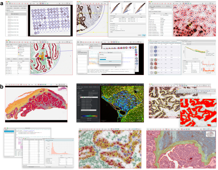
QuPath is new bioimage analysis software designed to meet the growing need for a user-friendly, extensible, open-source solution for digital pathology and whole slide image analysis. In addition to offering a comprehensive panel of tumor identification and high-throughput biomarker evaluation tools, QuPath provides researchers with powerful batch-processing and scripting functionality, and an extensible platform with which to develop and share new algorithms to analyze complex tissue images. Furthermore, QuPath's flexible design makes it suitable for a wide range of additional image analysis applications across biomedical research.
Overview
The claims and statistics of this paper haven’t been checked yet.
Run the check — free, no account needed. It reads the open-access text and checks the sources it cites.
Integrity signals
Registry
Cites
Statistics
Figures
Verification
Claims the paper makes
The claims and statistics of this paper haven’t been checked yet.
Run the check — free, no account needed. It reads the open-access text and checks the sources it cites.
Claims other papers make about this one
Each is checked against this paper’s own text: does it say what the citing paper says it does?
The source backs part of the claim, not all of it — or backs it for a different population, condition or comparison.
In the citing paper
“On the software side, frameworks such as TensorFlow and PyTorch support AI model development, while tools like QuPath enable image annotation and analysis [44].”
From Artificial Intelligence in Dermatopathology: An Update and Review of the Current Literature (2026) · cites this paper as [44]
Experimental — accuracy not yet measuredThe cited source says what the claim attributes to it.
In the citing paper
“Emerging solutions include standardized open-source tools (e.g., QuPath) [44], collaborative multi-institutional annotation efforts, and AI-assisted annotation workflows that reduce manual burden [45].”
From Artificial Intelligence in Dermatopathology: An Update and Review of the Current Literature (2026) · cites this paper as [44]
The cited source says what the claim attributes to it.
In the citing paper
“The microglial ultrastructural analyses was performed using QuPath (v.0.4.4) [71] on images blinded to both animal and group, following an adaptation from our previous work [56, 64].”
From Sex chromosomes and sex hormones differently shape microglial properties during normal physiological conditions in the adult mouse hippocampus (2025) · cites this paper as [71]
Self-check
Percentages, sums and reported differences re-computed from the paper’s own numbers.
The self-check needs the paper’s full text.
Citations
Cites
Breiman, L. (2001) · Machine Learning · doi:10.1023/a:1010933404324 · Cited by 133,538
Fiji: an open-source platform for biological-image analysis
Schindelin, J., Arganda-Carreras, I., Frise, E., Kaynig, V., Longair, M., Pietzsch, T., Preibisch, S., Rueden, C., Saalfeld, S., Schmid, B., et al. (2012) · Nature Methods · doi:10.1038/nmeth.2019 · Cited by 66,890
NIH Image to ImageJ: 25 years of image analysis
Schneider, C. A., Rasband, W. S., & Eliceiri, K. W. (2012) · Nature Methods · doi:10.1038/nmeth.2089 · Cited by 64,638
PD-1 and PD-L1 Immune Checkpoint Blockade to Treat Breast Cancer
Hartkopf, A. D., Taran, F., Wallwiener, M., Walter, C. B., Krämer, B., Grischke, E., & Brucker, S. Y. (2016) · Breast Care · doi:10.1159/000453569 · Cited by 30,989
Textural Features for Image Classification
Haralick, R. M., Shanmugam, K., & Dinstein, I. (1973) · IEEE Transactions on Systems, Man, and Cybernetics · doi:10.1109/tsmc.1973.4309314 · Cited by 22,886
Adverse Renal Effects of Immune Checkpoint Inhibitors: A Narrative Review
Wanchoo, R., Karam, S., Uppal, N. N., Barta, V. S., Deray, G., Devoe, C., Launay‐Vacher, V., Jhaveri, K. D., & on behalf of Cancer and Kidney International Network Workgroup on Immune Checkpoint Inhibitors (2017) · American Journal of Nephrology · doi:10.1159/000455014 · Cited by 15,320
SLIC Superpixels Compared to State-of-the-Art Superpixel Methods
Achanta, R., Shaji, A., Smith, K., Lucchi, A., Fua, P., & Susstrunk, S. E. (2012) · IEEE Transactions on Pattern Analysis and Machine Intelligence · doi:10.1109/tpami.2012.120 · Cited by 9,451
Type, Density, and Location of Immune Cells Within Human Colorectal Tumors Predict Clinical Outcome
Galon, J., Costes, A., Sanchez-Cabo, F., Kirilovsky, A., Mlecnik, B., Lagorce-Pagès, C., Tosolini, M., Camus, M., Berger, A., Wind, P., et al. (2006) · Science · doi:10.1126/science.1129139 · Cited by 6,516
Pembrolizumab for the Treatment of Non–Small-Cell Lung Cancer
Garon, E. B., Rizvi, N. A., Hui, R., Leighl, N., Balmanoukian, A. S., Eder, J. P., Patnaik, A., Aggarwal, C., Gubens, M., Horn, L., et al. (2015) · New England Journal of Medicine · doi:10.1056/nejmoa1501824 · Cited by 6,198
Quantification of histochemical staining by color deconvolution.
Ruifrok, A. C. C., & Johnston, D. A. (2001) · PubMed · Cited by 2,029
Cited by
Sorted by citations
Items 701–800 of 1,045
Levy, J., Azizgolshani, N., Andersen, M. J., Suriawinata, A. A., Liu, X., Lisovsky, M., Ren, B., Bobak, C. A., Christensen, B. C., & Vaickus, L. J. (2020) · Modern Pathology · doi:10.1038/s41379-020-00718-1 · Cited by 56
TIMP1 expression underlies sex disparity in liver metastasis and survival in pancreatic cancer
Hermann, C. D., Schoeps, B., Eckfeld, C., Munkhbaatar, E., Kniep, L., Prokopchuk, O., Wirges, N., Steiger, K., Häußler, D., Knolle, P. A., et al. (2021) · The Journal of Experimental Medicine · doi:10.1084/jem.20210911 · Cited by 55
Subtyping non-small cell lung cancer by histology-guided spatial metabolomics
Neumann, J. M., Freitag, H., Hartmann, J. S., Niehaus, K., Galanis, M., Grießhammer, M., Kellner, U., & Bednarz, H. (2021) · Journal of Cancer Research and Clinical Oncology · doi:10.1007/s00432-021-03834-w · Cited by 55
Wedge, M.-È., Jennings, V. A., Crupi, M. J. F., Poutou, J., Jamieson, T. R., Pelin, A., Pugliese, G., de Souza, C. T., Petryk, J., Laight, B. J., et al. (2022) · Nature Communications · doi:10.1038/s41467-022-29526-8 · Cited by 55
Applications of Artificial Intelligence in Breast Pathology
Liu, Y., Han, D., Parwani, A. V., & Li, Z. (2023) · Archives of Pathology & Laboratory Medicine · doi:10.5858/arpa.2022-0457-ra · Cited by 55
Pielawski, N., Andersson, A., Avenel, C., Beháňová, A., Chelebian, E., Klemm, A. H., Nysjö, F., Solorzano, L., & Wählby, C. (2023) · Heliyon · doi:10.1016/j.heliyon.2023.e15306 · Cited by 55
Drumm, M. R., Wang, W., Sears, T. K., Burdett, K. B., Javier, R., Cotton, K. Y., Webb, B., Byrne, K., Unruh, D. J., Thirunavu, V. M., et al. (2023) · Journal of Clinical Investigation · doi:10.1172/jci168035 · Cited by 58
Blanchi, B., Taurand, M., Sauty-Colace, C., Thomaidou, S., Audeoud, C., Fantuzzi, F., Sawatani, T., Gheibi, S., Sabadell‐Basallote, J., Boot, F., et al. (2023) · Molecular Metabolism · doi:10.1016/j.molmet.2023.101772 · Cited by 55
Therapy-induced senescence as a component of tumor biology: Evidence from clinical cancer
Saleh, T., Bloukh, S., Hasan, M., & Shboul, S. A. (2023) · Biochimica et Biophysica Acta (BBA) - Reviews on Cancer · doi:10.1016/j.bbcan.2023.188994 · Cited by 55
Dorner, H., Stolzer, I., Mattner, J., Kaminski, S., Leistl, S., Edrich, L.-M., Schwendner, R., Hobauer, J., Sebald, A., Leikam, S., et al. (2024) · Gastroenterology · doi:10.1053/j.gastro.2024.06.032 · Cited by 55
The spatial landscape of glial pathology and T cell response in Parkinson’s disease substantia nigra
Ma, M., Paryani, F., Jakubiak, K. A., Xia, S., Antoku, S., Kannan, A., Lee, J., Madden, N., Kumar, S. S., Li, J., et al. (2025) · Nature Communications · doi:10.1038/s41467-025-62478-3 · Cited by 61
Quantitative patterns of motor cortex proteinopathy across ALS genotypes
Nolan, M., Scott, C., Pallebage‐Gamarallage, M. M., Lunn, D., Carpenter, K., McDonough, E., Meyer, D. E., Kaanumalle, S., Santamaría-Pang, A., Turner, M. R., et al. (2020) · Acta Neuropathologica Communications · doi:10.1186/s40478-020-00961-2 · Cited by 55
Goyal, L., Sirard, C. A., Schrag, M. L., Kagey, M. H., Eads, J. R., Stein, S., El-Khoueiry, A. B., Manji, G. A., Abrams, T. A., Khorana, A. A., et al. (2020) · Clinical Cancer Research · doi:10.1158/1078-0432.ccr-20-1310 · Cited by 55
Nguyen, C. T., Caruso, S., Maille, P., Bèaufrere, A., Augustin, J., Favre, L., Pujals, A., Boulagnon‐Rombi, C., Rhaiem, R., Amaddeo, G., et al. (2021) · Clinical Cancer Research · doi:10.1158/1078-0432.ccr-21-1219 · Cited by 55
Hoye, M. L., Calviello, L., Poff, A., Ejimogu, E., Newman, C. R., Montgomery, M. D., Ou, J., Floor, S. N., & Silver, D. L. (2022) · eLife · doi:10.7554/elife.78203 · Cited by 55
Multifaceted modes of action of the anticancer probiotic Enterococcus hirae
Goubet, A.-G., Wheeler, R., Fluckiger, A., Qu, B., Lemaître, F., Iribarren, K., Mondragón, L., Tidjani Alou, M., Pizzato, E., Durand, S., et al. (2021) · Cell Death & Differentiation · doi:10.1038/s41418-021-00753-8 · Cited by 54
Roehlen, N., Saviano, A., El Saghire, H., Crouchet, E., Nehme, Z., Del Zompo, F., Jühling, F., Oudot, M. A., Durand, S. C., Duong, F. H. T., et al. (2022) · Science Translational Medicine · doi:10.1126/scitranslmed.abj4221 · Cited by 54
A thalamo-preoptic pathway promotes social grooming in rodents
Keller, D., Láng, T., Cservenák, M., Puska, G., Barna, J., Csillag, V., Farkas, I., Zelena, D., Dóra, F., Küppers, S., et al. (2022) · Current Biology · doi:10.1016/j.cub.2022.08.062 · Cited by 54
Bórquez, J. C., Díaz-Castro, F., La Fuente, F. P.-D., Espinoza, K., Figueroa, A. M., Martínez-Ruíz, I., Hernández, V., López-Soldado, I., Ventura, R., Domingo, J. C., et al. (2024) · Metabolism · doi:10.1016/j.metabol.2023.155765 · Cited by 54
Martins, T. A., Kaymak, D., Tatari, N., Gerster, F., Hogan, S., Ritz, M.-F., Sabatino, V., Wieboldt, R., Bartoszek, E. M., McDaid, M., et al. (2024) · Nature Communications · doi:10.1038/s41467-024-54129-w · Cited by 54
Tirado‐González, I., Descot, A., Soetopo, D., Nevmerzhitskaya, A., Schäffer, A., Kur, I.-M., Członka, E., Wachtel, C., Tsoukala, I., Müller, L., et al. (2021) · Cancer Discovery · doi:10.1158/2159-8290.cd-20-1378 · Cited by 54
Lorusso, G., Wyss, C. B., Kuonen, F., Vannini, N., Billottet, C., Duffey, N., Pineau, R., Lan, Q., Wirapati, P., Barras, D., et al. (2022) · Science Translational Medicine · doi:10.1126/scitranslmed.aax8933 · Cited by 54
Powell, L. C., Cullen, J. K., Boyle, G. M., Ridder, T. R. D., Yap, P.-Y., Xue, W., Pierce, C. J., Pritchard, M., Menzies, G. E., Abdulkarim, M., et al. (2022) · Science Translational Medicine · doi:10.1126/scitranslmed.abn3758 · Cited by 54
Bronsard, J., Savary, C. C., Massart, J., Viel, R., Moutaux, L., Catheline, D., Rioux, V., Clément, B., Corlu, A., Fromenty, B., et al. (2023) · Toxicology in Vitro · doi:10.1016/j.tiv.2023.105728 · Cited by 54
Pregnancy-responsive pools of adult neural stem cells for transient neurogenesis in mothers
Chaker, Z., Segalada, C., Kretz, J. A., Acar, İ. E., Delgado, A. C., Crotet, V., Moor, A. E., & Doetsch, F. K. (2023) · Science · doi:10.1126/science.abo5199 · Cited by 54
Priego, N., de Pablos-Aragoneses, A., Perea-García, M., Pieri, V., Hernández-Oliver, C., Álvaro‐Espinosa, L., Rojas, A., Sánchez, O., Steindl, A., Caleiras, E. J., et al. (2024) · Cancer Discovery · doi:10.1158/2159-8290.cd-24-0134 · Cited by 54
Midkine as a driver of age-related changes and increase in mammary tumorigenesis
Yan, P., Rojas-Jiménez, E. A., Li, Z., Bui, T. M., Seehawer, M., Nishida, J., Foidart, P., Stevens, L. E., Xie, Y., Gomez, M. M., et al. (2024) · Cancer Cell · doi:10.1016/j.ccell.2024.09.002 · Cited by 54
Maiques, Ó., Sallan, M. C., Laddach, R., Pandya, P., Varela-Vázquez, A., Crosas‐Molist, E., Barcelo, J., Courbot, O., Liu, Y., Graziani, V., et al. (2025) · Nature Communications · doi:10.1038/s41467-025-56299-7 · Cited by 54
Hörst, F., Rempe, M., Becker, H., Heine, L., Keyl, J., & Kleesiek, J. (2026) · Computer Methods and Programs in Biomedicine · doi:10.1016/j.cmpb.2025.109206 · Cited by 54
Kriegsmann, K., Zgorzelski, C., Kazdal, D. N., Cremer, M., Muley, T., Winter, H., Longuespée, R., Kriegsmann, J., Warth, A., & Kriegsmann, M. (2018) · Applied immunohistochemistry & molecular morphology · doi:10.1097/pai.0000000000000715 · Cited by 54
Ahtiainen, M., Wirta, E., Kuopio, T., Seppälä, T. T., Rantala, J., Mecklin, J., & Böhm, J. (2019) · Modern Pathology · doi:10.1038/s41379-019-0219-7 · Cited by 54
Hamaïdia, M., Gazon, H., Hoyos, C., Hoffmann, G. B., Louis, R. E., Duysinx, B., & Willems, L. (2019) · JCI Insight · doi:10.1172/jci.insight.128474 · Cited by 54
Murphy, K. J., Reed, D. A., Vennin, C., Conway, J. R. W., Nobis, M., Yin, J. X. M., Chambers, C. R., Pereira, B., Lee, V., Filipe, E. C., et al. (2021) · Science Advances · doi:10.1126/sciadv.abh0363 · Cited by 54
AMPK activation protects against prostate cancer by inducing a catabolic cellular state
Penfold, L., Woods, A., Pollard, A., Arizanova, J., Pascual-Navarro, E., Muckett, P. J., Dore, M. H., Montoya, A., Whilding, C., Fets, L., et al. (2023) · Cell Reports · doi:10.1016/j.celrep.2023.112396 · Cited by 54
Engineered human osteoarthritic cartilage organoids
Dönges, L., Damle, A., Mainardi, A., Bock, T., Schönenberger, M., Martín, I., & Barbero, A. (2024) · Biomaterials · doi:10.1016/j.biomaterials.2024.122549 · Cited by 53
Remke, M., Groll, T., Metzler, T., Urbauer, E., Kövilein, J., Schnalzger, T., Ruland, J., Haller, D., & Steiger, K. (2024) · Experimental and Molecular Pathology · doi:10.1016/j.yexmp.2024.104938 · Cited by 53
Ribonucleotide incorporation into mitochondrial DNA drives inflammation
Bahat, A., Milenkovic, D., Cors, E., Barnett, M., Niftullayev, S., Katsalifis, A., Schwill, M., Kirschner, P., MacVicar, T., Giavalisco, P., et al. (2025) · Nature · doi:10.1038/s41586-025-09541-7 · Cited by 53
Wu, R., Young, I.-C., Chen, Y.-F., Chuang, S.-T., Toubaji, A., & Wu, M. (2019) · Nature Communications · doi:10.1038/s41467-019-12184-8 · Cited by 53
Zhang, Y., Ware, M. B., Zaidi, M. Y., Ruggieri, A. N., Olson, B. M., Komar, H. M., Farren, M. R., Nagaraju, G. P., Zhang, C., Chen, Z., et al. (2020) · Molecular Cancer Therapeutics · doi:10.1158/1535-7163.mct-19-0911 · Cited by 53
Osteoblast-derived EGFL6 couples angiogenesis to osteogenesis during bone repair
Chen, K., Liao, S., Li, Y., Jiang, H., Liu, Y., Wang, C., Kuek, V., Kenny, J., Li, B., Huang, Q., et al. (2021) · Theranostics · doi:10.7150/thno.60902 · Cited by 53
Xu, H., Cha, Y. J., Clemenceau, J. R. V., Choi, J., Lee, S. H., Kang, J., & Hwang, T. H. (2022) · The Journal of Pathology Clinical Research · doi:10.1002/cjp2.273 · Cited by 53
Liu, X., Rulina, A., Choi, M. H., Pedersen, L., Lepland, J., Takle, S. T., Madeleine, N., Peters, S. D., Wogsland, C. E., Grøndal, S. M., et al. (2022) · Nature Communications · doi:10.1038/s41467-021-27734-2 · Cited by 53
An “eat me” combinatory nano-formulation for systemic immunotherapy of solid tumors
Abdel‐Bar, H. M., Walters, A. A., Lim, Y. M., Rouatbi, N., Qin, Y., Gheidari, F., Han, S., Osman, R., Wang, J. T.-W., & Al‐Jamal, K. T. (2021) · Theranostics · doi:10.7150/thno.56936 · Cited by 52
Modeling of the Senescence-Associated Phenotype in Human Skin Fibroblasts
Gerasymchuk, M., Robinson, G. I., Kovalchuk, O., & Kovalchuk, I. (2022) · International Journal of Molecular Sciences · doi:10.3390/ijms23137124 · Cited by 52
Chatziioannou, E., Roßner, J., Aung, T. N., Rimm, D. L., Niessner, H., Keim, U., Serna-Higuita, L. M., Bonzheim, I., Cuellar, L. K., Westphal, D., et al. (2023) · EBioMedicine · doi:10.1016/j.ebiom.2023.104644 · Cited by 52
Pekayvaz, K., Gold, C., Hoseinpour, P., Engel, A., Martinez-Navarro, A., Eivers, L., Coletti, R., Joppich, M., Dionísio, F., Kaiser, R. W. J., et al. (2023) · Immunity · doi:10.1016/j.immuni.2023.08.002 · Cited by 52
Human APOBEC3B promotes tumor development in vivo including signature mutations and metastases
Durfee, C. C., Temiz, N. A., Levin-Klein, R., Argyris, P. P., Alsøe, L., Carracedo, S., de la Vega, A. A., Proehl, J., Holzhauer, A. M., Seeman, Z. J., et al. (2023) · Cell Reports Medicine · doi:10.1016/j.xcrm.2023.101211 · Cited by 52
Zhang, K., Zakeri, A., Alban, T. J., Dong, J., Ta, H. M., Zalavadia, A. H., Branicky, A., Zhao, H., Jurić, I., Husic, H., et al. (2024) · Cell Reports · doi:10.1016/j.celrep.2023.113661 · Cited by 52
Theobald, H., Bejarano, D. A., Katzmarski, N., Haub, J., Schulte-Schrepping, J., Yu, J., Bassler, K. E., Ament, A. L., Osei-Sarpong, C., Piattini, F., et al. (2024) · Nature Immunology · doi:10.1038/s41590-024-01830-z · Cited by 54
Braun, M., Sevao, M., Keil, S. A., Gino, E., Wang, M. X., Lee, S. J., Haveliwala, M. A., Klein, E., Agarwal, S., Pedersen, T. J., et al. (2024) · Brain · doi:10.1093/brain/awae065 · Cited by 52
Interneuron diversity in the human dorsal striatum
Garma, L. D., Harder, L., Barba-Reyes, J. M., Salas, S. M., Díez-Salguero, M., Nilsson, M. F., Serrano‐Pozo, A., Hyman, B. T., & Muñoz‐Manchado, A. B. (2024) · Nature Communications · doi:10.1038/s41467-024-50414-w · Cited by 56
Myeloid progenitor dysregulation fuels immunosuppressive macrophages in tumours
Hegde, S., Giotti, B., Soong, B. Y., Halász, L., Bérichel, J. L., Schaefer, M. M., Kloeckner, B., Mattiuz, R., Park, M. D., Magen, A., et al. (2025) · Nature · doi:10.1038/s41586-025-09493-y · Cited by 52
Pauli, J., Garger, D., Peymani, F., Wettich, J. L., Sachs, N., Wirth, J., Steiger, K., Hillig, C., Zhang, H., Tabas, I. A., et al. (2025) · Nature Communications · doi:10.1038/s41467-025-67679-4 · Cited by 52
Innervation of papillary thyroid cancer and its association with extra-thyroidal invasion
Rowe, C. W., Dill, T., Griffin, N., Jobling, P., Faulkner, S., Paúl, J., King, S., Smith, R., & Hondermarck, H. (2020) · Scientific Reports · doi:10.1038/s41598-020-58425-5 · Cited by 52
Zhu, L., Nazeri, A., Pacia, C. P., Yue, Y., & Chen, H. (2020) · PLoS ONE · doi:10.1371/journal.pone.0234182 · Cited by 52
A new tool for technical standardization of the Ki67 immunohistochemical assay
Aung, T. N., Ács, B., Warrell, J., Bai, Y., Gaule, P., Martínez-Morilla, S., Vathiotis, I. A., Shafi, S., Moutafi, M. K., Gerstein, M., et al. (2021) · Modern Pathology · doi:10.1038/s41379-021-00745-6 · Cited by 52
Artificial Intelligence in Pathology: From Prototype to Product
Homeyer, A., Lotz, J., Schwen, L. O., Weiss, N., Romberg, D., Höfener, H., Zerbe, N., & Hufnagl, P. (2021) · Journal of Pathology Informatics · doi:10.4103/jpi.jpi_84_20 · Cited by 52
Tryptophan metabolism is inversely regulated in the tumor and blood of patients with glioblastoma
Panitz, V., Končarević, S., Sadik, A., Friedel, D., Bausbacher, T., Trump, S., Farztdinov, V. M., Schulz, S., Sievers, P., Schmidt, S., et al. (2021) · Theranostics · doi:10.7150/thno.60679 · Cited by 52
Klempner, S. J., Bendell, J. C., Villaflor, V. M., Tenner, L. L., Stein, S., Rottman, J. B., Naik, G. S., Sirard, C. A., Kagey, M. H., Chaney, M. F., et al. (2021) · Molecular Cancer Therapeutics · doi:10.1158/1535-7163.mct-21-0273 · Cited by 52
Predicting biochemical recurrence of prostate cancer with artificial intelligence
Pinckaers, H., van Ipenburg, J., Melamed, J., Marzo, A. M. D., Platz, E. A., van Ginneken, B., van der Laak, J. A. W. M., & Litjens, G. J. S. (2022) · Communications Medicine · doi:10.1038/s43856-022-00126-3 · Cited by 51
Zhang, Q., Fei, L., Han, R., Huang, R., Wang, Y., Chen, H., Yao, B., Qiao, N., Wang, Z., Ma, Z., et al. (2022) · Cell Discovery · doi:10.1038/s41421-022-00459-2 · Cited by 51
A pan-cancer analysis shows immunoevasive characteristics in NRF2 hyperactive squamous malignancies
Härkönen, J., Pölönen, P., Deen, A. J., Selvarajan, I., Teppo, H., Dimova, E. Y., Kietzmann, T., Ahtiainen, M., Väyrynen, J. P., Väyrynen, S. A., et al. (2023) · Redox Biology · doi:10.1016/j.redox.2023.102644 · Cited by 51
Microglia directly associate with pericytes in the central nervous system
Morris, G. P., Foster, C. G., Courtney, J., Collins, J. M., Cashion, J. M., Brown, L. S., Howells, D. W., DeLuca, G. C., Canty, A. J., King, A. E., et al. (2023) · Glia · doi:10.1002/glia.24371 · Cited by 52
Prospective on Imaging Mass Spectrometry in Clinical Diagnostics
Moore, J. L., Patterson, N. H., Norris, J. L., & Caprioli, R. M. (2023) · Molecular & Cellular Proteomics · doi:10.1016/j.mcpro.2023.100576 · Cited by 53
An interpretable decision-support model for breast cancer diagnosis using histopathology images
Krishna, S., Suganthi, S. S., Bhavsar, A. V., Yesodharan, J., & Krishnamoorthy, S. (2023) · Journal of Pathology Informatics · doi:10.1016/j.jpi.2023.100319 · Cited by 51
Dashti, A., Sukkestad, S., Horner, A. M., Neja, M., Siddiqi, Z., Waller, C., Goldy, J. N., Monroe, D., Lin, A., Schoof, N., et al. (2023) · Nature Medicine · doi:10.1038/s41591-023-02570-7 · Cited by 51
MAPS: pathologist-level cell type annotation from tissue images through machine learning
Shaban, M., Bai, Y., Qiu, H., Mao, S., Yeung, J., Yeo, Y. Y., Shanmugam, V., Chen, H. M., Zhu, B., Weirather, J. L., et al. (2024) · Nature Communications · doi:10.1038/s41467-023-44188-w · Cited by 51
Library size confounds biology in spatial transcriptomics data
Bhuva, D. D., Tan, C. W., Salim, A., Marceaux, C., Pickering, M. A., Chen, J., Kharbanda, M., Jin, X., Liu, N., Feher, K., et al. (2024) · Genome biology · doi:10.1186/s13059-024-03241-7 · Cited by 56
Li, C., Qian, W., Wei, X., Narasimhan, H., Wu, Y., Arish, M., Cheon, I. S., Tang, J., Santos, G. D. A., Li, Y., et al. (2024) · Science Translational Medicine · doi:10.1126/scitranslmed.adn0136 · Cited by 51
Klein, L., Tu, M., Krebs, N., Urbach, L., Grimm, D. T., Latif, M. U., Penz, F., Blandau, A., Wu, X., Samuel, R. D., et al. (2025) · Nature Communications · doi:10.1038/s41467-024-55330-7 · Cited by 51
Lubrano, S., Cervantes‐Villagrana, R. D., Faraji, F., Ramirez, S. I., Sato, K., Adame-Garcia, S. R., Officer, A., Arang, N., Rigiracciolo, D. C., Quiroz, P. Y. A., et al. (2025) · Cancer Cell · doi:10.1016/j.ccell.2025.02.001 · Cited by 51
Yao, J., Chakhoyan, A., Nathanson, D. A., Yong, W. H., Salamon, N., Raymond, C., Mareninov, S., Lai, A., Nghiemphu, P. L., Prins, R. M., et al. (2019) · Neuro-Oncology · doi:10.1093/neuonc/noz078 · Cited by 51
Tsakiroglou, A. M., Fergie, M., Oguejiofor, K., Linton, K., Thomson, D. J., Stern, P. L., Astley, S., Byers, R. J., & West, C. (2019) · British Journal of Cancer · doi:10.1038/s41416-019-0634-z · Cited by 51
PyHIST: A Histological Image Segmentation Tool
Muñoz-Aguirre, M., Ntasis, V. F., Rojas, S., & Guigó, R. (2020) · PLoS Computational Biology · doi:10.1371/journal.pcbi.1008349 · Cited by 51
Cancer Cells Retrace a Stepwise Differentiation Program during Malignant Progression
Saghafinia, S., Homicskó, K., Domenico, A. D., Wullschleger, S., Perren, A. A., Marinoni, I., Ciriello, G., Michael, I. P., & Hanahan, D. (2021) · Cancer Discovery · doi:10.1158/2159-8290.cd-20-1637 · Cited by 51
Aboulgheit, A., Karbasiafshar, C., Zhang, Z., Sabra, M., Shi, G., Tucker, A., Sodha, N. R., Abid, M. R., & Sellke, F. W. (2021) · American Journal of Physiology-Heart and Circulatory Physiology · doi:10.1152/ajpheart.00278.2021 · Cited by 51
Highly multiplexed immunofluorescence of the human kidney using co-detection by indexing
Neumann, E. K., Patterson, N. H., Rivera, E. S., Allen, J. L., Brewer, M., deCaestecker, M. P., Caprioli, R. M., Fogo, A. B., & Spraggins, J. M. (2021) · Kidney International · doi:10.1016/j.kint.2021.08.033 · Cited by 51
Viiklepp, K., Nissinen, L. M., Ojalill, M., Riihilä, P., Kallajoki, M. A., Meri, S., Heino, J., & Kähäri, V. (2021) · Journal of Investigative Dermatology · doi:10.1016/j.jid.2021.10.008 · Cited by 51
Cobb, D. A., de Rossi, J., Liu, L., An, E., & Lee, D. W. (2022) · Journal for ImmunoTherapy of Cancer · doi:10.1136/jitc-2021-003816 · Cited by 51
el Maaty, M. A. A., Terzic, J., Keime, C., Rovito, D., Lutzing, R., Yanushko, D., Parisotto, M., Grelet, É., Namer, I. J., Lindner, V., et al. (2022) · Science Advances · doi:10.1126/sciadv.abo2295 · Cited by 52
Muhuri, M., Zhan, W., Maeda, Y., Li, J., Lotun, A., Chen, J., Sylvia, K., Dasgupta, I., Arjomandnejad, M., Nixon, T., et al. (2021) · Frontiers in Immunology · doi:10.3389/fimmu.2021.674242 · Cited by 50
Kriegsmann, K., Lobers, F., Zgorzelski, C., Kriegsmann, J., Janßen, C., Meliß, R. R., Muley, T., Sack, U., Steinbuss, G., & Kriegsmann, M. (2022) · Frontiers in Oncology · doi:10.3389/fonc.2022.1022967 · Cited by 50
Crum, R. J., Hall, K., Molina, C. P., Hussey, G. S., Graham, E., Li, H., & Badylak, S. F. (2022) · npj Regenerative Medicine · doi:10.1038/s41536-022-00208-9 · Cited by 50
Sabe, S. A., Xu, C., Sabra, M., Harris, D. D., Malhotra, A., Aboulgheit, A., Stanley, M. E., Abid, M. R., & Sellke, F. W. (2022) · Journal of the American Heart Association · doi:10.1161/jaha.122.028623 · Cited by 50
Indian Hedgehog release from TNF-activated renal epithelia drives local and remote organ fibrosis
O'Sullivan, E. D., Mylonas, K. J., Xin, C., Baird, D., Carvalho, C., Docherty, M.-H., Campbell, R., Matchett, K. P., Waddell, S. H., Walker, A. D., et al. (2023) · Science Translational Medicine · doi:10.1126/scitranslmed.abn0736 · Cited by 50
Jacobs, C. L., Shah, S., Lu, W., Ray, H., Wang, J., Hockaden, N., Sandusky, G. E., Nephew, K. P., Lü, X., Cao, S., et al. (2023) · Cancer Research · doi:10.1158/0008-5472.can-23-0902 · Cited by 50
Spence, H., Waldron, F. M., Saleeb, R. S., Brown, A., Rifai, O. M., Gilodi, M., Read, F. L., Roberts, K., Milne, G., Wilkinson, D., et al. (2024) · Acta Neuropathologica · doi:10.1007/s00401-024-02705-1 · Cited by 50
Pervasive environmental chemicals impair oligodendrocyte development
Cohn, E. F., Clayton, B. L. L., Madhavan, M., Lee, K. A., Yacoub, S. G., Federov, Y., Scavuzzo, M. A., Friedman, K. P., Shafer, T. J., & Tesar, P. J. (2024) · Nature Neuroscience · doi:10.1038/s41593-024-01599-2 · Cited by 51
Mori, Y., Okimoto, Y., Sakai, H., Kanda, Y., Ohata, H., Shiokawa, D., Suzuki, M., Yoshida, H., Ueda, H., Sekizuka, T., et al. (2024) · Cell Reports Medicine · doi:10.1016/j.xcrm.2024.101532 · Cited by 50
Spatial multiplexing and omics
Carstens, J. L., Krishnan, S., Rao, A. U. K., Sorace, A. G., Seeley, E. H., Borgogno, S. F., & Burks, J. K. (2024) · Nature Reviews Methods Primers · doi:10.1038/s43586-024-00330-6 · Cited by 50
Ben‐Shmuel, A., Gruper, Y., Halperin, C., Levi-Galibov, O., Rosenberg-Fogler, H., Barki, D., Carradori, G., Stein, Y., Yagel, G., Naumova, M., et al. (2025) · Cancer Discovery · doi:10.1158/2159-8290.cd-24-0131 · Cited by 54
Humphries, M. P., McQuaid, S., Craig, S. G., Bingham, V., Maxwell, P., Maurya, M., McLean, F., Sampson, J., Higgins, P., Greene, C., et al. (2018) · Journal of Thoracic Oncology · doi:10.1016/j.jtho.2018.09.025 · Cited by 50
Self-citationRobertson, S., Ács, B., Lippert, M. F., & Hartman, J. (2020) · Breast Cancer Research and Treatment · doi:10.1007/s10549-020-05752-w · Cited by 50
NCLX prevents cell death during adrenergic activation of the brown adipose tissue
Assali, E. A., Jones, A. E., Veliova, M., Acín‐Pérez, R., Taha, M., Miller, N., Shum, M., de Oliveira, M. F., Las, G., Liesa, M., et al. (2020) · Nature Communications · doi:10.1038/s41467-020-16572-3 · Cited by 50
Ghasemi, F., Tessier, T. M., Gameiro, S. F., Maciver, A. H., Cecchini, M. J., & Mymryk, J. S. (2020) · Scientific Reports · doi:10.1038/s41598-020-71775-4 · Cited by 50
Alame, M., Cornillot, E., Cacheux, V., Rigau, V., Costes‐Martineau, V., Szablewski, V., & Colinge, J. (2021) · Theranostics · doi:10.7150/thno.54343 · Cited by 51
Jacobs, S., Wesseling, P., de Keizer, B., Tolboom, N., Ververs, T. F., Krijger, G. C., Westerman, B. A., Snijders, T. J., Robe, P. A. J. T., & van der Kolk, A. G. (2021) · European Journal of Nuclear Medicine and Molecular Imaging · doi:10.1007/s00259-021-05196-4 · Cited by 50
Kelly, S. B., Stojanovska, V., Zahra, V. A., Moxham, A. M., Miller, S., Moss, T. J. M., Hooper, S. B., Nold, M. F., Nold‐Petry, C. A., Dean, J. M., et al. (2021) · Journal of Neuroinflammation · doi:10.1186/s12974-021-02238-4 · Cited by 50
Marian, O. C., Teo, J. D., Lee, J. Y., Song, H., Kwok, J. B., Landin-Romero, R., Halliday, G., & Don, A. S. (2023) · Acta Neuropathologica Communications · doi:10.1186/s40478-023-01544-7 · Cited by 50
Distinct Nrf2 Signaling Thresholds Mediate Lung Tumor Initiation and Progression
DeBlasi, J. M., Falzone, A., Caldwell, S., Prieto-Farigua, N., Prigge, J. R., Schmidt, E. E., Chio, I. I. C., Karreth, F. A., & DeNicola, G. M. (2023) · Cancer Research · doi:10.1158/0008-5472.can-22-3848 · Cited by 49
“Slides were scanned with theAperio imagerat20 (cid:2) andtheH-score ofatleast fi verepresentative regions/mouse was analyzed with QuPath software (42).”
Metadata
- Authors
- Bankhead, P., Loughrey, M. B., Fernández, J. A., Dombrowski, Y., McArt, D. G., Dunne, P. D., McQuaid, S., Gray, R. T., Murray, L. J., Coleman, H. G., James, J. A., Salto-Tellez, M., & Hamilton, P. W.
- Affiliations
- Queen's University Belfast (GB)
- Corresponding author
- Hamilton, Peter W.
- ORCID
- Venue
- Scientific Reports
- Published
- 2017-12-04
- Volume
- 7
- Issue
- 1
- Pages
- 16878-16878
- ISSN
- 2045-2322 (ISSN-L)
- PMC
- PMC5715110
- OpenAlex
- W2952481429
- Cited by
- 8,908
- References
- 37
- Citations by year
- 2026: 1616 · 2025: 1896 · 2024: 1681 · 2023: 1359 · 2022: 1023 · 2021: 704 · 2020: 396 · 2019: 171 · 2018: 49 · 2017: 4
- FWCI
- 99.74
- Type
- journal article
- Language
- en
- Open access
- gold
- doi.org
- Locations
- Scientific Reports (publishedVersion, cc-by)
- bioRxiv (Cold Spring Harbor Laboratory) (acceptedVersion, cc-by-nc-nd)
- Europe PMC (PubMed Central) (publishedVersion, cc-by)
- PubMed Central (publishedVersion, cc-by)
- PubMed (publishedVersion)
- DOAJ (DOAJ: Directory of Open Access Journals) (submittedVersion)
- Edinburgh Research Explorer (University of Edinburgh) (submittedVersion, cc-by)
- Funders
- Invest Northern Ireland (RDO0712612)
- Friends of the Cancer Centre
- Cancer Research UK (20256, A15333, C37703/A15333, C11512, A20256, C11512/A20256, C50104/A17592, 15333)
- Public Health Agency (SPI/3315/06, EAT/4905/13)
- Health and Social Care Research and Development Division
- Topics
- Physical Sciences › Computer Science › Artificial Intelligence › AI in cancer detection
- Life Sciences › Biochemistry, Genetics and Molecular Biology › Biophysics › Cell Image Analysis Techniques
- Health Sciences › Medicine › Radiology, Nuclear Medicine and Imaging › Radiomics and Machine Learning in Medical Imaging
- Keywords
- QuPath, digital pathology, whole slide image analysis, open source software, tumor detection, biomarker analysis, batch processing, scripting, tissue image analysis
- MeSH terms
- Algorithms, Colonic Neoplasms, Humans, Image Interpretation, Computer-Assisted, Biomarkers, Tumor, User-Computer Interface, Kaplan-Meier Estimate, Programmed Cell Death 1 Ligand 2 Protein
About these checks
Registry, retraction, Cites and statistics results are computed, not judged, with one exception: a printed reference that no index lists is read once by a model before it counts as a citation error. Claims are extracted by a model and kept only when their quote appears verbatim in the text; each cited source is then checked, with a model’s help, for whether it says what the claim relies on. Those verdicts can be wrong — read the claim’s quote and the cited source before relying on one.
Statistics and arithmetic are recomputed, not judged.
Confidence bands are shown only for fields calibrated on our annotated test set. None is calibrated yet, so no band is shown.
What counts as a claim ›How we measure confidence ›How we check this ›
Are you an author, or do you see a problem with this page? Request a review
