

The DOI matches its Crossref record; 26 of the 27 references it cites were found in a registry or an index.
Definition of Fiducial Points in the Normal Seismocardiogram
Open accessAbstract
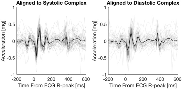
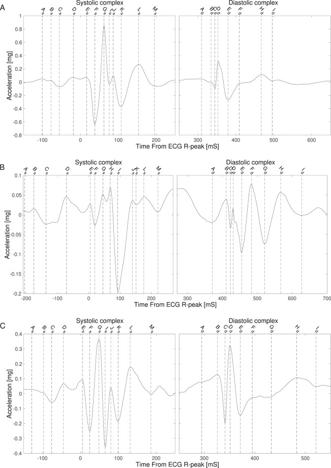
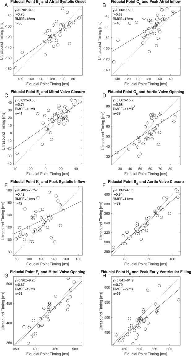
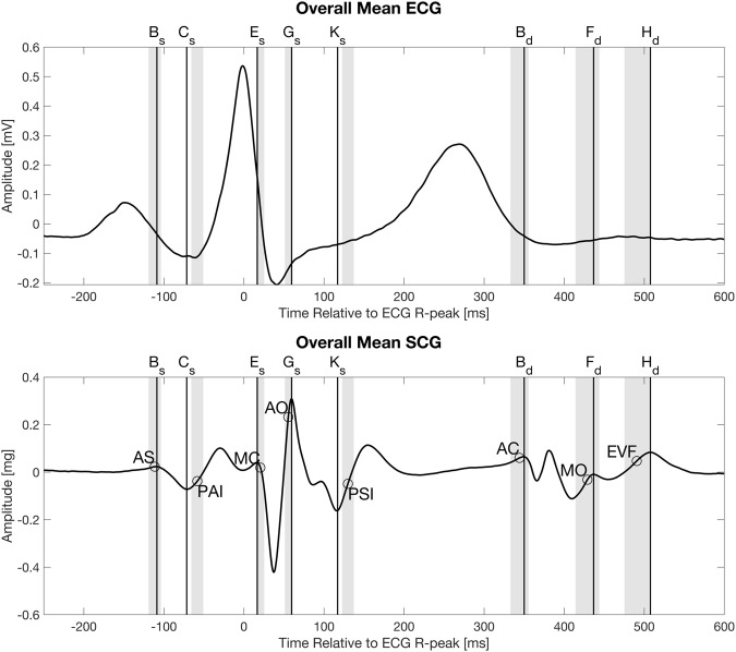
The purpose of this work is to define fiducial points in the seismocardiogram (SCG) and to correlate them with physiological events identified in ultrasound images. For 45 healthy subjects the SCG and the electrocardiogram (ECG) were recorded simultaneously at rest. Immediately following the SCG and ECG recordings ultrasound images of the heart were also obtained at rest. For all subjects a mean SCG signal was calculated and all fiducial points (peaks and valleys) were identified and labeled in the same way across all signals. Eight physiologic events, including the valve openings and closings, were annotated from ultrasound as well and the fiducial points were correlated with those physiologic events. A total of 42 SCG signals were used in the data analysis. The smallest mean differences (±SD) between the eight events found in the ultrasound images and the fiducial points, together with their correlation coefficients (r) were: atrial systolic onset: −2 (±16) ms, r = 0.75 (p < 0.001); peak atrial inflow: 13 (±19) ms, r = 0.63 (p < 0.001); mitral valve closure: 4 (±11) ms, r = 0.71 (p < 0.01); aortic valve opening: −3 (±11) ms, r = 0.60 (p < 0.001); peak systolic outflow: 13 (±23) ms, r = 0.42 (p < 0.01); aortic valve closure: −5 (±12) ms, r = 0.94 (p < 0.001); mitral valve opening: −7 (±19) ms, r = 0.87 (p < 0.001) and peak early ventricular filling: −18 (±28 ms), r = 0.79 (p < 0.001). In conclusion eight physiologic events characterizeing the cardiac cycle, are associated with reproducible, well-defined fiducial points in the SCG.
Overview
The claims and statistics of this paper haven’t been checked yet.
Run the check — free, no account needed. It reads the open-access text and checks the sources it cites.
Integrity signals
Registry
Cites
Statistics
Figures
Verification
Claims the paper makes
The claims and statistics of this paper haven’t been checked yet.
Run the check — free, no account needed. It reads the open-access text and checks the sources it cites.
Claims other papers make about this one
Each is checked against this paper’s own text: does it say what the citing paper says it does?
The source backs part of the claim, not all of it — or backs it for a different population, condition or comparison.
In the citing paper
“This result agrees with previous literature. [38,39]”
From A Chest‐Conformable, Wireless Electro‐Mechanical E‐Tattoo for Measuring Multiple Cardiac Time Intervals (2023) · cites this paper as [39]
Experimental — accuracy not yet measuredThe source backs part of the claim, not all of it — or backs it for a different population, condition or comparison.
In the citing paper
“the AC is near the beginning of the first major minima of the second heart sound.”
From A Chest‐Conformable, Wireless Electro‐Mechanical E‐Tattoo for Measuring Multiple Cardiac Time Intervals (2023) · cites this paper as [39]
Experimental — accuracy not yet measuredThe check wasn’t confident either way, so it gives no verdict — never disputed.
In the citing paper
“the AO clearly corresponds to the first major peak of the first heart sound”
From A Chest‐Conformable, Wireless Electro‐Mechanical E‐Tattoo for Measuring Multiple Cardiac Time Intervals (2023) · cites this paper as [39]
Experimental — accuracy not yet measured
Self-check
Percentages, sums and reported differences re-computed from the paper’s own numbers.
The self-check needs the paper’s full text.
Citations
Cites
Toward ubiquitous blood pressure monitoring via pulse transit time: Theory and practice
Mukkamala, R., Hahn, J.-O., Inan, O. T., Mestha, L. K., Kim, C.-S., Toreyin, H., & Kyal, S. (2015) · IEEE Transactions on Biomedical Engineering · doi:10.1109/tbme.2015.2441951 · Cited by 935
Ballistocardiography and Seismocardiography: A Review of Recent Advances
Inan, O. T., Migeotte, P.-F., Park, K.-S., Etemadi, M., Tavakolian, K., Casanella, R., Zanetti, J., Tank, J., Funtova, I., Prisk, G. K., et al. (2015) · IEEE Journal of Biomedical and Health Informatics · doi:10.1109/jbhi.2014.2361732 · Cited by 775
Relationship between Seismocardiogram and Echocardiogram for Events in the Cardiac Cycle
Crow, R. S., Hannan, P., Jacobs, D., Hedquist, L., & Salerno, D. M. (2017) · American Journal of Noninvasive Cardiology · doi:10.1159/000470156 · Cited by 186
Di Rienzo, M., Vaini, E., Castiglioni, P., Merati, G., Meriggi, P., Parati, G., Faini, A., & Rizzo, F. (2013) · Autonomic Neuroscience · doi:10.1016/j.autneu.2013.04.005 · Cited by 163
Seismocardiography: Past, present and future
Zanetti, J. M., & Tavakolian, K. (2013) · Annual International Conference of the IEEE Engineering in Medicine and Biology Society (EMBC) · doi:10.1109/embc.2013.6611170 · Cited by 152
Salerno, D. M., & Zanetti, J. M. (1990) · Cited by 123
Seismocardiography: a technique for recording precordial acceleration
Zanetti, J. M., & Salerno, D. M. (2002) · doi:10.1109/cbms.1991.128936 · Cited by 103
Automatic Annotation of Seismocardiogram With High-Frequency Precordial Accelerations
Khosrow-Khavar, F., Tavakolian, K., Blaber, A. P., Zanetti, J. M., Fazel-Rezai, R., & Menon, C. (2014) · IEEE Journal of Biomedical and Health Informatics · doi:10.1109/jbhi.2014.2360156 · Cited by 91
THE MASS-MOVEMENTS OF THE CIRCULATION AS SHOWN BY A RECOIL CURVE
Henderson, Y. (1905) · American Journal of Physiology-Legacy Content · doi:10.1152/ajplegacy.1905.14.3.287 · Cited by 74
Seismocardiography: waveform identification and noise analysis
Zanetti, J. M., Poliac, M. O., & Crow, R. S. (2002) · Computers in cardiology · doi:10.1109/cic.1991.169042 · Cited by 62
Research works printed but not linked: 2
- Baevsky, R., Egorov, A. & Kazarian, L. Metodika seismokardiografii. Kardiologia 18, 87–89 (1964).
- Otto, Catherine M. Textbook of Clinical Echocardiography: Expert Consult - Online and Print, 4e. (Saunders, 2009).
Cited by
Sorted by citations
Contactless seismocardiography via deep learning radars
Ha, U., Assana, S., & Adib, F. (2020) · ACM Annual International Conference on Mobile Computing and Networking (MobiCom) · doi:10.1145/3372224.3419982 · Cited by 203
Wearable sensors and devices for real-time cardiovascular disease monitoring
Lin, J., Fu, R., Zhong, X., Yu, P., Tan, G., Li, W., Zhang, H., Li, Y., Zhou, L., & Ning, C. (2021) · Cell Reports Physical Science · doi:10.1016/j.xcrp.2021.100541 · Cited by 169
Flexible sensors and machine learning for heart monitoring
Kwon, S. H., & Dong, L. (2022) · Nano Energy · doi:10.1016/j.nanoen.2022.107632 · Cited by 99
Wearable Sensors for the Monitoring of Maternal Health—A Systematic Review
Alim, A., & Imtiaz, M. H. (2023) · Sensors · doi:10.3390/s23052411 · Cited by 89
Dehkordi, P., Khosrow-Khavar, F., Rienzo, M. D., Inan, O. T., Schmidt, S. E., Blaber, A. P., Sørensen, K., Struijk, J. J., Zakeri, V., Lombardi, P., et al. (2019) · Frontiers in Physiology · doi:10.3389/fphys.2019.01057 · Cited by 82
Self-citation
Metadata
- Authors
- Sørensen, K., Schmidt, S. E., Jensen, A. S., Søgaard, P., & Struijk, J. J.
- Affiliations
- Aalborg University (DK)
- Aalborg University Hospital (DK)
- Corresponding author
- Sørensen, Kasper
- Venue
- Scientific Reports
- Published
- 2018-10-18
- Volume
- 8
- Issue
- 1
- Pages
- 15455-15455
- ISSN
- 2045-2322 (ISSN-L)
- PMC
- PMC6193995
- OpenAlex
- W2896674935
- Cited by
- 111
- References
- 27
- Citations by year
- 2026: 13 · 2025: 17 · 2024: 8 · 2023: 14 · 2022: 20 · 2021: 11 · 2020: 22 · 2019: 6
- FWCI
- 7.35
- Type
- journal article
- Language
- en
- Update notices
- correction (2020-09-04)
- Open access
- gold
- doi.org
- Locations
- Scientific Reports (publishedVersion, cc-by)
- Europe PMC (PubMed Central) (publishedVersion, other-oa)
- PubMed Central (publishedVersion, cc-by)
- PubMed (publishedVersion)
- VBN Forskningsportal (Aalborg Universitet) (publishedVersion, cc-by)
- DOAJ (DOAJ: Directory of Open Access Journals) (submittedVersion)
- Topics
- Health Sciences › Medicine › Pulmonary and Respiratory Medicine › Phonocardiography and Auscultation Techniques
- Physical Sciences › Engineering › Biomedical Engineering › Non-Invasive Vital Sign Monitoring
- Health Sciences › Medicine › Cardiology and Cardiovascular Medicine › ECG Monitoring and Analysis
- Keywords
- seismocardiogram, fiducial points, mitral valve closure, aortic valve closure, valve opening, echocardiography, electrocardiogram
- MeSH terms
- Adult, Aged, Aged, 80 and over, Diastole, Echocardiography, Electrocardiography, Heart Function Tests, Heart Valves, Humans, Image Processing, Computer-Assisted, Middle Aged, Myocardial Contraction, Reference Values, Signal Processing, Computer-Assisted, Systole, Vibration, Reproducibility of Results, Young Adult, Fiducial Markers, Accelerometry
About these checks
Registry, retraction, Cites and statistics results are computed, not judged, with one exception: a printed reference that no index lists is read once by a model before it counts as a citation error. Claims are extracted by a model and kept only when their quote appears verbatim in the text; each cited source is then checked, with a model’s help, for whether it says what the claim relies on. Those verdicts can be wrong — read the claim’s quote and the cited source before relying on one.
Statistics and arithmetic are recomputed, not judged.
Confidence bands are shown only for fields calibrated on our annotated test set. None is calibrated yet, so no band is shown.
What counts as a claim ›How we measure confidence ›How we check this ›
Are you an author, or do you see a problem with this page? Request a review
