

The DOI matches its Crossref record; 43 of the 43 references it cites were found in a registry or an index; 8 of the 23 sources its claims cite were checked for support.
Sigma-2 Receptor/TMEM97 and PGRMC-1 Increase the Rate of Internalization of LDL by LDL Receptor through the Formation of a Ternary Complex
Open accessAbstract
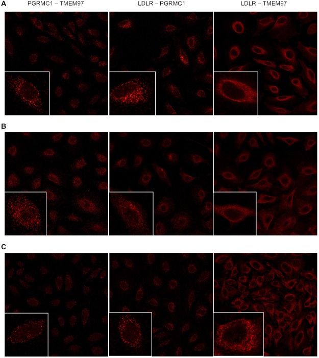
CRISPR/Cas gene studies were conducted in HeLa cells where either PGRMC1, TMEM97 or both proteins were removed via gene editing. A series of radioligand binding studies, confocal microscopy studies, and internalization of radiolabeled or fluorescently tagged LDL particles were then conducted in these cells. The results indicate that PGRMC1 knockout (KO) did not reduce the density of binding sites for the sigma-2 receptor (σ2R) radioligands, [ 125 I] RHM-4 or [ 3 H]DTG, but a reduction in the receptor affinity of both radioligands was observed. TMEM97 KO resulted in a complete loss of binding of [ 125 I] RHM-4 and a significant reduction in binding of [ 3 H]DTG. TMEM97 KO and PGRMC1 KO resulted in an equal reduction in the rate of uptake of fluorescently-tagged or 3 H-labeled LDL, and knocking out both proteins did not result in a further rate of reduction of LDL uptake. Confocal microscopy and Proximity Ligation Assay studies indicated a clear co-localization of LDLR, PGRMC1 and TMEM97. These data indicate that the formation of a ternary complex of LDLR-PGRMC1-TMEM97 is necessary for the rapid internalization of LDL by LDLR.
Overview
AI snapshot
Written from the paper’s full text.
TL;DR
CRISPR knockout of TMEM97/σ2R or PGRMC1 in HeLa cells equally slows LDL internalization, and double knockout is not additive, indicating both proteins are needed in a ternary complex with LDLR for rapid LDL uptake. Radioligand binding shows TMEM97 is required for [125I]RHM-4 binding and most [3H]DTG binding, while PGRMC1 loss lowers σ2R ligand affinity without changing Bmax.
Key findings
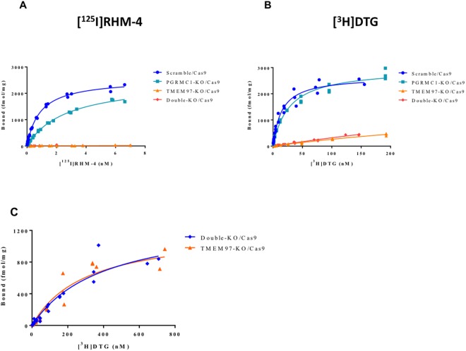
- PGRMC1 KO did not reduce Bmax for [125I]RHM-4 or [3H]DTG but increased Kd: [3H]DTG Kd rose from 14.8 ± 1.8 nM to 27.5 ± 2.5 nM, and [125I]RHM-4 Kd rose from 0.88 ± 0.04 nM to 2.66 ± 0.12 nM.
- TMEM97 KO eliminated [125I]RHM-4 binding and reduced [3H]DTG binding; residual [3H]DTG binding had Kd 302 ± 80 nM and Bmax 1218 ± 148 fmol/mg, with similar values in double KO (Kd 402.6 ± 109.8 nM; Bmax 1374 ± 192 fmol/mg).
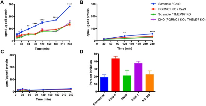
- LDL uptake was significantly decreased in PGRMC1 KO, TMEM97 KO, and double KO cells, with no further reduction in double KO; LDL binding at 4 °C was similar across cell lines, and somatostatin/insulin uptake was unaffected.
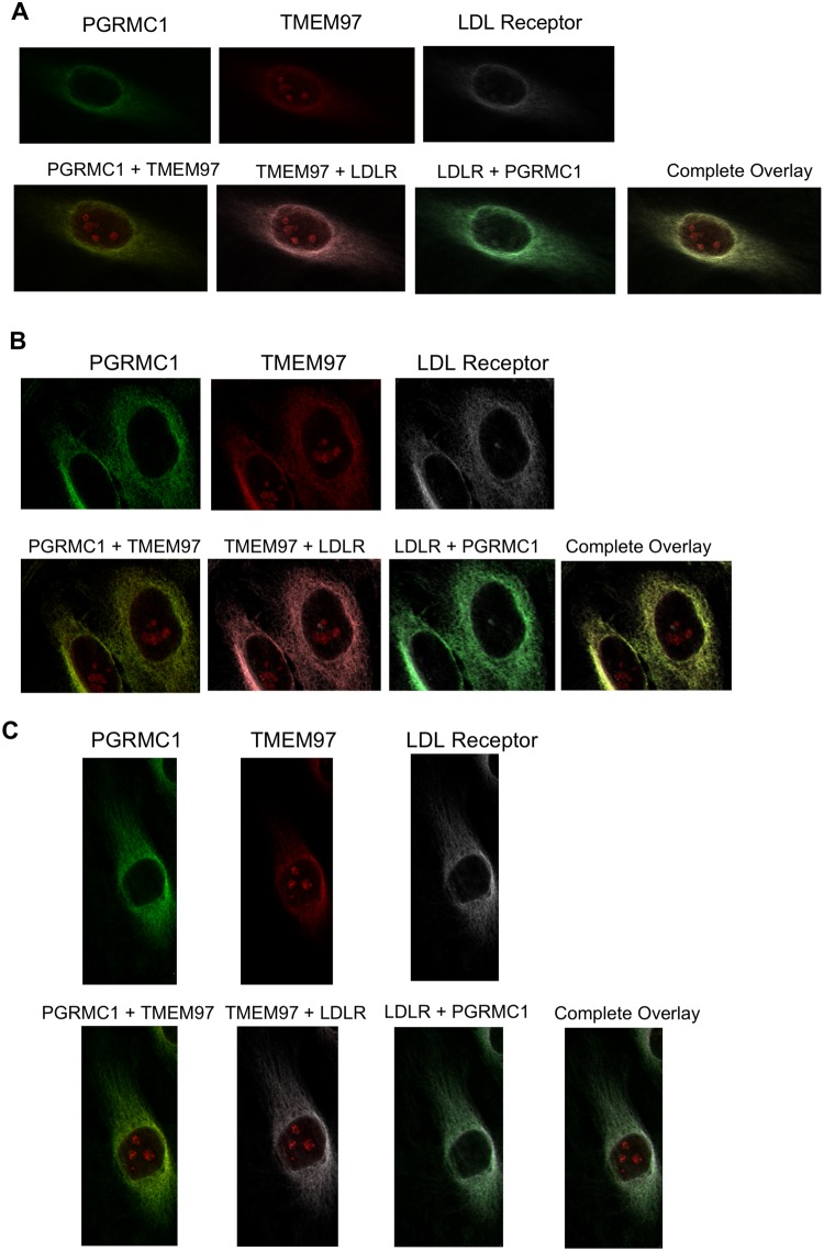
- Confocal microscopy and proximity ligation assays showed co-localization/interaction of LDLR, PGRMC1, and TMEM97, supporting a ternary complex required for rapid LDL internalization.
Methods
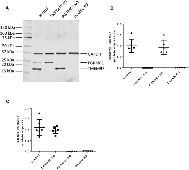
- HeLa cells were gene-edited with CRISPR/Cas9 to generate control, TMEM97 KO, PGRMC1 KO, and TMEM97/PGRMC1 double KO lines; knockout was confirmed by western blot.
- Saturation radioligand binding used [3H]DTG and [125I]RHM-4 with σ1R masking; LDL uptake was measured with [3H]cholesterol-labeled LDL or DiI-LDL, plus [125I]somatostatin and [125I]insulin uptake assays.
- Confocal microscopy and proximity ligation assays assessed LDLR, PGRMC1, and TMEM97 localization/interaction; pharmacological inhibitors included RHM-4, RHM-1, siramesine, SW43, and AG-205.
Limitations
- Stated: residual [3H]DTG binding in TMEM97 KO and double KO cells was not attributed to σ1R or PGRMC1; the authors state whether it represents a third, uncharacterized sigma receptor or another off-target site remains to be determined.
- Stated: a population of PGRMC1 was not co-localized with LDLR and TMEM97, consistent with PGRMC1 also associating with other membrane-bound receptors.
- Evident, not stated as a limitation: findings are from HeLa cells and in vitro assays, with no in vivo validation or testing in other cell types reported.
Model-written summary. Check it against the paper before relying on it.
Integrity signals
Registry
Cites
Statistics
Figures
Verification
Claims the paper makes
33 Own result · 15 Own interpretation · 9 Own method · 5 Not checked yet · 3 Supported so far · 2 Partly supported · 2 No full text of the sources · 2 No basis given · 1 Supported · 1 Sources not found · accuracy not yet measured
Every source the paper cites for this claim was checked and supports it.
In the paper
“In 2011, we reported a series of experimental results suggesting that the σ2R was a binding site in a protein complex containing PGRMC18.”
§ Sigma-2 Receptor/TMEM97 and PGRMC-1 Increase the Rate of Internalization of LDL by LDL Receptor through the Formation of a Ternary Complex > Discussion
What the sources say
- Supportedcites [8] Xu, J. et al. Identification of the PGRMC1 protein complex as the putative sigma-2 receptor binding site. Nature Communications 2, 380, https://doi.org/10.1038/ncomms1386 (2011). source
The Discussion states that the study’s results place the putative sigma-2 receptor ligand-binding site within the PGRMC1 protein complex. The citing sentence’s cautious wording, “suggesting,” is consistent with that conclusion. (LLM, conf=0.94)
hedgedcorrelational- Asserted: passes — The authors put it forward as true.
- Checkable: passes — It can be checked against evidence.
- Load-bearing: unclear — It is unclear whether the paper’s conclusions depend on it.
- Based: passes — The paper gives a reason why it holds.
- Supported
The cited sources support only part of this claim — read each source’s verdict below.
In the paper
“Subsequent studies using cancer cells having a stable knockdown or overexpression of PGRMC1 challenged these conclusions since neither condition resulted in a change in binding of the σ2R radioligand, [3H]DTG17,18.”
§ Sigma-2 Receptor/TMEM97 and PGRMC-1 Increase the Rate of Internalization of LDL by LDL Receptor through the Formation of a Ternary Complex > Discussion
What the sources say
- Supportedcites [17] Abate, C., Niso, M., Infantino, V., Menga, A. & Berardi, F. Elements in support of the ‘non-identity’ of the PGRMC1 protein with the sigma2 receptor. European journal of pharmacology 758, 16–23, https://doi.org/10.1016/j.ejphar.2015.03.067 (2015). source
The Methods (section 2.6, pp. 2–3) describe establishing PGRMC1-silenced MCF7 cancer-cell lines. The Discussion (p. 6) reports that both PGRMC1-overexpressing and silenced MCF7 cells had [3H]-DTG saturation curves, Bmax, and Kd values very similar to those of unmodified MCF7 cells, challenging the proposed σ2 receptor–PGRMC1 identity. (LLM, conf=0.92)
- Partly supportedcites [18] Chu, U. B. et al. The Sigma-2 Receptor and Progesterone Receptor Membrane Component 1 are Different Binding Sites Derived From Independent Genes. EBioMedicine 2, 1806–1813, https://doi.org/10.1016/j.ebiom.2015.10.017 (2015). sourceExperimental — accuracy not yet measured
The Results section reports that PGRMC1 knockout and overexpression did not significantly change [3H]-DTG binding, supporting the central finding. But this paper tested knockout rather than stable knockdown, and its binding assays used NSC34 cells rather than cancer cells; the Methods section identifies those cells and conditions. Page numbers are unavailable in our copy. (LLM, conf=0.88)
plain- Asserted: passes — The authors put it forward as true.
- Checkable: passes — It can be checked against evidence.
- Load-bearing: unclear — It is unclear whether the paper’s conclusions depend on it.
- Based: passes — The paper gives a reason why it holds.
- Supported
A result the paper reports from its own data — it cites no other source for it, so there is none to check. The quote shows it is in the paper.
In the paper
“However LDLR and PGRMC1 were shown to interact in the TMEM97 KO, and between LDLR and TMEM97 in the PGRMC1 KO, indicating that the LDLR interacts with TMEM97 and PGRMC1 despite the absence of the other protein.”
§ Sigma-2 Receptor/TMEM97 and PGRMC-1 Increase the Rate of Internalization of LDL by LDL Receptor through the Formation of a Ternary Complex > Results > Proximity Ligation Assays
hedgedcorrelational- Asserted: passes — The authors put it forward as true.
- Checkable: passes — It can be checked against evidence.
- Load-bearing: passes — The paper’s conclusions depend on it.
- Based: passes — The paper gives a reason why it holds.
- Rests on an assumption: The antibodies used in confocal microscopy and proximity ligation assays specifically recognize LDLR, PGRMC1, and TMEM97.
A result the paper reports from its own data — it cites no other source for it, so there is none to check. The quote shows it is in the paper.
In the paper
“and a significant reduction in binding of [3H]DTG.”
- Also stated in: § In vitro Binding Studies · plain
“We also observed a large, but incomplete, reduction of [3H]DTG binding to TMEM97 KO cells (Fig. 2B).”
- Also stated in: § In vitro Binding Studies · plain
“(B) [3H]DTG binding was significantly reduced in the TMEM97 KO and double KO cells, but some residual specific binding was observed.”
§ Sigma-2 Receptor/TMEM97 and PGRMC-1 Increase the Rate of Internalization of LDL by LDL Receptor through the Formation of a Ternary Complex > Abstract
plaindirect- Asserted: passes — The authors put it forward as true.
- Checkable: passes — It can be checked against evidence.
- Load-bearing: passes — The paper’s conclusions depend on it.
- Based: passes — The paper gives a reason why it holds.
- Rests on an assumption: The CRISPR/Cas9 knockout cell lines do not carry off-target mutations or compensatory changes that could account for the observed reductions in LDL uptake and radioligand binding.
- Rests on an assumption: The σ2R radioligand binding assays measure specific binding to σ2R/TMEM97 under the conditions used, with 1 μM (+)-pentazocine masking σ1R.
- Also stated in: § In vitro Binding Studies · plain
A result the paper reports from its own data — it cites no other source for it, so there is none to check. The quote shows it is in the paper.
In the paper
“All compounds resulted in a reduction of LDL uptake, with the classical σ2R antagonists having a greater effect than both the σ2R agonists and the PGRMC1 inhibitor.”
§ Sigma-2 Receptor/TMEM97 and PGRMC-1 Increase the Rate of Internalization of LDL by LDL Receptor through the Formation of a Ternary Complex > Results > Interactions between TMEM97, PGRMC1, and the LDL Receptor
plaindirect- Asserted: passes — The authors put it forward as true.
- Checkable: passes — It can be checked against evidence.
- Load-bearing: passes — The paper’s conclusions depend on it.
- Based: passes — The paper gives a reason why it holds.
- Rests on an assumption: The pharmacological inhibitors used (RHM-4, RHM-1, Siramesine, SW43, AG-205) act selectively on their intended targets at the concentrations used.
A result the paper reports from its own data — it cites no other source for it, so there is none to check. The quote shows it is in the paper.
In the paper
“However LDLR and PGRMC1 were shown to interact in the TMEM97 KO, and between LDLR and TMEM97 in the PGRMC1 KO, indicating that the LDLR interacts with TMEM97 and PGRMC1 despite the absence of the other protein.”
§ Sigma-2 Receptor/TMEM97 and PGRMC-1 Increase the Rate of Internalization of LDL by LDL Receptor through the Formation of a Ternary Complex > Results > Proximity Ligation Assays
boostedcorrelational- Asserted: passes — The authors put it forward as true.
- Checkable: passes — It can be checked against evidence.
- Load-bearing: passes — The paper’s conclusions depend on it.
- Based: passes — The paper gives a reason why it holds.
- Rests on an assumption: The antibodies used in confocal microscopy and proximity ligation assays specifically recognize LDLR, PGRMC1, and TMEM97.
A result the paper reports from its own data — it cites no other source for it, so there is none to check. The quote shows it is in the paper.
In the paper
“Under serum-starved conditions, there was an asymmetrical distribution of LDLR in the plasma membrane of HeLa cells.”
§ Sigma-2 Receptor/TMEM97 and PGRMC-1 Increase the Rate of Internalization of LDL by LDL Receptor through the Formation of a Ternary Complex > Results > Confocal Microscopy Studies
plain- Asserted: passes — The authors put it forward as true.
- Checkable: passes — It can be checked against evidence.
- Load-bearing: unclear — It is unclear whether the paper’s conclusions depend on it.
- Based: passes — The paper gives a reason why it holds.
- Rests on an assumption: The antibodies used in confocal microscopy and proximity ligation assays specifically recognize LDLR, PGRMC1, and TMEM97.
A result the paper reports from its own data — it cites no other source for it, so there is none to check. The quote shows it is in the paper.
In the paper
“This decreased LDL uptake was also observed in experiments using fluorescently tagged DiI-LDL (Supplementary Fig. 5).”
- Also stated in: § Abstract · plain
“fluorescently-tagged or 3H-labeled LDL”
§ Sigma-2 Receptor/TMEM97 and PGRMC-1 Increase the Rate of Internalization of LDL by LDL Receptor through the Formation of a Ternary Complex > Results > Interactions between TMEM97, PGRMC1, and the LDL Receptor
plaindirect- Asserted: passes — The authors put it forward as true.
- Checkable: passes — It can be checked against evidence.
- Load-bearing: unclear — It is unclear whether the paper’s conclusions depend on it.
- Based: passes — The paper gives a reason why it holds.
- Rests on an assumption: The CRISPR/Cas9 knockout cell lines do not carry off-target mutations or compensatory changes that could account for the observed reductions in LDL uptake and radioligand binding.
- Also stated in: § Abstract · plain
A result the paper reports from its own data — it cites no other source for it, so there is none to check. The quote shows it is in the paper.
In the paper
“TMEM97 KO resulted in a complete loss of binding of [125I]RHM-4”
- Also stated in: § In vitro Binding Studies · plain
“Knocking out TMEM97 resulted in a complete reduction in binding of [125I]RHM-4 to the HeLa cells (Fig. 2B).”
- Also stated in: § In vitro Binding Studies · plain
“(A) [125I]RHM-4 binding was completely eliminated in the TMEM97 KO and double KO cells.”
§ Sigma-2 Receptor/TMEM97 and PGRMC-1 Increase the Rate of Internalization of LDL by LDL Receptor through the Formation of a Ternary Complex > Abstract
plaindirect- Asserted: passes — The authors put it forward as true.
- Checkable: passes — It can be checked against evidence.
- Load-bearing: passes — The paper’s conclusions depend on it.
- Based: passes — The paper gives a reason why it holds.
- Rests on an assumption: The CRISPR/Cas9 knockout cell lines do not carry off-target mutations or compensatory changes that could account for the observed reductions in LDL uptake and radioligand binding.
- Rests on an assumption: The σ2R radioligand binding assays measure specific binding to σ2R/TMEM97 under the conditions used, with 1 μM (+)-pentazocine masking σ1R.
- Also stated in: § In vitro Binding Studies · plain
A result the paper reports from its own data — it cites no other source for it, so there is none to check. The quote shows it is in the paper.
In the paper
“We observed no significant difference in the [3H]DTG Bmax and Kd between the TMEM97 KO and the TMEM97/PGRMC1 double KO cells (Table 1)”
- Also stated in: § Discussion · plain
“Since this residual binding was also observed in the double KO cells, it does not represent binding to the low affinity DTG binding site identified previously in the PGRMC1 dimer.”
§ Sigma-2 Receptor/TMEM97 and PGRMC-1 Increase the Rate of Internalization of LDL by LDL Receptor through the Formation of a Ternary Complex > Results > In vitro Binding Studies
plain- Asserted: passes — The authors put it forward as true.
- Checkable: passes — It can be checked against evidence.
- Load-bearing: passes — The paper’s conclusions depend on it.
- Based: passes — The paper gives a reason why it holds.
- Rests on an assumption: The σ2R radioligand binding assays measure specific binding to σ2R/TMEM97 under the conditions used, with 1 μM (+)-pentazocine masking σ1R.
- Also stated in: § Discussion · plain
- Probably not a substantial claim. 13
- It may state two separate claims as one. 19
The sources with a verdict support this claim; the rest weren’t checked yet, have no full text we could read, or their verdicts aren’t rated yet.
In the paper
“an increase in cholesterol uptake has been demonstrated for proliferating cells36–38”
§ Sigma-2 Receptor/TMEM97 and PGRMC-1 Increase the Rate of Internalization of LDL by LDL Receptor through the Formation of a Ternary Complex > Discussion
What the sources say
- Supportedcites [36] Gabitova, L., Gorin, A. & Astsaturov, I. Molecular pathways: sterols and receptor signaling in cancer. Clinical cancer research: an official journal of the American Association for Cancer Research 20, 28–34, https://doi.org/10.1158/1078-0432.ccr-13-0122 (2014). source
The Background section, under “Maintenance of high sterol levels in cancer cells,” explicitly states that proliferating cells increase cholesterol uptake. This directly supports the attributed claim. (LLM, conf=0.95)
- Not checked — no full text of the sourcecites [37] Martínez-botas, j, suárez, y, ferruelo, aj, gómez-coronado, d & lasunción, ma Cholesterol starvation decreases P34cdc2 kinase activity and arrests the cell cycle at G2. The FASEB Journal 13, 1359–1370, https://doi.org/10.1096/fasebj.13.11.1359 (1999). source
- Not checked — no full text of the sourcecites [38] Martinez-Botas, J. et al. Dose-dependent effects of lovastatin on cell cycle progression. Distinct requirement of cholesterol and non-sterol mevalonate derivatives. Biochimica et biophysica acta 1532, 185–194 (2001). source
boostedcorrelational- Asserted: passes — The authors put it forward as true.
- Checkable: passes — It can be checked against evidence.
- Load-bearing: fails — The paper’s conclusions may not depend on it.
- Based: passes — The paper gives a reason why it holds.
Probably not a substantial claim. The paper’s conclusions may not depend on it.
- Supported
The cited sources support only part of this claim — read each source’s verdict below.
In the paper
“most cancer cells are characterized by an upregulation of the various pathways responsible for its biosynthesis36,39”
§ Sigma-2 Receptor/TMEM97 and PGRMC-1 Increase the Rate of Internalization of LDL by LDL Receptor through the Formation of a Ternary Complex > Discussion
What the sources say
- Supportedcites [36] Gabitova, L., Gorin, A. & Astsaturov, I. Molecular pathways: sterols and receptor signaling in cancer. Clinical cancer research: an official journal of the American Association for Cancer Research 20, 28–34, https://doi.org/10.1158/1078-0432.ccr-13-0122 (2014). source
The Abstract describes accelerated cholesterol metabolism as a hallmark of cancer and says cancer cells depend on high-intensity endogenous cholesterol biosynthesis. The Background section, “Maintenance of high sterol levels in cancer cells,” likewise describes accelerated endogenous cholesterol production as a mechanism cancer cells use to maintain high cholesterol levels. (LLM, conf=0.88)
- Partly supportedcites [39] Guo, D. et al. An LXR Agonist Promotes Glioblastoma Cell Death through Inhibition of an EGFR/AKT/SREBP-1/LDLR–Dependent Pathway. Cancer Discovery, https://doi.org/10.1158/2159-8290.cd-11-0102 (2011). sourceExperimental — accuracy not yet measured
The Introduction says SREBPs promote genes involved in cholesterol synthesis, and the Discussion reports an activated SREBP-1 pathway in glioblastoma. But the reported finding concerns increased LDLR expression and uptake of extracellular cholesterol; the paper does not establish that most cancer cells upregulate cholesterol-biosynthesis pathways. (LLM, conf=0.84)
plaincorrelational- Asserted: passes — The authors put it forward as true.
- Checkable: passes — It can be checked against evidence.
- Load-bearing: fails — The paper’s conclusions may not depend on it.
- Based: passes — The paper gives a reason why it holds.
Probably not a substantial claim. The paper’s conclusions may not depend on it.
- Supported
The sources with a verdict support this claim; the rest weren’t checked yet, have no full text we could read, or their verdicts aren’t rated yet.
In the paper
“These data are consistent with previous reports that PGRMC1 may be associated with other membrane-bound receptors such as the epidermal growth factor receptor (EGFR) and insulin receptor (IR)22,35.”
§ Sigma-2 Receptor/TMEM97 and PGRMC-1 Increase the Rate of Internalization of LDL by LDL Receptor through the Formation of a Ternary Complex > Discussion
What the sources say
- Not checked — no full text of the sourcecites [22] Bartz, F. et al. Identification of cholesterol-regulating genes by targeted RNAi screening. Cell metabolism 10, 63–75, https://doi.org/10.1016/j.cmet.2009.05.009 (2009). source
- Supportedcites [35] Hampton, K. K., Frazier, H., Anderson, K., Thibault, O. & Craven, R. J. Insulin receptor plasma membrane levels increased by the progesterone receptor membrane component 1. Molecular pharmacology, https://doi.org/10.1124/mol.117.110510 (2018). source
The Discussion states that PGRMC1 is associated with EGFR and reports that this study found PGRMC1 also associates with IR. The results section further reports that PGRMC1 and IR coprecipitated in two cell lines. Page numbers are not provided in our copy. (LLM, conf=0.94)
hedgedcorrelational- Asserted: passes — The authors put it forward as true.
- Checkable: passes — It can be checked against evidence.
- Load-bearing: fails — The paper’s conclusions may not depend on it.
- Based: passes — The paper gives a reason why it holds.
Probably not a substantial claim. The paper’s conclusions may not depend on it.
- Rests on an assumption: The antibodies used in confocal microscopy and proximity ligation assays are specific for LDLR, TMEM97, and PGRMC1.
The sources with a verdict support this claim; the rest weren’t checked yet, have no full text we could read, or their verdicts aren’t rated yet.
In the paper
“Cholesterol is a key component of the cell membrane and an essential requirement for cell division”
§ Sigma-2 Receptor/TMEM97 and PGRMC-1 Increase the Rate of Internalization of LDL by LDL Receptor through the Formation of a Ternary Complex > Discussion
What the sources say
- Supportedcites [36] Gabitova, L., Gorin, A. & Astsaturov, I. Molecular pathways: sterols and receptor signaling in cancer. Clinical cancer research: an official journal of the American Association for Cancer Research 20, 28–34, https://doi.org/10.1158/1078-0432.ccr-13-0122 (2014). source
The Background section, “Synthesis of cholesterol and its intermediates,” calls cholesterol a crucial component of cell membranes. The Background section, “Maintenance of high sterol levels in cancer cells,” states that cholesterol composition of cellular membranes is an essential metabolic requirement for cell divisions. (LLM, conf=0.94)
- Not checked — no full text of the sourcecites [37] Martínez-botas, j, suárez, y, ferruelo, aj, gómez-coronado, d & lasunción, ma Cholesterol starvation decreases P34cdc2 kinase activity and arrests the cell cycle at G2. The FASEB Journal 13, 1359–1370, https://doi.org/10.1096/fasebj.13.11.1359 (1999). source
- Not checked — no full text of the sourcecites [38] Martinez-Botas, J. et al. Dose-dependent effects of lovastatin on cell cycle progression. Distinct requirement of cholesterol and non-sterol mevalonate derivatives. Biochimica et biophysica acta 1532, 185–194 (2001). source
plain- Asserted: passes — The authors put it forward as true.
- Checkable: passes — It can be checked against evidence.
- Load-bearing: fails — The paper’s conclusions may not depend on it.
- Based: passes — The paper gives a reason why it holds.
Probably not a substantial claim. The paper’s conclusions may not depend on it. It may state two separate claims as one.
- Supported
A result the paper reports from its own data — it cites no other source for it, so there is none to check. The quote shows it is in the paper.
In the paper
“TMEM97 KO and PGRMC1 KO resulted in an equal reduction in the rate of uptake of fluorescently-tagged or 3H-labeled LDL, and knocking out both proteins did not result in a further rate of reduction of LDL uptake.”
- Also stated in: § Interactions between TMEM97, PGRMC1, and the LDL Receptor · plain
“Moreover, this decrease in LDL internalization was not a stepwise reduction with the loss of each protein but rather a complete diminished capability, as PGRMC1 KO and TMEM97 KO cells had the same decreased internalization of radiolabeled LDL as the double KO cells (Fig. 3A).”
- Also stated in: § Discussion · hedged
“Knocking out both TMEM97 and PGRMC1 did not lead to a further reduction in 3H-LDL uptake, indicating that the effect was not additive, and loss of one protein has the same effect as losing both proteins.”
§ Sigma-2 Receptor/TMEM97 and PGRMC-1 Increase the Rate of Internalization of LDL by LDL Receptor through the Formation of a Ternary Complex > Abstract
plain- Asserted: passes — The authors put it forward as true.
- Checkable: passes — It can be checked against evidence.
- Load-bearing: passes — The paper’s conclusions depend on it.
- Based: passes — The paper gives a reason why it holds.
It may state two separate claims as one.
- Rests on an assumption: The CRISPR/Cas9 knockout cell lines do not carry off-target mutations or compensatory changes that could account for the observed reductions in LDL uptake and radioligand binding.
- Also stated in: § Interactions between TMEM97, PGRMC1, and the LDL Receptor · plain
A result the paper reports from its own data — it cites no other source for it, so there is none to check. The quote shows it is in the paper.
In the paper
“The interactions between LDLR and TMEM97 and between TMEM97 and PGRMC1 were not observed in the TMEM97 KO and double KO cell lines (Supplementary Fig. 10), as expected.”
§ Sigma-2 Receptor/TMEM97 and PGRMC-1 Increase the Rate of Internalization of LDL by LDL Receptor through the Formation of a Ternary Complex > Results > Proximity Ligation Assays
plain- Asserted: passes — The authors put it forward as true.
- Checkable: passes — It can be checked against evidence.
- Load-bearing: passes — The paper’s conclusions depend on it.
- Based: passes — The paper gives a reason why it holds.
It may state two separate claims as one.
- Rests on an assumption: The antibodies used in confocal microscopy and proximity ligation assays specifically recognize LDLR, PGRMC1, and TMEM97.
A result the paper reports from its own data — it cites no other source for it, so there is none to check. The quote shows it is in the paper.
In the paper
“Similarly, the interactions between LDLR and PGRMC1 and between TMEM97 and PGRMC1 were not observed in the PGRMC1 KO and double KO cell lines.”
§ Sigma-2 Receptor/TMEM97 and PGRMC-1 Increase the Rate of Internalization of LDL by LDL Receptor through the Formation of a Ternary Complex > Results > Proximity Ligation Assays
plain- Asserted: passes — The authors put it forward as true.
- Checkable: passes — It can be checked against evidence.
- Load-bearing: passes — The paper’s conclusions depend on it.
- Based: passes — The paper gives a reason why it holds.
It may state two separate claims as one.
- Rests on an assumption: The antibodies used in confocal microscopy and proximity ligation assays specifically recognize LDLR, PGRMC1, and TMEM97.
A result the paper reports from its own data — it cites no other source for it, so there is none to check. The quote shows it is in the paper.
In the paper
“This pharmacologic inhibition of LDL uptake was observed in control cells only, but did not further decrease uptake in the knockout cells lines, further suggesting that PGRMC1 and TMEM97 must both be present and functional for effective LDL-LDLR complex internalization (Supplementary Fig. 7).”
§ Sigma-2 Receptor/TMEM97 and PGRMC-1 Increase the Rate of Internalization of LDL by LDL Receptor through the Formation of a Ternary Complex > Results > Interactions between TMEM97, PGRMC1, and the LDL Receptor
plain- Asserted: passes — The authors put it forward as true.
- Checkable: passes — It can be checked against evidence.
- Load-bearing: passes — The paper’s conclusions depend on it.
- Based: passes — The paper gives a reason why it holds.
It may state two separate claims as one.
- Rests on an assumption: The CRISPR/Cas9 knockout cell lines do not carry off-target mutations or compensatory changes that could account for the observed reductions in LDL uptake and radioligand binding.
- Rests on an assumption: The pharmacological inhibitors used (RHM-4, RHM-1, Siramesine, SW43, AG-205) act selectively on their intended targets at the concentrations used.
A result the paper reports from its own data — it cites no other source for it, so there is none to check. The quote shows it is in the paper.
In the paper
“The data showed that TMEM97 KO did not affect PRGMC1 protein level and vice versa (Fig. 1B,C).”
- Also stated in: § CRISPR Gene Editing of PGRMC1 and TMEM97 · plain
“The data showed that TMEM97 KO did not affect the PRGMC1 protein level and vice versa.”
§ Sigma-2 Receptor/TMEM97 and PGRMC-1 Increase the Rate of Internalization of LDL by LDL Receptor through the Formation of a Ternary Complex > Results > CRISPR Gene Editing of PGRMC1 and TMEM97
plainn = 6- Asserted: passes — The authors put it forward as true.
- Checkable: passes — It can be checked against evidence.
- Load-bearing: unclear — It is unclear whether the paper’s conclusions depend on it.
- Based: passes — The paper gives a reason why it holds.
It may state two separate claims as one.
- Rests on an assumption: The CRISPR/Cas9 knockout cell lines do not carry off-target mutations or compensatory changes that could account for the observed reductions in LDL uptake and radioligand binding.
- Also stated in: § CRISPR Gene Editing of PGRMC1 and TMEM97 · plain
A result the paper reports from its own data — it cites no other source for it, so there is none to check. The quote shows it is in the paper.
In the paper
“Internalization of LDL by LDLR can occur in the absence of this protein complex, but at a much slower rate than what is observed with the ternary complex.”
§ Sigma-2 Receptor/TMEM97 and PGRMC-1 Increase the Rate of Internalization of LDL by LDL Receptor through the Formation of a Ternary Complex > Discussion
plaindirect- Asserted: passes — The authors put it forward as true.
- Checkable: passes — It can be checked against evidence.
- Load-bearing: passes — The paper’s conclusions depend on it.
- Based: passes — The paper gives a reason why it holds.
It may state two separate claims as one.
- Rests on an assumption: TMEM97 is the sigma-2 receptor (σ2R).
- Rests on an assumption: CRISPR/Cas9 knockout of PGRMC1 and TMEM97 is specific and does not produce off-target effects that affect the results.
- Rests on an assumption: The antibodies used in confocal microscopy and proximity ligation assays are specific for LDLR, TMEM97, and PGRMC1.
- Rests on an assumption: The radiolabeled LDL uptake assay measures LDL internalization specifically via LDLR.
A conclusion the paper draws from its own results — it cites no other source for it, so there is none to check. The quote shows it is in the paper.
In the paper
“further suggesting that PGRMC1 and TMEM97 must both be present and functional for effective LDL-LDLR complex internalization”
§ Sigma-2 Receptor/TMEM97 and PGRMC-1 Increase the Rate of Internalization of LDL by LDL Receptor through the Formation of a Ternary Complex > Results > Interactions between TMEM97, PGRMC1, and the LDL Receptor
hedgeddirect- Asserted: passes — The authors put it forward as true.
- Checkable: passes — It can be checked against evidence.
- Load-bearing: passes — The paper’s conclusions depend on it.
- Based: passes — The paper gives a reason why it holds.
It may state two separate claims as one.
- Rests on an assumption: The pharmacological inhibitors used (RHM-4, RHM-1, Siramesine, SW43, AG-205) act selectively on their intended targets at the concentrations used.
What the paper did — it cites no source for this, so there is none to check. The quote shows it is in the paper.
In the paper
“Under these conditions the excess LDL competes with radiolabeled LDL for binding with LDLR, resulting in significantly decreased internalization (Fig. 3B). These data confirm that the LDL-LDLR pathway is responsible for internalization in these cells.”
§ Sigma-2 Receptor/TMEM97 and PGRMC-1 Increase the Rate of Internalization of LDL by LDL Receptor through the Formation of a Ternary Complex > Results > Interactions between TMEM97, PGRMC1, and the LDL Receptor
boosteddirect- Asserted: passes — The authors put it forward as true.
- Checkable: passes — It can be checked against evidence.
- Load-bearing: passes — The paper’s conclusions depend on it.
- Based: passes — The paper gives a reason why it holds.
It may state two separate claims as one.
- Rests on an assumption: Excess unlabeled LDL competes with radiolabeled LDL for binding to LDLR.
None of the sources cited for this claim have been checked yet.
In the paper
“Previous studies from our group have shown that the density of σ2Rs is higher in proliferating breast cancer cells relative to cancer cells driven into quiescence (i.e., G0) by prolonged serum starvation28,29.”
§ Sigma-2 Receptor/TMEM97 and PGRMC-1 Increase the Rate of Internalization of LDL by LDL Receptor through the Formation of a Ternary Complex > Discussion
What the sources say
- Not checked — no full text of the sourcecites [28] Mach, R. H. et al. Sigma-2 receptors as potential biomarkers of proliferation in breast cancer. Cancer Res 57, 156–161 (1997).
- Not checked yetcites [29] Wheeler, K. T. et al. Sigma-2 receptors as a biomarker of proliferation in solid tumours. Br J Cancer 82, 1223–1232 (2000). source
Checked before — its earlier verdict is held back until a new check runs.
plaincorrelational- Asserted: passes — The authors put it forward as true.
- Checkable: passes — It can be checked against evidence.
- Load-bearing: fails — The paper’s conclusions may not depend on it.
- Based: passes — The paper gives a reason why it holds.
Probably not a substantial claim. The paper’s conclusions may not depend on it.
A conclusion the paper draws from its own results — it cites no other source for it, so there is none to check. The quote shows it is in the paper.
In the paper
“Their similar capacity to bind LDL further suggests that both PGRMC1 and TMEM97 are involved in the internalization of the LDL-LDLR complex, and they are not involved in the initial binding of LDL to LDLR.”
§ Sigma-2 Receptor/TMEM97 and PGRMC-1 Increase the Rate of Internalization of LDL by LDL Receptor through the Formation of a Ternary Complex > Results > Interactions between TMEM97, PGRMC1, and the LDL Receptor
hedgeddirectn = 4- Asserted: passes — The authors put it forward as true.
- Checkable: passes — It can be checked against evidence.
- Load-bearing: passes — The paper’s conclusions depend on it.
- Based: passes — The paper gives a reason why it holds.
It may state two separate claims as one.
What the paper did — it cites no source for this, so there is none to check. The quote shows it is in the paper.
In the paper
“Western blot showed that TMEM97 or/and PGRMC1 were completely removed in the knockout cell lines (Fig. 1A).”
- Also stated in: § Interactions between TMEM97, PGRMC1, and the LDL Receptor · plain
“TMEM97 was not detected in the TMEM97 KO and double KO cell lines, and PGRMC1 was not detected in the PGRMC1 KO cells and double KO cell lines, verifying the knockout of the representative proteins (Fig. 1; Supplementary Fig. 3).”
§ Sigma-2 Receptor/TMEM97 and PGRMC-1 Increase the Rate of Internalization of LDL by LDL Receptor through the Formation of a Ternary Complex > Results > CRISPR Gene Editing of PGRMC1 and TMEM97
plainn = 6- Asserted: passes — The authors put it forward as true.
- Checkable: passes — It can be checked against evidence.
- Load-bearing: passes — The paper’s conclusions depend on it.
- Based: passes — The paper gives a reason why it holds.
It may state two separate claims as one.
- Rests on an assumption: The CRISPR/Cas9 knockout cell lines do not carry off-target mutations or compensatory changes that could account for the observed reductions in LDL uptake and radioligand binding.
- Also stated in: § Interactions between TMEM97, PGRMC1, and the LDL Receptor · plain
None of the sources cited for this claim have been checked yet.
In the paper
“PET imaging studies with a σ2R -selective radiotracer, [18F]ISO-1, have shown a strong correlation between radiotracer uptake and the density of σ2R in a murine model of breast cancer30, and an excellent correlation between radiotracer uptake and Ki-67 score in a mixed population of patients diagnosed with lymphoma, breast, and head & neck cancer31.”
§ Sigma-2 Receptor/TMEM97 and PGRMC-1 Increase the Rate of Internalization of LDL by LDL Receptor through the Formation of a Ternary Complex > Discussion
What the sources say
- Not checked yetcites [30] Shoghi, K. I. et al. Quantitative receptor-based imaging of tumor proliferation with the sigma-2 ligand [(18)F]ISO-1. PloS one 8, e74188, https://doi.org/10.1371/journal.pone.0074188 (2013). source
Checked before — its earlier verdict is held back until a new check runs.
- Not checked yetcites [31] Dehdashti, F. et al. Assessment of cellular proliferation in tumors by PET using 18F-ISO-1. Journal of nuclear medicine: official publication, Society of Nuclear Medicine 54, 350–357, https://doi.org/10.2967/jnumed.112.111948 (2013). source
Checked before — its earlier verdict is held back until a new check runs.
boostedcorrelational- Asserted: passes — The authors put it forward as true.
- Checkable: passes — It can be checked against evidence.
- Load-bearing: passes — The paper’s conclusions depend on it.
- Based: passes — The paper gives a reason why it holds.
It may state two separate claims as one.
- Not checked yet
A conclusion the paper draws from its own results — it cites no other source for it, so there is none to check. The quote shows it is in the paper.
In the paper
“Taken together this data indicated that the LDLR, TMEM97, and PGRMC1 proteins are associated within close proximity of each other and form a complex.”
§ Sigma-2 Receptor/TMEM97 and PGRMC-1 Increase the Rate of Internalization of LDL by LDL Receptor through the Formation of a Ternary Complex > Results > Proximity Ligation Assays
hedgedcorrelational- Asserted: passes — The authors put it forward as true.
- Checkable: passes — It can be checked against evidence.
- Load-bearing: passes — The paper’s conclusions depend on it.
- Based: passes — The paper gives a reason why it holds.
It may state two separate claims as one.
- Rests on an assumption: The antibodies used in confocal microscopy and proximity ligation assays specifically recognize LDLR, PGRMC1, and TMEM97.
What the paper did — it cites no source for this, so there is none to check. The quote shows it is in the paper.
In the paper
“The nonspecific binding signal for both groups was defined with 10 μM DTG, and 1 μM of (+)−pentazocine was added for [3H]DTG binding to mask the σ1R binding site.”
§ Sigma-2 Receptor/TMEM97 and PGRMC-1 Increase the Rate of Internalization of LDL by LDL Receptor through the Formation of a Ternary Complex > Materials and Methods > Saturation binding assays
plain- Asserted: passes — The authors put it forward as true.
- Checkable: passes — It can be checked against evidence.
- Load-bearing: fails — The paper’s conclusions may not depend on it.
- Based: passes — The paper gives a reason why it holds.
Probably not a substantial claim. The paper’s conclusions may not depend on it. It may state two separate claims as one.
What the paper did — it cites no source for this, so there is none to check. The quote shows it is in the paper.
In the paper
“PGRMC1 knockout for each clone was examined by western blot analysis.”
§ Sigma-2 Receptor/TMEM97 and PGRMC-1 Increase the Rate of Internalization of LDL by LDL Receptor through the Formation of a Ternary Complex > Materials and Methods > PGRMC1 knockout in HeLa cells
plain- Asserted: passes — The authors put it forward as true.
- Checkable: passes — It can be checked against evidence.
- Load-bearing: passes — The paper’s conclusions depend on it.
- Based: passes — The paper gives a reason why it holds.
Probably not a substantial claim.
A conclusion the paper draws from its own results — it cites no other source for it, so there is none to check. The quote shows it is in the paper.
In the paper
“Addition of LDL to HeLa cells after a brief period of serum starvation resulted in the internalization and likely dissociation of the LDL-LDLR-TMEM97-PGRMC1 tetrameric complex in the acidic environment of the lysosomes”
§ Sigma-2 Receptor/TMEM97 and PGRMC-1 Increase the Rate of Internalization of LDL by LDL Receptor through the Formation of a Ternary Complex > Discussion
hedgedconditional- Asserted: passes — The authors put it forward as true.
- Checkable: passes — It can be checked against evidence.
- Load-bearing: passes — The paper’s conclusions depend on it.
- Based: passes — The paper gives a reason why it holds.
It may state two separate claims as one.
- Rests on an assumption: The antibodies used in confocal microscopy and proximity ligation assays are specific for LDLR, TMEM97, and PGRMC1.
- Rests on an assumption: The radiolabeled LDL uptake assay measures LDL internalization specifically via LDLR.
A conclusion the paper draws from its own results — it cites no other source for it, so there is none to check. The quote shows it is in the paper.
In the paper
“These data indicate that [125I]RHM-4 binds selectively to TMEM97, whereas there is some residual binding of [3H]DTG to a site not associated with TMEM97.”
§ Sigma-2 Receptor/TMEM97 and PGRMC-1 Increase the Rate of Internalization of LDL by LDL Receptor through the Formation of a Ternary Complex > Discussion
hedgedcorrelational- Asserted: passes — The authors put it forward as true.
- Checkable: passes — It can be checked against evidence.
- Load-bearing: unclear — It is unclear whether the paper’s conclusions depend on it.
- Based: passes — The paper gives a reason why it holds.
It may state two separate claims as one.
In the paper
“Another protein that has been strongly linked to cholesterol trafficking is PGRMC1, and in a recent review by Cahill and Medlock, a theoretical mechanism showing a role of PGRMC1 in the membrane trafficking of LDLR was proposed32.”
§ Sigma-2 Receptor/TMEM97 and PGRMC-1 Increase the Rate of Internalization of LDL by LDL Receptor through the Formation of a Ternary Complex > Discussion
What the sources say
- Not checked — no full text of the sourcecites [32] Cahill, M. A. & Medlock, A. E. Thoughts on interactions between PGRMC1 and diverse attested and potential hydrophobic ligands. J Steroid Biochem Mol Biol 171, 11–33, https://doi.org/10.1016/j.jsbmb.2016.12.020 (2017). source
boostedcorrelational- Asserted: passes — The authors put it forward as true.
- Checkable: passes — It can be checked against evidence.
- Load-bearing: passes — The paper’s conclusions depend on it.
- Based: passes — The paper gives a reason why it holds.
Probably not a substantial claim. It may state two separate claims as one.
What the paper did — it cites no source for this, so there is none to check. The quote shows it is in the paper.
In the paper
“Care was taken not to vortex LDL to avoid aggregation of particles.”
§ Sigma-2 Receptor/TMEM97 and PGRMC-1 Increase the Rate of Internalization of LDL by LDL Receptor through the Formation of a Ternary Complex > Materials and Methods > Preparation of Radiolabelled LDL
plain- Asserted: passes — The authors put it forward as true.
- Checkable: passes — It can be checked against evidence.
- Load-bearing: fails — The paper’s conclusions may not depend on it.
- Based: passes — The paper gives a reason why it holds.
Probably not a substantial claim. The paper’s conclusions may not depend on it.
What the paper did — it cites no source for this, so there is none to check. The quote shows it is in the paper.
In the paper
“Single cell clones were isolated for all cell lines.”
§ Sigma-2 Receptor/TMEM97 and PGRMC-1 Increase the Rate of Internalization of LDL by LDL Receptor through the Formation of a Ternary Complex > Materials and Methods > PGRMC1 and TMEM97 double knockout in HeLa cells
plain- Asserted: passes — The authors put it forward as true.
- Checkable: passes — It can be checked against evidence.
- Load-bearing: fails — The paper’s conclusions may not depend on it.
- Based: passes — The paper gives a reason why it holds.
Probably not a substantial claim. The paper’s conclusions may not depend on it.
In the paper
“The formation of a trimeric LDLR-PGRMC1-TMEM97 complex that results in the rapid internalization and trafficking of LDL is consistent with the conclusion we stated in our 2011 paper, that the σ2R represents a binding site in the protein complex containing PGRMC1.”
§ Sigma-2 Receptor/TMEM97 and PGRMC-1 Increase the Rate of Internalization of LDL by LDL Receptor through the Formation of a Ternary Complex > Discussion
What the sources say
- Not matchedcites 2011 paper
Couldn’t be matched to a paper in Crossref, so it can’t be checked.
plaindirect- Asserted: passes — The authors put it forward as true.
- Checkable: passes — It can be checked against evidence.
- Load-bearing: passes — The paper’s conclusions depend on it.
- Based: passes — The paper gives a reason why it holds.
It may state two separate claims as one.
- Rests on an assumption: TMEM97 is the sigma-2 receptor (σ2R).
- Not matched
What the paper did — it cites no source for this, so there is none to check. The quote shows it is in the paper.
In the paper
“TMEM97 deletion was confirmed by western blot analysis.”
§ Sigma-2 Receptor/TMEM97 and PGRMC-1 Increase the Rate of Internalization of LDL by LDL Receptor through the Formation of a Ternary Complex > Materials and Methods > TMEM97 knockout in HeLa cells
plain- Asserted: passes — The authors put it forward as true.
- Checkable: passes — It can be checked against evidence.
- Load-bearing: passes — The paper’s conclusions depend on it.
- Based: passes — The paper gives a reason why it holds.
Probably not a substantial claim.
A conclusion the paper draws from its own results — it cites no other source for it, so there is none to check. The quote shows it is in the paper.
In the paper
“These results are consistent with an interaction between TMEM97 and PGRMC1, with the knocking out of PGRMC1 resulting in a small change in the 3-dimensional structure of TMEM97 leading to a reduction in affinity of σ2R radioligands binding to this site.”
§ Sigma-2 Receptor/TMEM97 and PGRMC-1 Increase the Rate of Internalization of LDL by LDL Receptor through the Formation of a Ternary Complex > Discussion
hedgeddirect- Asserted: passes — The authors put it forward as true.
- Checkable: passes — It can be checked against evidence.
- Load-bearing: passes — The paper’s conclusions depend on it.
- Based: passes — The paper gives a reason why it holds.
It may state two separate claims as one.
In the paper
“TMEM97 is thought to be involved in cholesterol trafficking through its association with lysosomal cholesterol transporter NPC1, a protein whose loss results in a fatal lysosomal storage disorder, Niemann–Pick disease type C119.”
§ Sigma-2 Receptor/TMEM97 and PGRMC-1 Increase the Rate of Internalization of LDL by LDL Receptor through the Formation of a Ternary Complex > Discussion
What the sources say
- Not checked yetcites [19] Alon, A. et al. Identification of the gene that codes for thesigma2 receptor. Proceedings of the National Academy of Sciences 114, 7160–7165, https://doi.org/10.1073/pnas.1705154114 (2017). source
hedgedcorrelational- Asserted: passes — The authors put it forward as true.
- Checkable: passes — It can be checked against evidence.
- Load-bearing: fails — The paper’s conclusions may not depend on it.
- Based: passes — The paper gives a reason why it holds.
Probably not a substantial claim. The paper’s conclusions may not depend on it.
The paper states this without a source or data behind it here, so there is nothing to check it against. The quote shows it is in the paper.
In the paper
“Since the demand for cholesterol to support cell proliferation is high”
§ Sigma-2 Receptor/TMEM97 and PGRMC-1 Increase the Rate of Internalization of LDL by LDL Receptor through the Formation of a Ternary Complex > Discussion
plain- Asserted: passes — The authors put it forward as true.
- Checkable: passes — It can be checked against evidence.
- Load-bearing: fails — The paper’s conclusions may not depend on it.
- Based: passes — The paper gives a reason why it holds.
Probably not a substantial claim. The paper’s conclusions may not depend on it.
The paper relies on this without support.
The CRISPR/Cas9 knockout cell lines do not carry off-target mutations or compensatory changes that could account for the observed reductions in LDL uptake and radioligand binding.
“All the four cell lines were generated using the same plasmid transfection and lentivirus-infection procedures. Single clones were isolated for all knockout cell lines and used throughout the current study.”
The paper relies on this without support.
The antibodies used in confocal microscopy and proximity ligation assays specifically recognize LDLR, PGRMC1, and TMEM97.
“To confirm that the observed associations between the protein was indeed a complex formed, proximity ligation assays were conducted pairwise between LDLR, PGRMC1, and TMEM97.”
The paper relies on this without support.
Excess unlabeled LDL competes with radiolabeled LDL for binding to LDLR.
“Under these conditions the excess LDL competes with radiolabeled LDL for binding with LDLR, resulting in significantly decreased internalization (Fig. 3B).”
The paper relies on this without support.
The σ2R radioligand binding assays measure specific binding to σ2R/TMEM97 under the conditions used, with 1 μM (+)-pentazocine masking σ1R.
“The in vitro binding studies were done in the presence of 1 μM (+)−pentazocine to mask σ1R.”
The paper relies on this without support.
The pharmacological inhibitors used (RHM-4, RHM-1, Siramesine, SW43, AG-205) act selectively on their intended targets at the concentrations used.
“Since TMEM97 is the protein which contains the σ2R, we used two ligands previously described selective σ2R antagonists RHM-4 (Kd ~ 0.2 nM) and RHM-1 (Kd ~ 0.66 nM); two ligands previously described as being σ2R agonists Siramesine (Kd ~ 1.6 nM) and SW43 (Kd ~ 12 nM). We also used AG-205 a PGRMC1 inhibitor (Kd ~ 1 μM against σ2R and Ki~1263 ± 325 to the σ2R for displacing DTG).”
The paper relies on this without support.
TMEM97 is the sigma-2 receptor (σ2R).
“The involvement of TMEM97/σ2R in the internalization and trafficking of cholesterol via LDL”
The paper relies on this without support.
CRISPR/Cas9 knockout of PGRMC1 and TMEM97 is specific and does not produce off-target effects that affect the results.
“PGRMC1 was knocked out in HeLa cells using CRISPR/Cas9 technology.”
The paper relies on this without support.
The antibodies used in confocal microscopy and proximity ligation assays are specific for LDLR, TMEM97, and PGRMC1.
“incubated with goat anti-PGRMC1 (Abcam ab48012) 1:200 in PBST +1% BSA overnight”
The paper relies on this without support.
The radiolabeled LDL uptake assay measures LDL internalization specifically via LDLR.
“Cells were incubated with radiolabeled LDL for 10–240 minutes at 37° for uptake studies”
The paper relies on this without support.
The σ2R ligands used (e.g., RHM-4) bind specifically to TMEM97/σ2R and not to other targets.
“with RHM-4, Siramesine, SW43, RHM-1, or AG-205 (1 nM - 500 nM)”
Claims other papers make about this one
Each is checked against this paper’s own text: does it say what the citing paper says it does?
The source backs part of the claim, not all of it — or backs it for a different population, condition or comparison.
In the citing paper
“this intact complex is required for efficient uptake of lipoproteins such as LDL and apolipoprotein E (apoE)”
From The Sigma-2 Receptor/TMEM97, PGRMC1, and LDL Receptor Complex Are Responsible for the Cellular Uptake of Aβ42 and Its Protein Aggregates (2020) · cites this paper as [8]
Experimental — accuracy not yet measuredThe source backs part of the claim, not all of it — or backs it for a different population, condition or comparison.
In the citing paper
“This was confirmed in pharmacological studies assessing the activity of S2R ligands where reduction, or ablation, of S2R activity was observed when TMEM97 expression was reduced, or absent, in calf liver cells [16,61] or human cervical cancer cells [66], respectively.”
From Sigma-2 Receptors—From Basic Biology to Therapeutic Target: A Focus on Age-Related Degenerative Diseases (2023) · cites this paper as [66]
Experimental — accuracy not yet measuredThe source backs part of the claim, not all of it — or backs it for a different population, condition or comparison.
In the citing paper
“One important function of S2R is regulation of cholesterol homeostasis by interacting with low-density lipoprotein receptor (LDLR), an apolipoprotein-E receptor, at the plasma membrane to transport apolipoprotein E into neurons [66] and with NPC1, intracellularly, to regulate LDL cholesterol transport out of lysosomes [93,94].”
From Sigma-2 Receptors—From Basic Biology to Therapeutic Target: A Focus on Age-Related Degenerative Diseases (2023) · cites this paper as [66]
Experimental — accuracy not yet measuredThe source backs part of the claim, not all of it — or backs it for a different population, condition or comparison.
In the citing paper
“In post synapses, we also observed FRET between Aβ and PrPc—which has also been observed to bind Aβ in model systems[62]—and between TMEM97 and PGRMC1 which are known to be binding partners in vitro and in human cases [55], and between TMEM97 and PrPc (Fig. 3).”
From Transmembrane protein 97 is a potential synaptic amyloid beta receptor in human Alzheimer’s disease (2024) · cites this paper as [55]
Experimental — accuracy not yet measuredThe cited source says what the claim attributes to it.
In the citing paper
“Our lab has recently shown that the Sigma-2 Receptor/Transmembrane Protein 97 (TMEM97) and Progesterone Receptor Membrane Component 1 (PGRMC1) form a complex with the Low Density Lipoprotein Receptor (LDLR)”
From The Sigma-2 Receptor/TMEM97, PGRMC1, and LDL Receptor Complex Are Responsible for the Cellular Uptake of Aβ42 and Its Protein Aggregates (2020) · cites this paper as [8]
The cited source says what the claim attributes to it.
In the citing paper
“previous studies characterized these cell lines and western blot data demonstrated a complete ablation of TMEM97 and/or PGRMC1 in the knockout cells [8, 39].”
From The Sigma-2 Receptor/TMEM97, PGRMC1, and LDL Receptor Complex Are Responsible for the Cellular Uptake of Aβ42 and Its Protein Aggregates (2020) · cites this paper as [8]
The cited source says what the claim attributes to it.
In the citing paper
“S2R has been shown to be closely associated and interact with key proteins to exert its functions, including PGRMC1 and LDLR [65,66].”
From Sigma-2 Receptors—From Basic Biology to Therapeutic Target: A Focus on Age-Related Degenerative Diseases (2023) · cites this paper as [66]
The cited source says what the claim attributes to it.
In the citing paper
“S2R is key in facilitating the uptake of LDL, as uptake of radiolabeled LDL was significantly decreased in PGRMC1 knockout, TMEM97 knockout, and double knockout cell lines [66,93].”
From Sigma-2 Receptors—From Basic Biology to Therapeutic Target: A Focus on Age-Related Degenerative Diseases (2023) · cites this paper as [66]
In the citing paper
“We found that disrupting this complex by genetically ablating or by pharmacologically targeting TMEM97 or PGRMC1 resulted in a decreased capacity for LDL internalization.”
From The Sigma-2 Receptor/TMEM97, PGRMC1, and LDL Receptor Complex Are Responsible for the Cellular Uptake of Aβ42 and Its Protein Aggregates (2020) · cites this paper as [8]
In the citing paper
“Of note, LPL is a key enzyme involved in lipid metabolism. By interacting with lipoprotein receptors, such as the direct interactor with S2R (TMEM97), LDLR [46], LPL promotes the internalization of lipoproteins, contributing to lipid homeostasis and receptor-mediated signaling in neurons. In addition, LPL maintains the balance of membrane composition and fluidity, which is crucial for proper”
In the citing paper
“Using these knockout cell lines, the same group demonstrated that both PGRMC1 and TMEM97 must be present and functional for effective LDL–LDLR complex internalization [66].”
From Sigma-2 Receptors—From Basic Biology to Therapeutic Target: A Focus on Age-Related Degenerative Diseases (2023) · cites this paper as [66]
In the citing paper
“Linking this function to Alzheimer’s, in cellular models, TMEM97 has recently been shown to form a ternary complex with Progesterone receptor membrane component 1 (PGRMC1) and LDLR [55] that may control the internalization of monomers and oligomers of Aβ [54].”
From Transmembrane protein 97 is a potential synaptic amyloid beta receptor in human Alzheimer’s disease (2024) · cites this paper as [55]
In the citing paper
“TMEM97 is thought to be an endo-lysosome-related protein which has itself been linked to cellular toxicity and is essential for the internalization of cholesterol molecules like LDL through the formation of a complex with PGRMC1 [4, 55]”
From Transmembrane protein 97 is a potential synaptic amyloid beta receptor in human Alzheimer’s disease (2024) · cites this paper as [55]
Self-check
Percentages, sums and reported differences re-computed from the paper’s own numbers.
Each contradiction between two passages listed here was confirmed by an independent check.
Rated minor: it would not touch a result — a value no result rests on, a p-value that stays on the same side of the significance level, or a conflict that is clerical, resolved elsewhere in the paper, or outside its own findings.
In the paper
§ Sigma-2 Receptor/TMEM97 and PGRMC-1 Increase the Rate of Internalization of LDL by LDL Receptor through the Formation of a Ternary Complex > Results > In vitro Binding Studies
“Knocking out TMEM97 resulted in a complete reduction in binding of [125I]RHM-4 to the HeLa cells (Fig. 2B).”
§ Sigma-2 Receptor/TMEM97 and PGRMC-1 Increase the Rate of Internalization of LDL by LDL Receptor through the Formation of a Ternary Complex > Results > In vitro Binding Studies
“(A) [125I]RHM-4 binding was completely eliminated in the TMEM97 KO and double KO cells. (B) [3H]DTG binding was significantly reduced in the TMEM97 KO and double KO cells”
Rated minor: it would not touch a result — a value no result rests on, a p-value that stays on the same side of the significance level, or a conflict that is clerical, resolved elsewhere in the paper, or outside its own findings.
In the paper
§ Sigma-2 Receptor/TMEM97 and PGRMC-1 Increase the Rate of Internalization of LDL by LDL Receptor through the Formation of a Ternary Complex > Materials and Methods > Radiolabeled LDL Uptake Assay
“Cells were incubated with radiolabeled LDL for 10–240 minutes at 37° for uptake studies (Fig. 3A,C,D) or 4 °C for the LDL surface binding study (Fig. 3B)”
§ Sigma-2 Receptor/TMEM97 and PGRMC-1 Increase the Rate of Internalization of LDL by LDL Receptor through the Formation of a Ternary Complex > Results > Interactions between TMEM97, PGRMC1, and the LDL Receptor
“(B) Uptake over time with the addition of excess unlabeled LDL (250 μg/mL). (C) Incubation at 4 °C to indicate similar capacity of LDL binding to LDL receptor.”
Citations
Cites
A JOURNAL OF PHARMACOLOGY AND EXPERIMENTAL THERAPEUTICS
Lindemann, L., Jaeschke, G., Michalon, A., Vieira, E., Honer, M., Spooren, W., Porter, R., Hartung, T., Kolczewski, S., Büttelmann, B., et al. (1909) · JAMA · doi:10.1001/jama.1909.02550020033007 · Cited by 7,889
Improved vectors and genome-wide libraries for CRISPR screening
Sanjana, N. E., Shalem, O., & Zhang, F. (2014) · Nature Methods · doi:10.1038/nmeth.3047 · Cited by 5,239
Martin, W. R., Eades, C. G., Thompson, J. A., Huppler, R. E., & Gilbert, P. E. (1976) · The Journal of Pharmacology and Experimental Therapeutics · doi:10.1016/s0022-3565(25)30536-7 · Cited by 2,992
Goldstein, J. L., & Brown, M. S. (2009) · Arteriosclerosis, Thrombosis, and Vascular Biology · doi:10.1161/atvbaha.108.179564 · Cited by 1,344
Purification, molecular cloning, and expression of the mammalian sigma1-binding site.
Hanner, M., Moebius, F. F., Flandorfer, A., Knaus, H. G., Striessnig, J., Kempner, E. S., & Glossmann, H. H. (1996) · Proceedings of the National Academy of Sciences · doi:10.1073/pnas.93.15.8072 · Cited by 914
Sigma receptors: biology and function.
Walker, J. M., Bowen, W. D., Walker, F. O., Matsumoto, R. R., de Costa, B., & Rice, K. C. (1990) · Pharmacological Reviews · doi:10.1016/s0031-6997(25)00047-x · Cited by 831
Crystal structure of the human σ1 receptor
Schmidt, H. R., Zheng, S., Gurpinar, E., Koehl, A., Manglik, A., & Kruse, A. C. (2016) · Nature · doi:10.1038/nature17391 · Cited by 482
Guo, D., Reinitz, F., Youssef, M., Hong, C., Nathanson, D., Akhavan, D., Kuga, D., Amzajerdi, A. N., Soto, H., Zhu, S., et al. (2011) · Cancer Discovery · doi:10.1158/2159-8290.cd-11-0102 · Cited by 452
THE MAMMALIAN LOW-DENSITY LIPOPROTEIN RECEPTOR FAMILY
Hussain, M. M., Strickland, D. K., & Bakillah, A. (1999) · Annual Review of Nutrition · doi:10.1146/annurev.nutr.19.1.141 · Cited by 391
Progesterone receptor membrane component 1: An integrative review
Cahill, M. A. (2007) · The Journal of Steroid Biochemistry and Molecular Biology · doi:10.1016/j.jsbmb.2007.02.002 · Cited by 363
Cited by
Sorted by citations
Items 101–154 of 154
The novel drug candidate S2/IAPinh improves survival in models of pancreatic and ovarian cancer
Hagi, T., Vangveravong, S., Takchi, R., Gong, Q., Goedegebuure, S. P., Tiriac, H., Tine, B. A. V., Powell, M. A., Hawkins, W. G., & Spitzer, D. (2024) · Scientific Reports · doi:10.1038/s41598-024-56928-z · Cited by 3
Zampieri, D., Romano, M., Fortuna, S., Amata, E., Dichiara, M., Cosentino, G., Marrazzo, A., & Mamolo, M. G. (2024) · Molecules · doi:10.3390/molecules29112697 · Cited by 3
Wang, J., Wang, T., Mou, T., Yang, T., Gao, X., An, X., Hu, B., Zhang, J., Zhang, X., Deuther‐Conrad, W., et al. (2024) · Journal of Medicinal Chemistry · doi:10.1021/acs.jmedchem.4c01315 · Cited by 3
Torres‐Gómez, H., Daniliuc, C. G., Schepmann, D., Laurini, E., Pricl, S., & Wünsch, B. (2021) · International Journal of Molecular Sciences · doi:10.3390/ijms22115685 · Cited by 2
Stathopoulos, A., Wang, J. J., Martin, S. F., & Zhang, S. X. (2025) · Biomolecules · doi:10.3390/biom15091228 · Cited by 2
Lizama, B. N., Reaver, A., Look, G. C., Caggiano, A. O., & Hamby, M. E. (2026) · Experimental Eye Research · doi:10.1016/j.exer.2026.110944 · Cited by 2
Harbert, D. H. (2026) · Frontiers in Pharmacology · doi:10.3389/fphar.2026.1830847 · Cited by 2
Niemann-Pick Type C Disease: At the Nexus of Neurodegenerative and Neurodevelopmental Disorders
(2020) · Journal of Parkinson’s Disease and Alzheimer’s Disease · doi:10.13188/2376-922x.1000029 · Cited by 2
Kotańska, M., Zadrożna, M., Kubacka, M., Mika, K., Szczepańska, K., Nowak, B., Alesci, A., Miller, A., Lauriano, E. R., & Kieć‐Kononowicz, K. (2024) · Pharmaceuticals · doi:10.3390/ph17070858 · Cited by 2
Mou, T., Wang, J., Hu, B., Gao, M., Yun, M., Gao, X., Tian, Y., Li, H., Jia, H., Zhang, X., et al. (2025) · Arteriosclerosis Thrombosis and Vascular Biology · doi:10.1161/atvbaha.125.322721 · Cited by 2
Sigma-2 Receptors: An Emerging Target for CNS PET Imaging Studies
Riad, A., Xu, J., & Mach, R. H. (2020) · Springer eBooks · doi:10.1007/978-3-030-53176-8_28 · Cited by 2
Self-citationDi Caro, V., Cho, E., Thiel, J., Lizama, B. N., Koel-Simmelink, M. J. A., Pandey, K., Duong, D., Seyfried, N. T., Grundman, M., Teunissen, C. E., et al. (2026) · Alzheimer's Research & Therapy · doi:10.1186/s13195-026-02025-4 · Cited by 1
Xing, B., Shen, X., Jia, X., Zhang, Y., Liu, Z., Guo, X., Li, X., & Liu, Z. (2026) · Pharmaceutics · doi:10.3390/pharmaceutics18020184 · Cited by 1
ST3GAL3-mediated selective α2,3-sialylation of LDLR promotes vesicular stomatitis virus entry
Isaji, T., Qi, F., Chien, Y.-C., Oyama, Y., Fukuda, T., Khoo, K., & Gu, J. (2026) · Biochimica et Biophysica Acta (BBA) - General Subjects · doi:10.1016/j.bbagen.2026.130921 · Cited by 1
TMEM97 regulates cholesterol biosynthesis and mitochondrial metabolism in gastric carcinoma
Ada, S. K., Yozbatıran, Y., Beyaz, B. Y., Sarı, C., Dolanbay, E. G., Saygı, H. İ., Sümer, C., Njume, C. M., Çakmak, A., Sarı, F. Z., et al. (2026) · iScience · doi:10.1016/j.isci.2026.116773 · Cited by 1
Molecular programs in human locus coeruleus link APOE and neuromelanin to Alzheimer’s vulnerability
Mulvey, B., Divecha, H. R., Tippani, M., Bach, S. V., Bharadwaj, R. A., Rosario, I. D., Maguire, S. E., Miller, R. A., Salisbury, A. J., Chandra, A., et al. (2026) · Acta Neuropathologica · doi:10.1007/s00401-026-03073-8 · Cited by 1
Bergkemper, M., Schepmann, D., & Wünsch, B. (2020) · ChemMedChem · doi:10.1002/cmdc.202000861 · Cited by 1
Emerging tools for studying receptor endocytosis and signaling
Pandita, P., Bhalla, R., Saini, A., & Mani, I. (2022) · Progress in molecular biology and translational science · doi:10.1016/bs.pmbts.2022.10.003 · Cited by 1
Yang, Z., Xu, J., Yang, X., Yi, P., Ruan, J., Wu, Y., Li, Y., Tian, G., Xu, F., Mi, J., et al. (2025) · Frontiers in Cardiovascular Medicine · doi:10.3389/fcvm.2025.1570729 · Cited by 1
Jin, J., Hand, R., Meltzer, M., Abate, C., Geva, M., Hayden, M. R., & Ross, C. A. (2025) · Molecular Neurobiology · doi:10.1007/s12035-025-05393-4 · Cited by 1
Catalano, S. M. (2022) · Cambridge University Press eBooks · doi:10.1017/9781108975759.047
Thejer, B. M., Adhikary, P. P., Teakel, S. L., Fang, J., Weston, P. A., Gurusinghe, S., Anwer, A. G., Gosnell, M. E., Jazayeri, J. A., Ludescher, M., et al. (2020) · Figshare · doi:10.6084/m9.figshare.12134406
Thejer, B. M., Adhikary, P. P., Teakel, S. L., Fang, J., Weston, P. A., Gurusinghe, S., Anwer, A. G., Gosnell, M. E., Jazayeri, J. A., Ludescher, M., et al. (2020) · Figshare · doi:10.6084/m9.figshare.12134418
Thejer, B. M., Adhikary, P. P., Teakel, S. L., Fang, J., Weston, P. A., Gurusinghe, S., Anwer, A. G., Gosnell, M. E., Jazayeri, J. A., Ludescher, M., et al. (2020) · Figshare · doi:10.6084/m9.figshare.12134451
Thejer, B. M., Adhikary, P. P., Teakel, S. L., Fang, J., Weston, P. A., Gurusinghe, S., Anwer, A. G., Gosnell, M. E., Jazayeri, J. A., Ludescher, M., et al. (2020) · Figshare · doi:10.6084/m9.figshare.12134430
Thejer, B. M., Adhikary, P. P., Teakel, S. L., Fang, J., Weston, P. A., Gurusinghe, S., Anwer, A. G., Gosnell, M. E., Jazayeri, J. A., Ludescher, M., et al. (2020) · Figshare · doi:10.6084/m9.figshare.12134412
Thejer, B. M., Adhikary, P. P., Teakel, S. L., Fang, J., Weston, P. A., Gurusinghe, S., Anwer, A. G., Gosnell, M. E., Jazayeri, J. A., Ludescher, M., et al. (2020) · Figshare · doi:10.6084/m9.figshare.12134391
Thejer, B. M., Adhikary, P. P., Teakel, S. L., Fang, J., Weston, P. A., Gurusinghe, S., Anwer, A. G., Gosnell, M. E., Jazayeri, J. A., Ludescher, M., et al. (2020) · Figshare · doi:10.6084/m9.figshare.12134421
Thejer, B. M., Adhikary, P. P., Teakel, S. L., Fang, J., Weston, P. A., Gurusinghe, S., Anwer, A. G., Gosnell, M. E., Jazayeri, J. A., Ludescher, M., et al. (2020) · Figshare · doi:10.6084/m9.figshare.12134373
Thejer, B. M., Adhikary, P. P., Teakel, S. L., Fang, J., Weston, P. A., Gurusinghe, S., Anwer, A. G., Gosnell, M. E., Jazayeri, J. A., Ludescher, M., et al. (2020) · Figshare · doi:10.6084/m9.figshare.12134442
Thejer, B. M., Adhikary, P. P., Teakel, S. L., Fang, J., Weston, P. A., Gurusinghe, S., Anwer, A. G., Gosnell, M. E., Jazayeri, J. A., Ludescher, M., et al. (2020) · Figshare · doi:10.6084/m9.figshare.12134400
Barata, I. S. (2022) · Portuguese National Funding Agency for Science, Research and Technology (RCAAP Project by FCT)
Elucidation of the role of sigma-2 receptors in the anticancer Activity of siramesine
Δημητρίου, Μ.-Χ. Ι., & Δημητρίου, Μ.-Χ. Ι. (2025) · University of Thessaly Institutional Repository (University of Thessaly)
Loraine, A., Farr, S. A., Niehoff, M. L., Larrea, I. G., Ganev, Y., Samanta, J., Rahman, K., Crider, A. M., Sandoval, K., & Witt, K. A. (2025) · Journal of Pharmacology and Experimental Therapeutics · doi:10.1016/j.jpet.2025.103795
Chaudhery, M., Burton, M. A., Sharkh, H. Y., Westbury, L. D., Dennison, E. M., Harvey, N. C., Cooper, C. C., Patel, H. P., Godfrey, K. M., Lillycrop, K. A., et al. (2026) · JCSM Communications · doi:10.1002/rco2.70034
Investigating the role of neuroglobin in cholesterol metabolism: a spotlight on brain-derived cells
Caputo, S., Parente, M., Colardo, M., Fiocchetti, M., Marino, M., Segatto, M., & Pallottini, V. (2026) · Lipids in Health and Disease · doi:10.1186/s12944-026-02985-4
Fitzgibbons, L., Zhang, Y., Fang, X., Gerger, M., Dinh, L., Wang, J., Rao, K., & Nie, D. (2026) · Kinases and Phosphatases · doi:10.3390/kinasesphosphatases4030023
Crystal Structure of the Sigma-2 Receptor Complexed with Investigational Drug Zervimesine (CT1812)
Look, G. C., Gilzer, D., Preuß, F., Wollenhaupt, J., Nitsche, J., Amaudrut, J., Malancona, S., Montalbetti, C., Caggiano, A. O., & Hamby, M. E. (2026) · ACS Medicinal Chemistry Letters · doi:10.1021/acsmedchemlett.6c00215
Li, J., Liu, W., Zhang, H., Zhang, J., & Li, F. (2026) · European Journal of Pharmacology · doi:10.1016/j.ejphar.2026.179260
Wang, Q., Wei, Y., Shen, C., Kang, W., & Wang, L. (2026) · Neurochemical Research · doi:10.1007/s11064-026-04864-x
Kumar, S., Aceves, M., Guerra, L., Granados, J., Novilla, E., Juarez, F., Oluwadairo, T., Leandro, A. C., Leandro, M., Peralta, J. M., et al. (2026) · Cells · doi:10.3390/cells15171592
Oulton, M. J., Kasheke, G. D. S., Meaney, K. A., Arseneault, R. A., Pushparaj, A. P., Dickinson, J. E., Hughes, C. S., & Robertson, G. S. (2026) · European Journal of Pharmacology · doi:10.1016/j.ejphar.2026.179427
BindingDB Entry 50019317: Discovery of WLB-89462, a New Drug-like and Highly Selective σ
BindingDB (2024) · UC San Diego · doi:10.7270/q29w0kt5
BindingDB Entry 50017610: Synthesis of tropane-based σ
BindingDB (2023) · UC San Diego · doi:10.7270/q21c21zf
BindingDB Entry 50015616: Tricyclic Triazoles as σ
BindingDB (2023) · UC San Diego · doi:10.7270/q2j1074m
BindingDB Entry 50019660: Targeting σ
BindingDB (2024) · UC San Diego · doi:10.7270/q2f76hxn
Cheng, Y., Zhang, X., Mou, T., Wang, J., Gao, X., Hu, B., Zhang, X., Deuther‐Conrad, W., Huang, Y. H., & Jia, H. (2026) · Acta Pharmaceutica Sinica B · doi:10.1016/j.apsb.2026.10.036
Metadata
- Authors
- Riad, A., Zeng, C., Weng, C.-C., Winters, H., Xu, K., Makvandi, M., Metz, T., Carlin, S., & Mach, R. H.
- Affiliations
- University of Pennsylvania (US)
- Corresponding author
- Mach, Robert H.
- ORCID
- Venue
- Scientific Reports
- Published
- 2018-11-15
- Volume
- 8
- Issue
- 1
- Pages
- 16845-16845
- ISSN
- 2045-2322 (ISSN-L)
- PMC
- PMC6238005
- OpenAlex
- W2899553700
- Cited by
- 147
- References
- 43
- Citations by year
- 2026: 16 · 2025: 14 · 2024: 18 · 2023: 16 · 2022: 19 · 2021: 24 · 2020: 33 · 2019: 7
- FWCI
- 11.44
- Type
- journal article
- Language
- en
- Open access
- gold
- doi.org
- Locations
- Scientific Reports (publishedVersion, cc-by)
- Europe PMC (PubMed Central) (publishedVersion, cc-by)
- PubMed Central (publishedVersion, cc-by)
- PubMed (publishedVersion)
- DOAJ (DOAJ: Directory of Open Access Journals) (submittedVersion)
- Topics
- Life Sciences › Pharmacology, Toxicology and Pharmaceutics › Pharmacology › Pharmacological Receptor Mechanisms and Effects
- Life Sciences › Biochemistry, Genetics and Molecular Biology › Biochemistry › Cholesterol and Lipid Metabolism
- Life Sciences › Biochemistry, Genetics and Molecular Biology › Molecular Biology › Lipid Membrane Structure and Behavior
- Keywords
- sigma-2 receptor, TMEM97, PGRMC1, LDL receptor, LDL internalization, ternary complex, receptor internalization, receptor binding affinity, protein colocalization, cholesterol absorption, DTG
- MeSH terms
- Gene Editing, Endocytosis, HeLa Cells, Humans, Insulin, Ligands, Membrane Proteins, Protein Binding, Receptors, LDL, Receptors, Progesterone, Somatostatin, Receptors, sigma, CRISPR-Cas Systems
About these checks
Registry, retraction, Cites and statistics results are computed, not judged, with one exception: a printed reference that no index lists is read once by a model before it counts as a citation error. Claims are extracted by a model and kept only when their quote appears verbatim in the text; each cited source is then checked, with a model’s help, for whether it says what the claim relies on. Those verdicts can be wrong — read the claim’s quote and the cited source before relying on one.
A language model drew the claims from the paper on 2026-11-10, under version 3 of the claim definition (model deepseek-v4.1-flash, prompt extract-v3).
A second model labelled each claim on 2026-11-10 (model jev-1.13-20260917, definition revision gold-guideline-v0.2).
Cited sources are checked with a language model’s help (gpt-6-sol).
Statistics and arithmetic are recomputed, not judged.
Confidence bands are shown only for fields calibrated on our annotated test set. None is calibrated yet, so no band is shown.
What counts as a claim ›How we measure confidence ›How we check this ›
Are you an author, or do you see a problem with this page? Request a review
