

The DOI matches its Crossref record; 35 of the 35 references it cites were found in a registry or an index.
Nonviral gene editing via CRISPR/Cas9 delivery by membrane-disruptive and endosomolytic helical polypeptide
Open accessAbstract
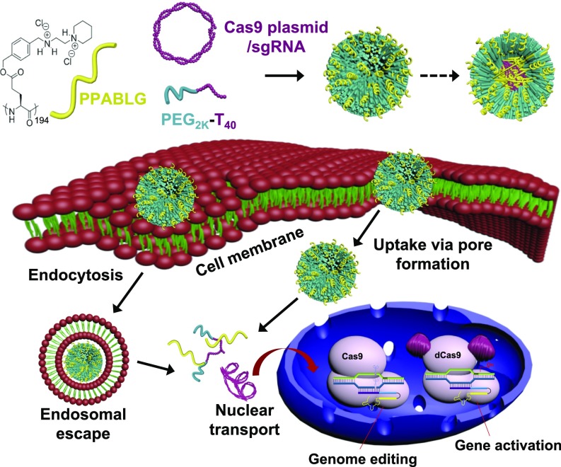
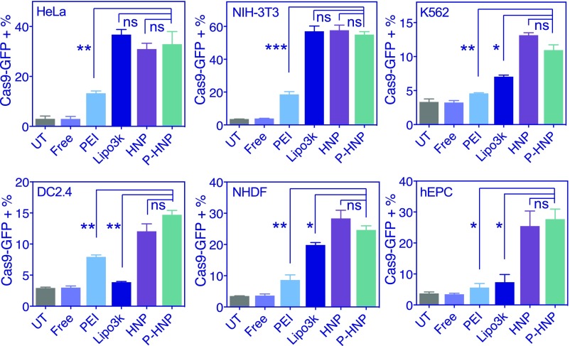
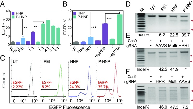
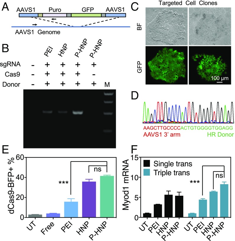
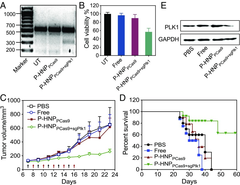
Effective and safe delivery of the CRISPR/Cas9 gene-editing elements remains a challenge. Here we report the development of PEGylated nanoparticles (named P-HNPs) based on the cationic α-helical polypeptide poly(γ-4-((2-(piperidin-1-yl)ethyl)aminomethyl)benzyl-l-glutamate) for the delivery of Cas9 expression plasmid and sgRNA to various cell types and gene-editing scenarios. The cell-penetrating α-helical polypeptide enhanced cellular uptake and promoted escape of pCas9 and/or sgRNA from the endosome and transport into the nucleus. The colloidally stable P-HNPs achieved a Cas9 transfection efficiency up to 60% and sgRNA uptake efficiency of 67.4%, representing an improvement over existing polycation-based gene delivery systems. After performing single or multiplex gene editing with an efficiency up to 47.3% in vitro, we demonstrated that P-HNPs delivering Cas9 plasmid/sgRNA targeting the polo-like kinase 1 (Plk1) gene achieved 35% gene deletion in HeLa tumor tissue to reduce the Plk1 protein level by 66.7%, thereby suppressing the tumor growth by >71% and prolonging the animal survival rate to 60% within 60 days. Capable of delivering Cas9 plasmids to various cell types to achieve multiplex gene knock-out, gene knock-in, and gene activation in vitro and in vivo, the P-HNP system offers a versatile gene-editing platform for biological research and therapeutic applications.
Overview
The claims and statistics of this paper haven’t been checked yet.
Run the check — free, no account needed. It reads the open-access text and checks the sources it cites.
Integrity signals
Registry
Cites
Statistics
Figures
Verification
Claims the paper makes
The claims and statistics of this paper haven’t been checked yet.
Run the check — free, no account needed. It reads the open-access text and checks the sources it cites.
Claims other papers make about this one
Each is checked against this paper’s own text: does it say what the citing paper says it does?
In the citing paper
“Similarly, Wang et al. synthesized a membrane-penetrating cationic α-helical polypeptide which enhanced cellular internalization of Cas9 plasmid and sgRNA.[228]”
From RNA delivery biomaterials for the treatment of genetic and rare diseases (2019) · cites this paper as [228]
Self-check
Percentages, sums and reported differences re-computed from the paper’s own numbers.
The self-check needs the paper’s full text.
Citations
Cites
Development and Applications of CRISPR-Cas9 for Genome Engineering
Hsu, P. D., Lander, E. S., & Zhang, F. (2014) · Cell · doi:10.1016/j.cell.2014.05.010 · Cited by 5,762
Non-viral vectors for gene-based therapy
Yin, H., Kanasty, R. L., Eltoukhy, A. A., Vegas, A. J., Dorkin, J. R., & Anderson, D. G. (2014) · Nature Reviews Genetics · doi:10.1038/nrg3763 · Cited by 3,123
Zuris, J. A., Thompson, D. B., Shu, Y., Guilinger, J. P., Bessen, J. L., Hu, J. H., Maeder, M. L., Joung, J. K., Chen, Z.-Y., & Liu, D. R. (2015) · Nature Biotechnology · doi:10.1038/nbt.3081 · Cited by 1,460
Therapeutic genome editing: prospects and challenges
Cox, D. B. T., Platt, R. J., & Zhang, F. (2015) · Nature Medicine · doi:10.1038/nm.3793 · Cited by 1,336
RNA-guided gene activation by CRISPR-Cas9-based transcription factors
Perez-Pinera, P., Kocak, D. D., Vockley, C. M., Adler, A. F., Kabadi, A. M., Polstein, L. R., Thakore, P. I., Glass, K. A., Ousterout, D. G., Leong, K. W., et al. (2013) · Nature Methods · doi:10.1038/nmeth.2600 · Cited by 1,328
Self-citationIversen, T., Skotland, T., & Sandvig, K. (2011) · Nano Today · doi:10.1016/j.nantod.2011.02.003 · Cited by 1,267
Identification of preexisting adaptive immunity to Cas9 proteins in humans
Charlesworth, C. T., Deshpande, P. S., Dever, D. P., Camarena, J., Lemgart, V. T., Cromer, M. K., Vakulskas, C. A., Collingwood, M. A., Zhang, L., Bode, N. M., et al. (2019) · Nature Medicine · doi:10.1038/s41591-018-0326-x · Cited by 1,093
Effect of genome size on AAV vector packaging
Wu, Z., Yang, H., & Colosi, P. (2010) · Molecular Therapy · doi:10.1038/mt.2009.255 · Cited by 1,029
Yin, H., Song, C.-Q., Dorkin, J. R., Zhu, L. J., Li, Y., Wu, Q., Park, A., Yang, J., Suresh, S., Bizhanova, A., et al. (2016) · Nature Biotechnology · doi:10.1038/nbt.3471 · Cited by 876
Rapid and highly efficient mammalian cell engineering via Cas9 protein transfection
Liang, X., Potter, J., Kumar, S., Zou, Y., Quintanilla, R., Sridharan, M., Carte, J., Chen, W., Roark, N., Ranganathan, S., et al. (2015) · Journal of Biotechnology · doi:10.1016/j.jbiotec.2015.04.024 · Cited by 733
Gene disruption by cell-penetrating peptide-mediated delivery of Cas9 protein and guide RNA
Ramakrishna, S., Kwaku Dad, A.-B., Beloor, J., Gopalappa, R., Lee, S.-K., & Kim, H. (2014) · Genome Research · doi:10.1101/gr.171264.113 · Cited by 655
Self‐assembled DNA nanoclews for the efficient delivery of CRISPR–Cas9 for genome editing
Sun, W., Ji, W., Hall, J. M., Hu, Q., Wang, C., Beisel, C. L., & Gu, Z. (2015) · Angewandte Chemie International Edition · doi:10.1002/anie.201506030 · Cited by 629
Efficient delivery of genome-editing proteins using bioreducible lipid nanoparticles
Wang, M., Zuris, J. A., Meng, F., Rees, H., Sun, S., Deng, P., Han, Y., Gao, X., Pouli, D., Wu, Q., et al. (2016) · Proceedings of the National Academy of Sciences · doi:10.1073/pnas.1520244113 · Cited by 624
Direct cytosolic delivery of CRISPR/Cas9-ribonucleoprotein for efficient gene editing
Mout, R., Ray, M., Yesilbag Tonga, G., Lee, Y.-W., Tay, T., Sasaki, K., & Rotello, V. M. (2017) · ACS Nano · doi:10.1021/acsnano.6b07600 · Cited by 579
Wang, H.-X., Li, M., Lee, C. M., Chakraborty, S., Kim, H.-W., Bao, G., & Leong, K. W. (2017) · Chemical Reviews · doi:10.1021/acs.chemrev.6b00799 · Cited by 560
Self-citationMiller, J. B., Zhang, S., Kos, P., Xiong, H., Zhou, K., Perelman, S. S., Zhu, H., & Siegwart, D. J. (2017) · Angewandte Chemie International Edition · doi:10.1002/anie.201610209 · Cited by 540
Gene Therapy Progress and Prospects: Electroporation and other physical methods
Wells, D. J. (2004) · Gene Therapy · doi:10.1038/sj.gt.3302337 · Cited by 373
Polo-like kinases and oncogenesis
Eckerdt, F., Yuan, J., & Strebhardt, K. (2005) · Oncogene · doi:10.1038/sj.onc.1208273 · Cited by 363
Exogenous siRNA delivery using peptide transduction domains/cell penetrating peptides
Meade, B. R., & Dowdy, S. F. (2007) · Advanced Drug Delivery Reviews · doi:10.1016/j.addr.2007.03.004 · Cited by 277
Gibbs, R. A., Nguyen, P. N., McBride, L. J., Koepf, S. M., & Caskey, C. T. (1989) · Proceedings of the National Academy of Sciences · doi:10.1073/pnas.86.6.1919 · Cited by 275
Ionic polypeptides with unusual helical stability
Lu, H., Wang, J., Bai, Y., Lang, J. W., Liu, S., Lin, Y., & Cheng, J. (2011) · Nature Communications · doi:10.1038/ncomms1209 · Cited by 272
Self-citationArtificial Virus Delivers CRISPR-Cas9 System for Genome Editing of Cells in Mice
Li, L., Song, L., Liu, X., Yang, X., Li, X., He, T., Wang, N., Yang, S., Yu, C., Yin, T., et al. (2017) · ACS Nano · doi:10.1021/acsnano.6b04261 · Cited by 244
Smith-Arica, J. R., Maguire, S. F., Davis, L. A., Alexander, M., Yang, F., Chandran, S., ffrench‐Constant, C., & Pedersen, R. A. (2007) · Stem Cells · doi:10.1634/stemcells.2007-0039 · Cited by 241
Macrophage-Specific in Vivo Gene Editing Using Cationic Lipid-Assisted Polymeric Nanoparticles
Luo, Y., Xu, C., Li, H., Cao, Z., Liu, J., Wang, J., Du, X., Yang, X., Gu, Z., & Wang, J. (2018) · ACS Nano · doi:10.1021/acsnano.7b07874 · Cited by 223
Wang, P., Zhang, L., Xie, Y., Wang, N., Tang, R., Zheng, W., & Jiang, X. (2017) · Advanced Science · doi:10.1002/advs.201700175 · Cited by 215
A CRISPR/Cas9-Based System for Reprogramming Cell Lineage Specification
Chakraborty, S., Ji, H., Kabadi, A. M., Gersbach, C. A., Christoforou, N., & Leong, K. W. (2014) · Stem Cell Reports · doi:10.1016/j.stemcr.2014.09.013 · Cited by 199
Self-citationKomin, A., Russell, L. M., Hristova, K. A., & Searson, P. C. (2016) · Advanced Drug Delivery Reviews · doi:10.1016/j.addr.2016.06.002 · Cited by 193
Comparative transfection of DNA into primary and transformed mammalian cells from different lineages
Maurisse, R., de Semir, D., Emamekhoo, H., Bedayat, B., Abdolmohammadi, A., Parsi, H., & Gruenert, D. C. (2010) · BMC Biotechnology · doi:10.1186/1472-6750-10-9 · Cited by 190
Reactive and Bioactive Cationic α‐Helical Polypeptide Template for Nonviral Gene Delivery
Gabrielson, N. P., Lu, H., Yin, L., Li, D., Wang, F., & Cheng, J. (2011) · Angewandte Chemie International Edition · doi:10.1002/anie.201104262 · Cited by 180
Self-citationYin, L., Song, Z., Kim, K. H., Zheng, N., Gabrielson, N. P., & Cheng, J. (2013) · Advanced Materials · doi:10.1002/adma.201205088 · Cited by 123
Self-citationHe, H., Zheng, N., Song, Z., Kim, K. H., Yao, C., Zhang, R., Zhang, C., Huang, Y., Uckun, F. M., Cheng, J., et al. (2016) · ACS Nano · doi:10.1021/acsnano.5b05470 · Cited by 121
Self-citationYin, L., Song, Z., Qu, Q., Kim, K. H., Zheng, N., Yao, C., Chaudhury, I., Tang, H., Gabrielson, N. P., Uckun, F. M., et al. (2013) · Angewandte Chemie International Edition · doi:10.1002/anie.201209991 · Cited by 92
Self-citationIdentification of Pre-Existing Adaptive Immunity to Cas9 Proteins in Humans
Charlesworth, C. T., Deshpande, P. S., Dever, D. P., Dejene, B., Gomez-Ospina, N., Mantri, S., Pavel-Dinu, M., Camarena, J., Weinberg, K. I., & Porteus, M. H. (2018) · doi:10.1101/243345 · Cited by 71
Zheng, N., Yin, L., Song, Z., Ma, L., Tang, H., Gabrielson, N. P., Lu, H., & Cheng, J. (2013) · Biomaterials · doi:10.1016/j.biomaterials.2013.09.090 · Cited by 56
Self-citationZheng, N., Song, Z., Yang, J., Liu, Y., Li, F., Cheng, J., & Yin, L. (2017) · Acta Biomaterialia · doi:10.1016/j.actbio.2017.05.001 · Cited by 30
Self-citation
Cited by
Sorted by citations
Li, H., Yang, Y., Hong, W., Huang, M., Wu, M., & Zhao, X. (2020) · Signal Transduction and Targeted Therapy · doi:10.1038/s41392-019-0089-y · Cited by 1,829
CRISPR technology: A decade of genome editing is only the beginning
Wang, J. Y., & Doudna, J. A. (2023) · Science · doi:10.1126/science.add8643 · Cited by 1,105
Recent progress in drug delivery
Li, C., Wang, J., Wang, Y., Gao, H., Wei, G., Huang, Y., Yu, H., Gan, Y., Wang, Y., Mei, L., et al. (2019) · Acta Pharmaceutica Sinica B · doi:10.1016/j.apsb.2019.08.003 · Cited by 818
Improving cancer immunotherapy using nanomedicines: progress, opportunities and challenges
Martin, J. D., Cabral, H., Stylianopoulos, T., & Jain, R. K. (2020) · Nature Reviews Clinical Oncology · doi:10.1038/s41571-019-0308-z · Cited by 691
Tissue Engineering and Regenerative Medicine: Achievements, Future, and Sustainability in Asia
Han, F., Wang, J., Ding, L., Hu, Y., Li, W., Yuan, Z., Guo, Q., Zhu, C., Yu, L., Wang, H., et al. (2020) · Frontiers in Bioengineering and Biotechnology · doi:10.3389/fbioe.2020.00083 · Cited by 382
Self-citation
Metadata
- Authors
- Wang, H.-X., Song, Z., Lao, Y.-H., Xu, X., Gong, J., Cheng, D., Chakraborty, S., Park, J. S., Li, M., Huang, D., Yin, L., Cheng, J., & Leong, K. W.
- Affiliations
- Department of Biomedical Engineering, Columbia University, New York, NY 10027;
- University of Illinois Urbana-Champaign (US)
- Columbia University (US)
- Soochow University (CN)
- Sun Yat-sen University (CN)
- Corresponding author
- Yin, Lichen
- Cheng, Jianjun
- Leong, Kam W.
- ORCID
- Venue
- Proceedings of the National Academy of Sciences
- Publisher
- National Academy of Sciences
- Published
- 2018-04-23
- Volume
- 115
- Issue
- 19
- Pages
- 4903-4908
- ISSN
- 0027-8424 (ISSN-L)
- 1091-6490
- PMC
- PMC5948953
- OpenAlex
- W2802061036
- Cited by
- 277
- References
- 34
- Citations by year
- 2026: 9 · 2025: 18 · 2024: 21 · 2023: 41 · 2022: 47 · 2021: 41 · 2020: 43 · 2019: 42 · 2018: 15
- FWCI
- 12.54
- Type
- journal article
- Language
- en
- Open access
- hybrid
- doi.org
- Licence
- Locations
- Proceedings of the National Academy of Sciences (publishedVersion, cc-by-nc-nd)
- Europe PMC (PubMed Central) (publishedVersion, cc-by-nc-nd)
- PubMed Central (publishedVersion, cc-by-nc-nd)
- PubMed (publishedVersion)
- Columbia Academic Commons (Columbia University)
- Funders
- National Natural Science Foundation of China (51573123, 51722305)
- Ministry of Science and Technology of the People's Republic of China (2016YFA0201200)
- Guangdong Innovative and Entrepreneurial Research Team Program (NO.2013S086)
- Global Research Laboratory Program Korean NSF (2015032163)
- Topics
- Life Sciences › Biochemistry, Genetics and Molecular Biology › Molecular Biology › RNA Interference and Gene Delivery
- Life Sciences › Biochemistry, Genetics and Molecular Biology › Molecular Biology › CRISPR and Genetic Engineering
- Physical Sciences › Materials Science › Biomaterials › Nanoparticle-Based Drug Delivery
- Keywords
- CRISPR/Cas9 delivery, PEGylated nanoparticles, cell-penetrating peptides, endosomal escape, multiplex genome editing, PLK1, tumor growth inhibition, gene knock-in, gene activation
- MeSH terms
- Gene Editing, Animals, HeLa Cells, Humans, Plasmids, Gene Transfer Techniques, K562 Cells, NIH 3T3 Cells, Mice, Nanoparticles, HEK293 Cells, Cell-Penetrating Peptides, CRISPR-Cas Systems
About these checks
Registry, retraction, Cites and statistics results are computed, not judged, with one exception: a printed reference that no index lists is read once by a model before it counts as a citation error. Claims are extracted by a model and kept only when their quote appears verbatim in the text; each cited source is then checked, with a model’s help, for whether it says what the claim relies on. Those verdicts can be wrong — read the claim’s quote and the cited source before relying on one.
Statistics and arithmetic are recomputed, not judged.
Confidence bands are shown only for fields calibrated on our annotated test set. None is calibrated yet, so no band is shown.
What counts as a claim ›How we measure confidence ›How we check this ›
Are you an author, or do you see a problem with this page? Request a review
