Cited by 93

The DOI matches its Crossref record; 1 of the 1 references it cites were found in a registry or an index.

Identification of an α Helical Motif Sufficient for Association with Papillomavirus E6

Chen, Jason J., Hong, Yihui, Rustamzadeh, Edward, Baleja, James D., Androphy, Elliot J.·1998·Journal of Biological Chemistry

Overview

The claims and statistics of this paper haven’t been checked yet.

Run the check — free, no account needed. It reads the open-access text and checks the sources it cites.

A few free checks a day · papers already verified open instantly · results are public

Integrity signals

Registry

Crossref · 10.1074/jbc.273.22.13537

Cites

1 of 1 references verified0 citation errors · 0 not checkable

Statistics

Figures

Image 1
CC-BY · From the PDF
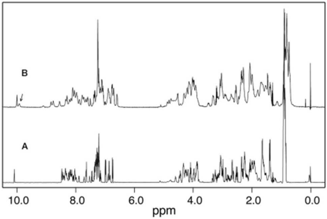
FIG. 2 Demonstration of calcium binding by E6 binding peptide by 1 H NMR spectroscopy. A , the metal-free peptide was at 1.0 mMconcentration in a solution containing 20 mM NaCl. B , addition of 80 mM calcium chloride (CaCl2 ) to the peptide induces chemical shift dispersion. The downfield-shifted resonance of glycine 17 is indicated with an arrow and is typical of a calcium-bound EF-hand. The addition of a small amount of TFE (5-10%) narrows some resonance line widths. Sample conditions were 1.0 mM peptide in a buffer containing 8% TFE, 20 mM NaCl, pH 6.0.
FIG. 2 Demonstration of calcium binding by E6 binding peptide by 1 H NMR spectroscopy. A , the metal-free peptide was at 1.0 mMconcentration in a solution containing 20 mM NaCl. B , addition of 80 mM calcium chloride (CaCl2 ) to the peptide induces chemical shift dispersion. The downfield-shifted resonance of glycine 17 is indicated with an arrow and is typical of a calcium-bound EF-hand. The addition of a small amount of TFE (5-10%) narrows some resonance line widths. Sample conditions were 1.0 mM peptide in a buffer containing 8% TFE, 20 mM NaCl, pH 6.0.CC-BY · From the PDF
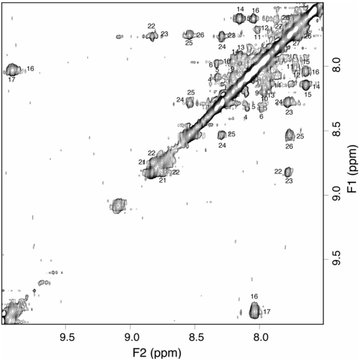
FIG. 3 The NH-NH region of the two-dimensional NOESY spectrum. Resonance assignments were accomplished by tracing connections between the NH, a , and b protons of a residue ( i ), and the NH of the next residue ( i 1 1). The helical nature of this peptide is illustrated by medium and strong NH to NH NOE cross-peaks between residues adjacent in sequence. Sample conditions are given in Fig. 3, and the NOE mixing time was 125 ms.
FIG. 3 The NH-NH region of the two-dimensional NOESY spectrum. Resonance assignments were accomplished by tracing connections between the NH, a , and b protons of a residue ( i ), and the NH of the next residue ( i 1 1). The helical nature of this peptide is illustrated by medium and strong NH to NH NOE cross-peaks between residues adjacent in sequence. Sample conditions are given in Fig. 3, and the NOE mixing time was 125 ms.CC-BY · From the PDF
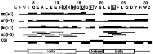
FIG. 4 Summaryof the conformational data. The sequence of the peptide, corresponding to residues 289-318 of E6BP, is renumbered as residues 1-31. The residues that ligate calcium are boxed . These residues form single ligands to calcium using their side chains, except for F18, which uses its backbone carbonyl, and E23, which contributes a double ligand. NOE cross-peaks describing NH-to-NH contacts, H a -NH contacts, H b -NH contacts, and H a to H b ( i 1 3) contacts are illustrated with the thickness of the filled-in bar corresponding to NOE intensity. The absence of a bar indicates that the NOE was not observed, whereas the presence of the dotted line indicates that the NOE was likely but was obscured by resonance overlap. A hollow bar indicates that the NOEinvolves a glycine residue and is not applicable. The chemical shift index ( CSI ) is positive if the difference in chemical shift between observed and a random coil peptide was greater than 0.1 ppm or negative if it was less than 0.1 ppm (61). A positive index indicates b sheet, whereas a negative index indicates a helix. A summary for the secondary structure of the peptide is shown under the conformational data.
FIG. 4 Summaryof the conformational data. The sequence of the peptide, corresponding to residues 289-318 of E6BP, is renumbered as residues 1-31. The residues that ligate calcium are boxed . These residues form single ligands to calcium using their side chains, except for F18, which uses its backbone carbonyl, and E23, which contributes a double ligand. NOE cross-peaks describing NH-to-NH contacts, H a -NH contacts, H b -NH contacts, and H a to H b ( i 1 3) contacts are illustrated with the thickness of the filled-in bar corresponding to NOE intensity. The absence of a bar indicates that the NOE was not observed, whereas the presence of the dotted line indicates that the NOE was likely but was obscured by resonance overlap. A hollow bar indicates that the NOEinvolves a glycine residue and is not applicable. The chemical shift index ( CSI ) is positive if the difference in chemical shift between observed and a random coil peptide was greater than 0.1 ppm or negative if it was less than 0.1 ppm (61). A positive index indicates b sheet, whereas a negative index indicates a helix. A summary for the secondary structure of the peptide is shown under the conformational data.CC-BY · From the PDF
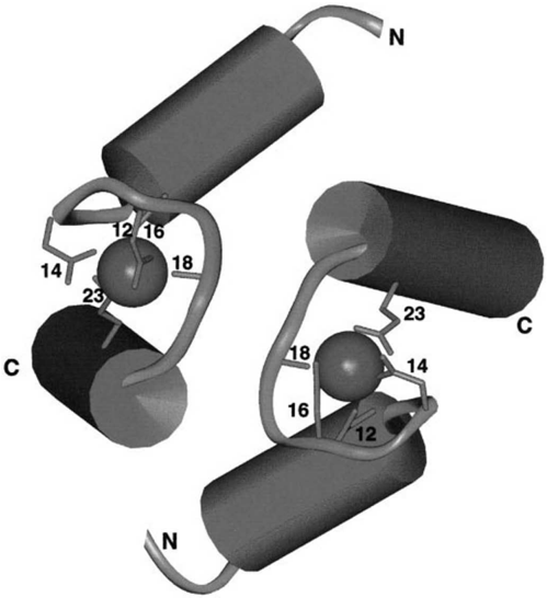
FIG. 5 Model for the E6bp peptide. The peptide was modeled using the x-ray structure of the calcium-bound motif from troponin C. The helical elements are shown as cylinders . The side chains and backbone carbonyl groups that ligate the calcium ions are shown.
FIG. 5 Model for the E6bp peptide. The peptide was modeled using the x-ray structure of the calcium-bound motif from troponin C. The helical elements are shown as cylinders . The side chains and backbone carbonyl groups that ligate the calcium ions are shown.CC-BY · From the PDF
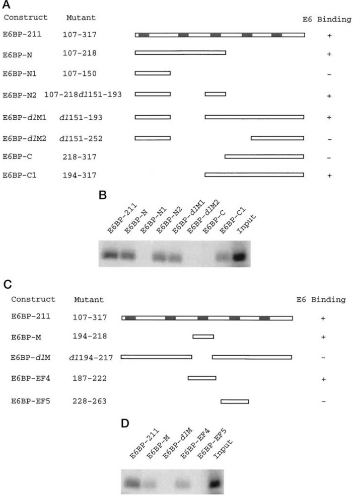
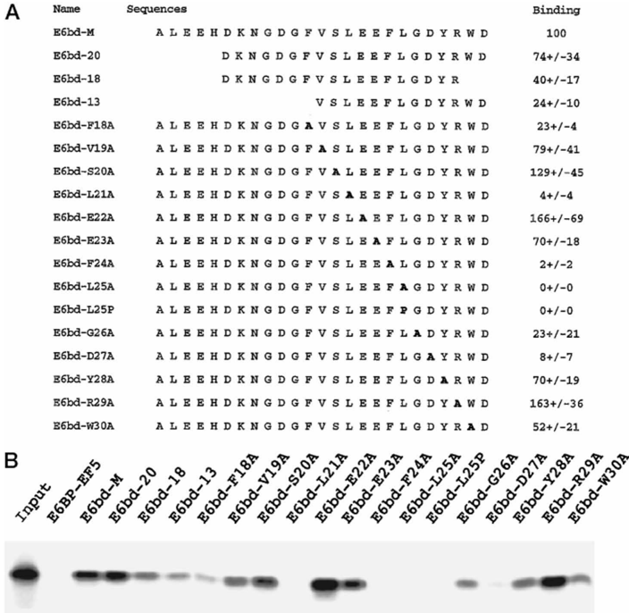
FIG. 6 An a helical motif is sufficient for association with E6. Glutathione-Sepharose beads containing GST-E6BP fusion proteins were combined with 35 S-labeled in vitro translated BPV-1 E6 proteins in LSAB. After incubation and washes, the bound products were separated by SDS-polyacrylamide gel electrophoresis. E6 binding was analyzed by Molecular Imager. A , construction of deletion and point mutations of E6bd GST fusion proteins. Numbers for binding are the percentage of association from each mutant normalized to the level seen with E6bd-M. The average of three independent experiments, including standard deviations, is given. B , an a helical motif is important for association with E6. The image of one representative experiment is shown. Sequences of the E6 binding domain (E6bd) 7 through 31 are given. Input was loaded directly into the well and represents 10% of the 35 S-labeled E6 used in each binding reaction.
FIG. 6 An a helical motif is sufficient for association with E6. Glutathione-Sepharose beads containing GST-E6BP fusion proteins were combined with 35 S-labeled in vitro translated BPV-1 E6 proteins in LSAB. After incubation and washes, the bound products were separated by SDS-polyacrylamide gel electrophoresis. E6 binding was analyzed by Molecular Imager. A , construction of deletion and point mutations of E6bd GST fusion proteins. Numbers for binding are the percentage of association from each mutant normalized to the level seen with E6bd-M. The average of three independent experiments, including standard deviations, is given. B , an a helical motif is important for association with E6. The image of one representative experiment is shown. Sequences of the E6 binding domain (E6bd) 7 through 31 are given. Input was loaded directly into the well and represents 10% of the 35 S-labeled E6 used in each binding reaction.CC-BY · From the PDF
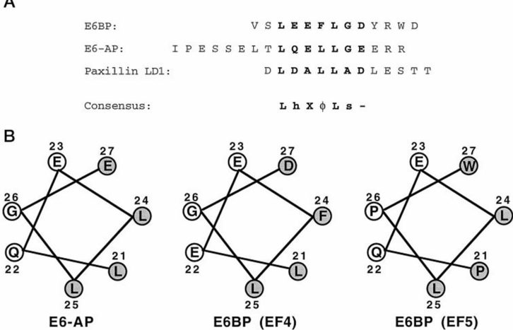
FIG. 7 An a helix is conserved among E6-binding proteins. A , comparison of E6 binding domains reveals a conserved motif. The E6 binding peptides of E6bd (residues 18-29), the 18 amino acids (amino acids 391-408) of E6AP (2), and the sequence of LD1 from paxillin (amino acids 3-15) (3) are aligned. h , amino acid that can form a double hydrogen bond, i.e. Asp, Glu, Gln. X , any amino acid. f , hydrophobic amino acid. s , small amino acid such as Gly or Ala. 2 , acidic residue. B , amino acids in the second helix of the helix-loop-helix that comprise the EF-hand in a helical wheel representation. In E6BP, replacement of either Leu-21, Phe-24, Leu-25, or Asp 27 by Ala reduced binding to E6 by at least 10-fold. Alanine replacements of the Gly-26 results in about a 5-fold loss in binding, whereas replacement of the other residues did not appreciably affect binding. Left , the motif for E6BP (EF-hand 4). Center , the motif for E6AP. Right , an example of a nonbinding E6 motif (the homologous residues from EF-hand 5 of E6BP).
FIG. 7 An a helix is conserved among E6-binding proteins. A , comparison of E6 binding domains reveals a conserved motif. The E6 binding peptides of E6bd (residues 18-29), the 18 amino acids (amino acids 391-408) of E6AP (2), and the sequence of LD1 from paxillin (amino acids 3-15) (3) are aligned. h , amino acid that can form a double hydrogen bond, i.e. Asp, Glu, Gln. X , any amino acid. f , hydrophobic amino acid. s , small amino acid such as Gly or Ala. 2 , acidic residue. B , amino acids in the second helix of the helix-loop-helix that comprise the EF-hand in a helical wheel representation. In E6BP, replacement of either Leu-21, Phe-24, Leu-25, or Asp 27 by Ala reduced binding to E6 by at least 10-fold. Alanine replacements of the Gly-26 results in about a 5-fold loss in binding, whereas replacement of the other residues did not appreciably affect binding. Left , the motif for E6BP (EF-hand 4). Center , the motif for E6AP. Right , an example of a nonbinding E6 motif (the homologous residues from EF-hand 5 of E6BP).CC-BY · From the PDF

Verification

Claims the paper makes

The claims and statistics of this paper haven’t been checked yet.

Run the check — free, no account needed. It reads the open-access text and checks the sources it cites.

A few free checks a day · papers already verified open instantly · results are public

Claims other papers make about this one

Each is checked against this paper’s own text: does it say what the citing paper says it does?

Self-check

Percentages, sums and reported differences re-computed from the paper’s own numbers.

The self-check needs the paper’s full text.

Citations

Cites

Show all 1 references

Research works printed but not linked: 58

  1. Chen, 1995, Science, 269, 529, 10.1126/science.7624774 · DOI · Paper page
  2. Huibregtse, 1993, Mol. Cell. Biol., 13, 4918
  3. Tong, 1997, J. Biol. Chem., 272, 33373, 10.1074/jbc.272.52.33373 · DOI
  4. zur Hausen, 1991, Virology, 184, 9, 10.1016/0042-6822(91)90816-T · DOI
  5. Schwarz, 1985, Nature, 314, 111, 10.1038/314111a0 · DOI
  6. Smotkin, 1986, Obstet. Gynecol., 68, 241
  7. Baker, 1987, J. Virol., 61, 962, 10.1128/jvi.61.4.962-971.1987 · DOI
  8. Schlegel, 1988, EMBO J., 7, 3181, 10.1002/j.1460-2075.1988.tb03185.x · DOI · Paper page
  9. Munger, 1989, J. Virol., 63, 4417, 10.1128/jvi.63.10.4417-4421.1989 · DOI · Paper page
  10. Hawley-Nelson, 1989, EMBO J., 8, 3905, 10.1002/j.1460-2075.1989.tb08570.x · DOI · Paper page
  11. Watanabe, 1989, J. Virol., 63, 965, 10.1128/jvi.63.2.965-969.1989 · DOI
  12. Kaur, 1989, Virology, 173, 302, 10.1016/0042-6822(89)90247-X · DOI
  13. Hudson, 1990, J. Virol., 64, 519, 10.1128/jvi.64.2.519-526.1990 · DOI · Paper page
  14. Storey, 1993, Oncogene, 8, 919
  15. Liu, 1994, Virology, 201, 388, 10.1006/viro.1994.1306 · DOI · Paper page
  16. Sedman, 1991, J. Virol., 65, 4860, 10.1128/jvi.65.9.4860-4866.1991 · DOI
  17. Band, 1991, J. Virol., 65, 6671, 10.1128/jvi.65.12.6671-6676.1991 · DOI · Paper page
  18. Sherman, 1996, J. Virol., 70, 3269, 10.1128/jvi.70.5.3269-3279.1996 · DOI
  19. Werness, 1990, Science, 248, 76, 10.1126/science.2157286 · DOI · Paper page
  20. Lechner, 1994, J. Virol., 68, 4262, 10.1128/jvi.68.7.4262-4273.1994 · DOI · Paper page
  21. Li, 1996, J. Virol., 70, 4509, 10.1128/jvi.70.7.4509-4516.1996 · DOI
  22. Scheffner, 1993, Cell, 75, 495, 10.1016/0092-8674(93)90384-3 · DOI · Paper page
  23. Schiller, 1984, Proc. Natl. Acad. Sci. U. S. A., 81, 7880, 10.1073/pnas.81.24.7880 · DOI
  24. Schiller, 1986, Cancer Cells ( Cold Spring Harbor ), 4, 571
  25. Steger, 1992, Arch. Virol., 125, 355, 10.1007/BF01309654 · DOI
  26. Band, 1993, EMBO J., 12, 1847, 10.1002/j.1460-2075.1993.tb05833.x · DOI
  27. Choo, 1993, Virology, 193, 1042, 10.1006/viro.1993.1224 · DOI
  28. Park, 1994, Oncogene, 9, 205
  29. Pim, 1994, Oncogene, 9, 1869
  30. Nakagawa, 1995, Virology, 212, 535, 10.1006/viro.1995.1511 · DOI
  31. Thomas, 1995, Oncogene, 10, 261
  32. Butz, 1995, Oncogene, 10, 927
  33. Pan, 1995, Genes Dev., 9, 2157, 10.1101/gad.9.17.2157 · DOI
  34. Xu, 1995, Proc. Natl. Acad. Sci. U. S. A., 92, 7829, 10.1073/pnas.92.17.7829 · DOI
  35. Klingelhutz, 1996, Nature, 380, 79, 10.1038/380079a0 · DOI · Paper page
  36. Weis, 1994, J. Biol. Chem., 269, 19142, 10.1016/S0021-9258(17)32286-X · DOI
  37. Androphy, 1985, Science, 230, 442, 10.1126/science.2996134 · DOI
  38. Tong, 1997, Proc. Natl. Acad. Sci. U. S. A., 94, 4412, 10.1073/pnas.94.9.4412 · DOI
  39. Tong, 1998, J. Virol., 72, 476, 10.1128/JVI.72.1.476-482.1998 · DOI
  40. Lee, 1997, Proc. Natl. Acad. Sci. U. S. A., 94, 6670, 10.1073/pnas.94.13.6670 · DOI · Paper page
  41. Kiyono, 1997, Proc. Natl. Acad. Sci. U. S. A., 94, 11612, 10.1073/pnas.94.21.11612 · DOI · Paper page
  42. Ikura, 1996, Trends Biochem. Sci., 21, 14, 10.1016/S0968-0004(06)80021-6 · DOI
  43. Schäfer, 1996, Trends. Biochem. Sci., 21, 134, 10.1016/S0968-0004(96)80167-8 · DOI
  44. Lin, 1997, Nat. Struct. Biol., 4, 538, 10.1038/nsb0797-539 · DOI
  45. Linse, 1997, Protein Sci., 6, 2385, 10.1002/pro.5560061112 · DOI
  46. Blanchard, 1997, Nat. Struct. Biol., 4, 532, 10.1038/nsb0797-532 · DOI
  47. Kay, 1991, Biochemistry, 30, 4324
  48. Crook, 1991, Cell, 67, 547, 10.1016/0092-8674(91)90529-8 · DOI
  49. Findlay, 1993, Biochemistry, 32, 3461, 10.1021/bi00064a033 · DOI
  50. Wagner, 1997, Nat. Struct. Biol., 4, 841
  51. Shaw, 1991, Biochemistry, 30, 8339, 10.1021/bi00098a009 · DOI
  52. WoÖjcik, 1997, Biochemistry, 36, 680, 10.1021/bi961821c · DOI
  53. Slupsky, 1995, Biochemistry, 34, 7365, 10.1021/bi00022a009 · DOI
  54. Herzberg, 1988, J. Mol. Biol., 203, 761, 10.1016/0022-2836(88)90208-2 · DOI
  55. Wishart, 1994, Comp. Appl. Biosci., 10, 121
  56. Ullman, 1996, Biochem. J., 319, 229, 10.1042/bj3190229 · DOI
  57. Doyle, 1996, Cell, 85, 1067, 10.1016/S0092-8674(00)81307-0 · DOI
  58. Wishart, 1991, J. Mol. Biol., 222, 311, 10.1016/0022-2836(91)90214-Q · DOI

Cited by

See all 93

Metadata

Authors
Chen, J. J., Hong, Y., Rustamzadeh, E., Baleja, J. D., & Androphy, E. J.
Venue
Journal of Biological Chemistry
Publisher
Elsevier BV
Published
1998-05
Volume
273
Issue
22
Pages
13537-13544
ISSN
0021-9258
Cited by
93
References
1
Language
en

About these checks

Registry, retraction, Cites and statistics results are computed, not judged, with one exception: a printed reference that no index lists is read once by a model before it counts as a citation error. Claims are extracted by a model and kept only when their quote appears verbatim in the text; each cited source is then checked, with a model’s help, for whether it says what the claim relies on. Those verdicts can be wrong — read the claim’s quote and the cited source before relying on one.

Statistics and arithmetic are recomputed, not judged.

Confidence bands are shown only for fields calibrated on our annotated test set. None is calibrated yet, so no band is shown.

What counts as a claim ›How we measure confidence ›How we check this ›

Are you an author, or do you see a problem with this page? Request a review

How to cite

Cite CoChat for the checks on this page, and the paper for its own findings.

This page
CoChat. (2026). Citation check: Identification of an α Helical Motif Sufficient for Association with Papillomavirus E6. https://app.cochat.ai/papers/10.1074/jbc.273.22.13537
The paper
Chen, J. J., Hong, Y., Rustamzadeh, E., Baleja, J. D., & Androphy, E. J. (1998). Identification of an α Helical Motif Sufficient for Association with Papillomavirus E6. Journal of Biological Chemistry. https://doi.org/10.1074/jbc.273.22.13537