Cited by 18

The DOI matches its Crossref record; 1 of the 1 references it cites were found in a registry or an index.

Enhanced macrophage delivery to the colon using magnetic lipoplexes with a magnetic field

Kono, Yusuke, Gogatsubo, Serika, Ohba, Takeshi, Fujita, Takuya·2019·Drug Delivery

Overview

The claims and statistics of this paper haven’t been checked yet.

Run the check — free, no account needed. It reads the open-access text and checks the sources it cites.

A few free checks a day · papers already verified open instantly · results are public

Integrity signals

Registry

Crossref · 10.1080/10717544.2019.1662515

Cites

1 of 1 references verified0 citation errors · 0 not checkable

Statistics

Figures

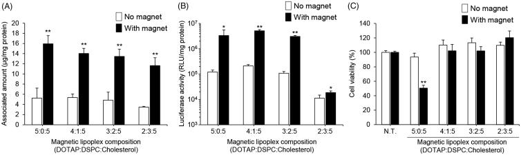
Figure 1. Effect of the lipid composition of the magnetic cationic liposomes on their cellular association, pDNA transfection efficiency, and cytotoxicity in RAW264 cells. Cellular association and/or uptake of magnetic cationic liposomes (A) and the level of luciferase expression following magnetic lipoplex administration (B). Magnetic lipoplexes prepared using various lipid compositions (DOTAP:DSPC:cholesterol = 5:0:5, 4:1:5, 3:2:5, or 2:3:5) and containing 0.1 mg/mL of SPIONs were added to each well (as 1 µg of pDNA), and incubated for 10 min at 37 °C in the presence or absence of a magnetic field. The mixing ratio of the magnetic cationic liposomes/pDNA was 10:1. Each value represents the mean + SD (n = 4). *p < .05; **p < .01, compared with no magnet. (C) Cell viability of RAW264 cells incubated with magnetic lipoplexes (as 1 µg of pDNA) for 10 min at 37 °C in the presence or absence of a magnetic field. Each value represents the mean + SD (n = 4). **p < .01, compared with non-treated (N.T).
Figure 1. Effect of the lipid composition of the magnetic cationic liposomes on their cellular association, pDNA transfection efficiency, and cytotoxicity in RAW264 cells. Cellular association and/or uptake of magnetic cationic liposomes (A) and the level of luciferase expression following magnetic lipoplex administration (B). Magnetic lipoplexes prepared using various lipid compositions (DOTAP:DSPC:cholesterol = 5:0:5, 4:1:5, 3:2:5, or 2:3:5) and containing 0.1 mg/mL of SPIONs were added to each well (as 1 µg of pDNA), and incubated for 10 min at 37 °C in the presence or absence of a magnetic field. The mixing ratio of the magnetic cationic liposomes/pDNA was 10:1. Each value represents the mean + SD (n = 4). *p < .05; **p < .01, compared with no magnet. (C) Cell viability of RAW264 cells incubated with magnetic lipoplexes (as 1 µg of pDNA) for 10 min at 37 °C in the presence or absence of a magnetic field. Each value represents the mean + SD (n = 4). **p < .01, compared with non-treated (N.T).CC BY · PubMed Central
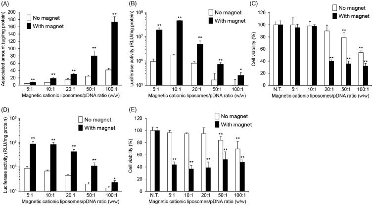
Figure 2. Effect of magnetic cationic liposome/pDNA ratio and SPION concentration on the cellular association, pDNA transfection efficiency, and cytotoxicity of magnetic lipoplexes in RAW264 cells. Cellular association and/or uptake of magnetic cationic liposomes containing 0.1 mg/mL of SPIONs (A) and the level of luciferase expression produced by magnetic lipoplexes containing 0.1 mg/mL (B) and 0.5 mg/mL (D) of SPIONs. Magnetic lipoplexes (DOTAP:DSPC:cholesterol = 4:1:5) were added to each well (as 1 µg of pDNA), and incubated for 10 min at 37 °C in the presence or absence of a magnetic field. Each value represents the mean + SD (n = 4). *p < .05; **p < .01, compared with no magnetic field. Cell viability of RAW264 cells incubated with magnetic lipoplexes containing 0.1 mg/mL (C) and 0.5 mg/mL (E) of SPIONs (as 1 µg of pDNA) for 10 min at 37 °C in the presence or absence of a magnetic field. Each value represents the mean + SD (n = 4). **p < .01, compared with N.T.
Figure 2. Effect of magnetic cationic liposome/pDNA ratio and SPION concentration on the cellular association, pDNA transfection efficiency, and cytotoxicity of magnetic lipoplexes in RAW264 cells. Cellular association and/or uptake of magnetic cationic liposomes containing 0.1 mg/mL of SPIONs (A) and the level of luciferase expression produced by magnetic lipoplexes containing 0.1 mg/mL (B) and 0.5 mg/mL (D) of SPIONs. Magnetic lipoplexes (DOTAP:DSPC:cholesterol = 4:1:5) were added to each well (as 1 µg of pDNA), and incubated for 10 min at 37 °C in the presence or absence of a magnetic field. Each value represents the mean + SD (n = 4). *p < .05; **p < .01, compared with no magnetic field. Cell viability of RAW264 cells incubated with magnetic lipoplexes containing 0.1 mg/mL (C) and 0.5 mg/mL (E) of SPIONs (as 1 µg of pDNA) for 10 min at 37 °C in the presence or absence of a magnetic field. Each value represents the mean + SD (n = 4). **p < .01, compared with N.T.CC BY · PubMed Central
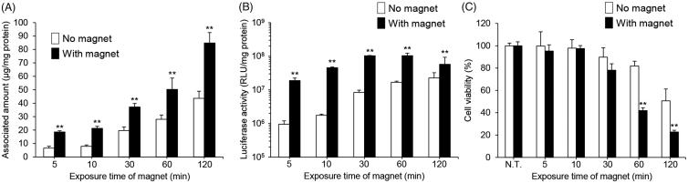
Figure 3. Effect of magnetic field exposure time on the cellular association, pDNA transfection efficiency, and cytotoxicity of magnetic lipoplexes in RAW264 cells. Cellular association and/or uptake of magnetic cationic liposomes (A) and the level of luciferase expression produced by magnetic lipoplexes (B). Magnetic lipoplexes (DOTAP:DSPC:cholesterol = 4:1:5) containing 0.1 mg/mL of SPIONs were added to each well (as 1 µg of pDNA), and incubated for 5–120 min at 37 °C in the presence or absence of a magnetic field. The mixing ratio of the magnetic cationic liposomes/pDNA was 10:1. Each value represents the mean + SD (n = 4). **p < .01, compared with no magnetic field. (B) Cell viability of RAW264 cells incubated with magnetic lipoplexes (as 1 µg of pDNA) for 5–120 min at 37 °C in the presence or absence of a magnetic field. Each value represents the mean + SD (n = 4). **p < .01, compared with N.T.
Figure 3. Effect of magnetic field exposure time on the cellular association, pDNA transfection efficiency, and cytotoxicity of magnetic lipoplexes in RAW264 cells. Cellular association and/or uptake of magnetic cationic liposomes (A) and the level of luciferase expression produced by magnetic lipoplexes (B). Magnetic lipoplexes (DOTAP:DSPC:cholesterol = 4:1:5) containing 0.1 mg/mL of SPIONs were added to each well (as 1 µg of pDNA), and incubated for 5–120 min at 37 °C in the presence or absence of a magnetic field. The mixing ratio of the magnetic cationic liposomes/pDNA was 10:1. Each value represents the mean + SD (n = 4). **p < .01, compared with no magnetic field. (B) Cell viability of RAW264 cells incubated with magnetic lipoplexes (as 1 µg of pDNA) for 5–120 min at 37 °C in the presence or absence of a magnetic field. Each value represents the mean + SD (n = 4). **p < .01, compared with N.T.CC BY · PubMed Central
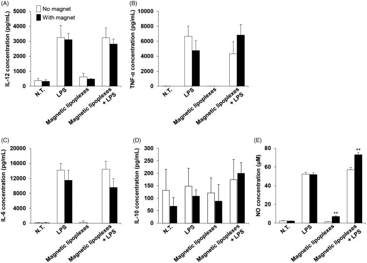
Figure 4. Cytokine and NO production in RAW264 cells treated with magnetic lipoplexes. Concentration of IL-12 (A), TNF-α (B), IL-6 (C), IL-10 (D), and NO (E) in the supernatant of RAW264 cells treated with or without magnetic lipoplexes (as 1 µg of pDNA) containing 0.1 mg/mL of SPIONs and LPS. The supernatants were collected 24 h after magnetic lipoplex treatment. Each value represents the mean + SD (n = 4). **p < .01, compared with N.T. macrophages.
Figure 4. Cytokine and NO production in RAW264 cells treated with magnetic lipoplexes. Concentration of IL-12 (A), TNF-α (B), IL-6 (C), IL-10 (D), and NO (E) in the supernatant of RAW264 cells treated with or without magnetic lipoplexes (as 1 µg of pDNA) containing 0.1 mg/mL of SPIONs and LPS. The supernatants were collected 24 h after magnetic lipoplex treatment. Each value represents the mean + SD (n = 4). **p < .01, compared with N.T. macrophages.CC BY · PubMed Central
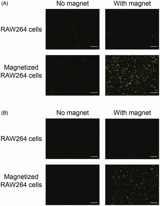
Figure 5. Adhesion of magnetized RAW264 cells to Caco-2 cell monolayers under a magnetic field. RAW264 cells treated with magnetic lipoplexes (DOTAP:DSPC:cholesterol = 4:1:5) containing 0.1 mg/mL of SPIONs were added to wells containing Caco-2 cell monolayers, and incubated for 5 min in an upright position (A) or for 30 min in an inverted position (B). RAW264 cells were fluorescently labeled with calcein-AM. Scale bar: 10 µm.
Figure 5. Adhesion of magnetized RAW264 cells to Caco-2 cell monolayers under a magnetic field. RAW264 cells treated with magnetic lipoplexes (DOTAP:DSPC:cholesterol = 4:1:5) containing 0.1 mg/mL of SPIONs were added to wells containing Caco-2 cell monolayers, and incubated for 5 min in an upright position (A) or for 30 min in an inverted position (B). RAW264 cells were fluorescently labeled with calcein-AM. Scale bar: 10 µm.CC BY · PubMed Central
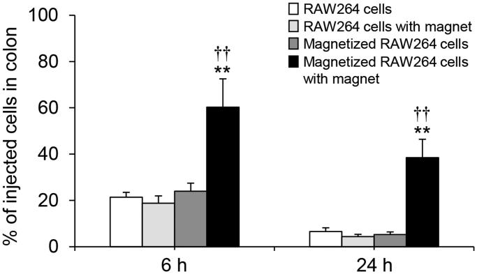
Figure 6. Adhesion of magnetized RAW264 cells to the colon under a magnetic field in mice. RAW264 cells treated with magnetic lipoplexes (DOTAP:DSPC:cholesterol = 4:1:5) containing 0.1 mg/mL of SPIONs were intrarectally injected into mice. The number of cells was measured at 6 and 24 h after the injection. Each value represents the mean + SD (n = 4). **p < .01, compared with RAW264 cells. ††p < .01, compared with magnetized RAW264 cells.
Figure 6. Adhesion of magnetized RAW264 cells to the colon under a magnetic field in mice. RAW264 cells treated with magnetic lipoplexes (DOTAP:DSPC:cholesterol = 4:1:5) containing 0.1 mg/mL of SPIONs were intrarectally injected into mice. The number of cells was measured at 6 and 24 h after the injection. Each value represents the mean + SD (n = 4). **p < .01, compared with RAW264 cells. ††p < .01, compared with magnetized RAW264 cells.CC BY · PubMed Central

Verification

Claims the paper makes

The claims and statistics of this paper haven’t been checked yet.

Run the check — free, no account needed. It reads the open-access text and checks the sources it cites.

A few free checks a day · papers already verified open instantly · results are public

Claims other papers make about this one

Each is checked against this paper’s own text: does it say what the citing paper says it does?

Self-check

Percentages, sums and reported differences re-computed from the paper’s own numbers.

The self-check needs the paper’s full text.

Citations

Cites

Research works printed but not linked: 34

  1. 10.1016/j.cellimm.2016.10.003 · DOI
  2. 10.1016/j.mam.2004.02.015 · DOI
  3. 10.1007/s11427-011-4215-5 · DOI
  4. 10.1021/jacs.8b10904 · DOI
  5. 10.1007/s00262-016-1824-7 · DOI
  6. 10.1039/C8NR05294H · DOI
  7. 10.1017/S1462399409001069 · DOI
  8. 10.1002/nbm.1260 · DOI
  9. 10.1053/j.gastro.2009.12.041 · DOI
  10. 10.1016/j.biomaterials.2015.05.028 · DOI
  11. 10.1016/j.arthro.2007.08.017 · DOI
  12. 10.1248/bpb.b17-00563 · DOI
  13. Kono Y, 2016, Pharmazie, 71, 619
  14. 10.1016/j.bbrc.2013.10.115 · DOI · Paper page
  15. 10.1016/j.jconrel.2006.04.014 · DOI · Paper page
  16. 10.1002/path.4133 · DOI · Paper page
  17. 10.1016/j.colsurfb.2008.09.017 · DOI
  18. 10.1016/j.nano.2014.12.002 · DOI
  19. 10.1038/gt.2008.57 · DOI
  20. 10.1248/bpb.32.1628 · DOI
  21. 10.1155/2018/8917804 · DOI · Paper page
  22. 10.1007/s00204-014-1210-1 · DOI
  23. 10.1002/nbm.2826 · DOI
  24. 10.1016/S0168-3659(99)00280-1 · DOI
  25. 10.1172/JCI59643 · DOI · Paper page
  26. 10.1097/MIB.0b013e3182a69dca · DOI
  27. 10.1016/j.ijpharm.2016.03.028 · DOI
  28. 10.1038/srep16632 · DOI
  29. 10.1089/hum.2009.106 · DOI
  30. 10.1189/jlb.0311124 · DOI
  31. 10.1186/s13045-017-0430-2 · DOI · Paper page
  32. 10.1007/s11051-009-9589-y · DOI
  33. 10.1016/j.jhep.2014.10.029 · DOI
  34. 10.1038/srep14273 · DOI

Cited by

Sorted by citations

See all 18

Metadata

Authors
Kono, Y., Gogatsubo, S., Ohba, T., & Fujita, T.
Affiliations
  • Ritsumeikan-Global Innovation Research Organization, Ritsumeikan University, Kusatsu, Japan;
  • Laboratory of Molecular Pharmacokinetics, College of Pharmaceutical Sciences, Ritsumeikan University, Kusatsu, Japan;
Venue
Drug Delivery
Published
2019-01-01
Volume
26
Issue
1
Pages
935-943
ISSN
  • 1071-7544
  • 1521-0464
Cited by
18
References
1
Language
en

About these checks

Registry, retraction, Cites and statistics results are computed, not judged, with one exception: a printed reference that no index lists is read once by a model before it counts as a citation error. Claims are extracted by a model and kept only when their quote appears verbatim in the text; each cited source is then checked, with a model’s help, for whether it says what the claim relies on. Those verdicts can be wrong — read the claim’s quote and the cited source before relying on one.

Statistics and arithmetic are recomputed, not judged.

Confidence bands are shown only for fields calibrated on our annotated test set. None is calibrated yet, so no band is shown.

What counts as a claim ›How we measure confidence ›How we check this ›

Are you an author, or do you see a problem with this page? Request a review