

The DOI matches its Crossref record; 47 of the 47 references it cites were found in a registry or an index.
Mitochondrial targeting and a novel transmembrane arrest of Alzheimer's amyloid precursor protein impairs mitochondrial function in neuronal cells
Open accessAbstract
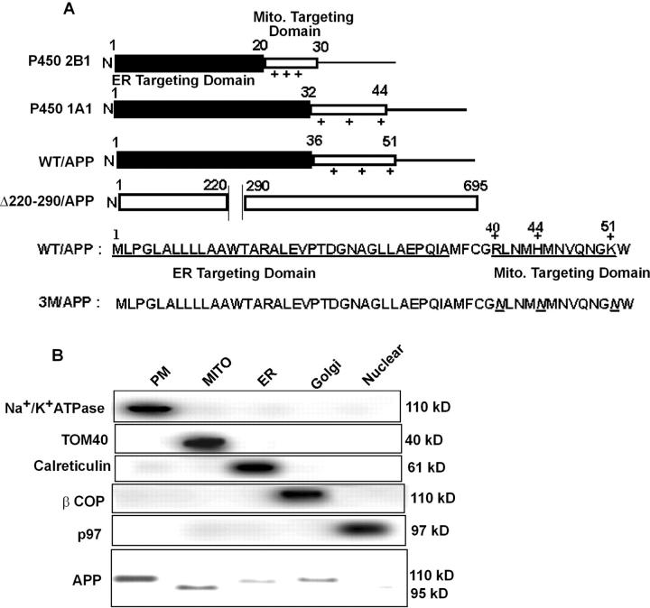
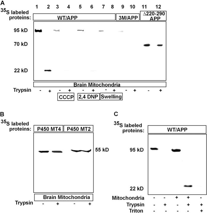
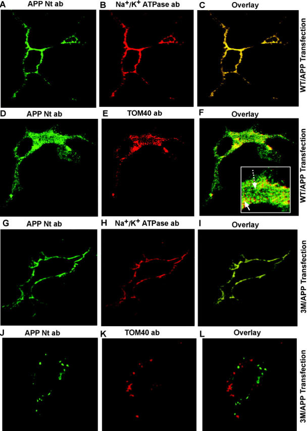
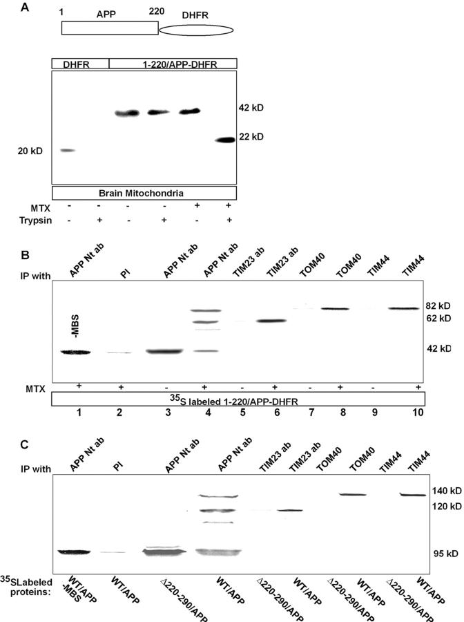
Alzheimer's amyloid precursor protein 695 (APP) is a plasma membrane protein, which is known to be the source of the toxic amyloid beta (Abeta) peptide associated with the pathogenesis of Alzheimer's disease (AD). Here we demonstrate that by virtue of its chimeric NH2-terminal signal, APP is also targeted to mitochondria of cortical neuronal cells and select regions of the brain of a transgenic mouse model for AD. The positively charged residues at 40, 44, and 51 of APP are critical components of the mitochondrial-targeting signal. Chemical cross-linking together with immunoelectron microscopy show that the mitochondrial APP exists in NH2-terminal inside transmembrane orientation and in contact with mitochondrial translocase proteins. Mutational studies show that the acidic domain, which spans sequence 220-290 of APP, causes the transmembrane arrest with the COOH-terminal 73-kD portion of the protein facing the cytoplasmic side. Accumulation of full-length APP in the mitochondrial compartment in a transmembrane-arrested form, but not lacking the acidic domain, caused mitochondrial dysfunction and impaired energy metabolism. These results show, for the first time, that APP is targeted to neuronal mitochondria under some physiological and pathological conditions.
Overview
The claims and statistics of this paper haven’t been checked yet.
Run the check — free, no account needed. It reads the open-access text and checks the sources it cites.
Integrity signals
Registry
Cites
Statistics
Figures
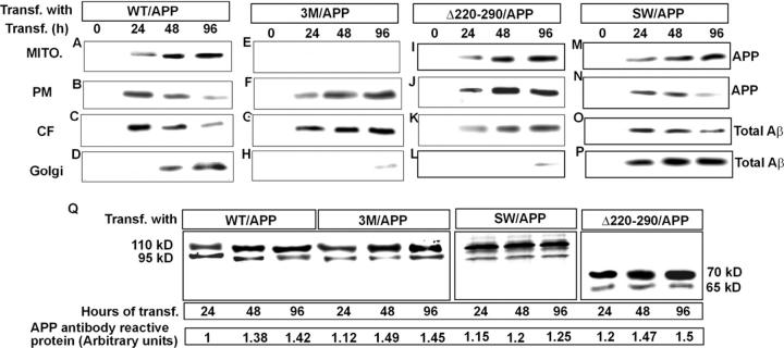
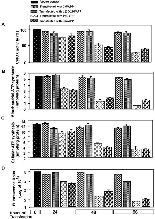
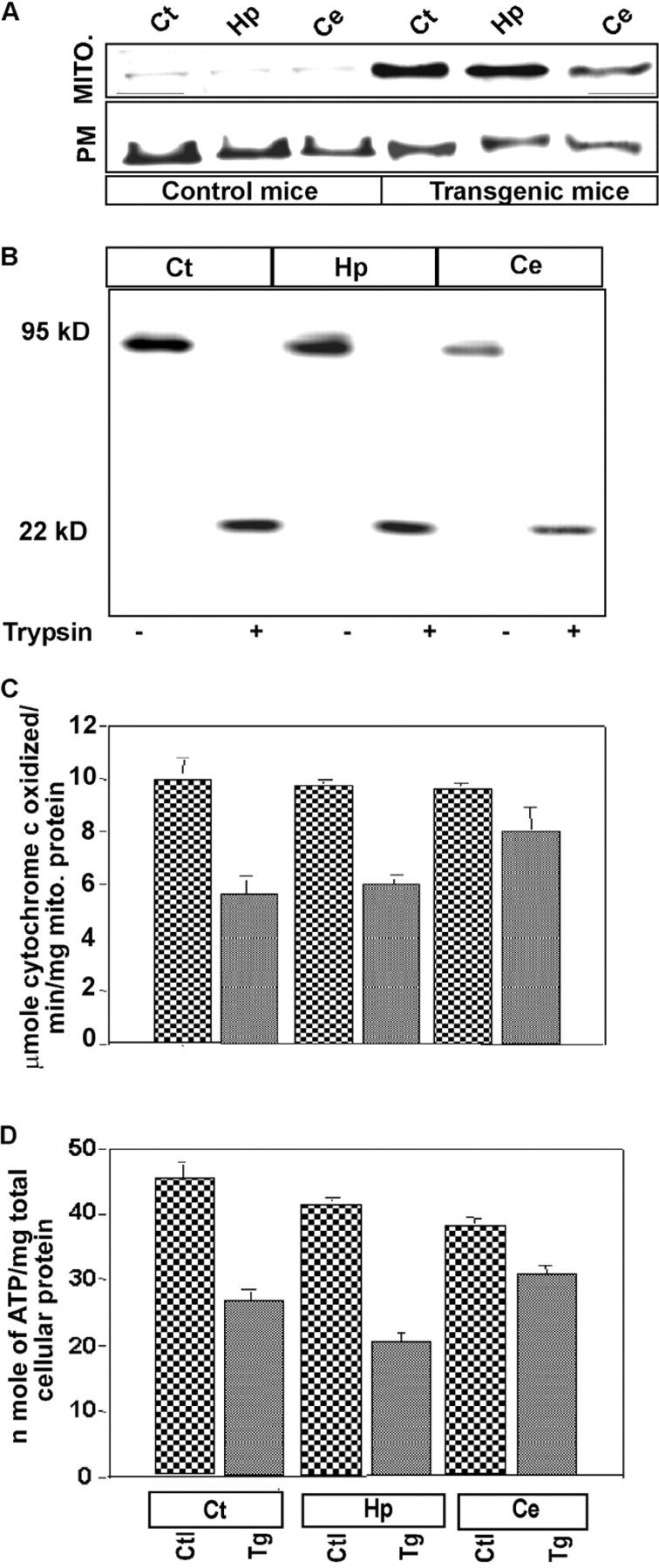
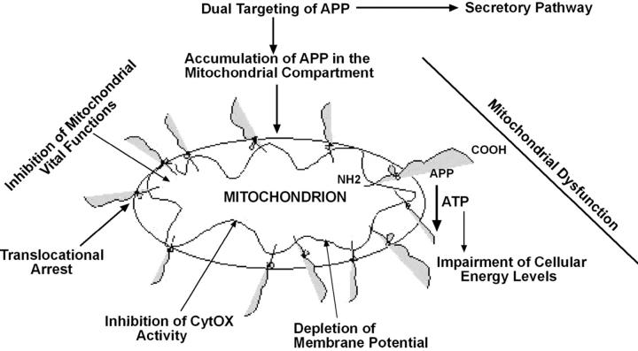
Verification
Claims the paper makes
The claims and statistics of this paper haven’t been checked yet.
Run the check — free, no account needed. It reads the open-access text and checks the sources it cites.
Claims other papers make about this one
Each is checked against this paper’s own text: does it say what the citing paper says it does?
The cited source says what the claim attributes to it.
In the citing paper
“We generated a mutant APP that does not localize to the mitochondria by deleting the mitochondrial targeting domain (residues 40–51) (42). We then compared the protection against mitophagy in Jurkat cells expressing either WT-APP or the mutant (Mut)-APP, which showed decreased localization to the mitochondria of the Jurkat cells than the WT-APP (Figure 3G, H).”
From Alzheimer's Disease–Associated Amyloid-β Precursor Protein Prevents Aging Stress–Induced Mitophagy and Fumarate Depletion to Improve Antitumor Immunity (2025) · cites this paper as [42]
In the citing paper
“accumulating data certainly do suggest APP and Aβ, as well as the entire γ-secretase complex, physically associate with mitochondria and can effect changes in mitochondrial function [58-67].”
From The Alzheimer’s disease mitochondrial cascade hypothesis: progress and perspectives (2014) · cites this paper as [61]
Self-check
Percentages, sums and reported differences re-computed from the paper’s own numbers.
The self-check needs the paper’s full text.
Citations
Cites
Translating cell biology into therapeutic advances in Alzheimer's disease
Selkoe, D. J. (1999) · Nature · doi:10.1038/399a023 · Cited by 1,166
Mitochondrial Dysfunction in Neurodegenerative Diseases
Johri, A., & Beal, M. F. (2012) · The Journal of Pharmacology and Experimental Therapeutics · doi:10.1124/jpet.112.192138 · Cited by 806
Binding of a specific ligand inhibits import of a purified precursor protein into mitochondria
Eilers, M., & Schatz, G. (1986) · Nature · doi:10.1038/322228a0 · Cited by 725
Mitochondrial dysfunction in neurodegenerative diseases
Beal, M. F. (1998) · Biochimica et Biophysica Acta (BBA) - Bioenergetics · doi:10.1016/s0005-2728(98)00114-5 · Cited by 611
Cytochrome oxidase deficiency in Alzheimer’s disease
Parker, W. D., Filley, C. M., & Parks, J. K. (1990) · Neurology · doi:10.1212/wnl.40.8.1302 · Cited by 598 · cited 1×
“…$8.00 The Journal of Cell Biology, Volume 161, Number 1, April 14, 2003 41–54 http://www.jcb.org/cgi/doi/10.1083/jcb.200207030 JCB Article 41 Mitochondrial targeting and a novel transmembrane arrest of Alzheimer’s amyloid precursor protein impairs mitochondrial function in neuronal cells”
BackgroundInfluentialMUTANT GENES IN FAMILIAL ALZHEIMER'S DISEASE AND TRANSGENIC MODELS
Price, D. L., & Sisodia, S. S. (1998) · Annual Review of Neuroscience · doi:10.1146/annurev.neuro.21.1.479 · Cited by 581
γ-Secretase, notch, Aβ and alzheimer's disease: Where do the presenilins fit in?
Sisodia, S. S., & St George-Hyslop, P. H. (2002) · Nature Reviews Neuroscience · doi:10.1038/nrn785 · Cited by 512
Regulation of APP cleavage by α‐, β‐ and γ‐secretases
Nunan, J., & Small, D. H. (2000) · FEBS Letters · doi:10.1016/s0014-5793(00)02076-7 · Cited by 509
A selective defect of cytochrome c oxidase is present in brain of Alzheimer disease patients
Maurer, I. (2000) · Neurobiology of Aging · doi:10.1016/s0197-4580(00)00112-3 · Cited by 475
Selkoe (1999) · doi:10.1038/19866 · Cited by 468
Research works printed but not linked: 1
- 1996, Int. J. Dev. Neurosci., 14, 61
Cited by
Sorted by citations
Items 401–500 of 570
Ma, G., Gao, J., Fu, Q., Jiang, L., Wang, R., Zhang, Y. E., & Liu, K. (2009) · Neurochemical Research · doi:10.1007/s11064-009-0007-8 · Cited by 17
Chua, L. M., Lim, M., & Wong, B. (2013) · Biochemical and Biophysical Research Communications · doi:10.1016/j.bbrc.2013.07.022 · Cited by 17
The Role of Oxidative Stress in Trisomy 21 Phenotype
Buczyńska, A., Sidorkiewicz, I., Kretowski, A. J., & Zbucka-Krętowska, M. (2023) · Cellular and Molecular Neurobiology · doi:10.1007/s10571-023-01417-6 · Cited by 21
Andrew, R. J., Fisher, K., Heesom, K. J., Kellett, K. A. B., & Hooper, N. M. (2019) · Journal of Neurochemistry · doi:10.1111/jnc.14666 · Cited by 16
Pharmacologic enrichment of exosome yields and mitochondrial cargo
Wang, X., Berkowicz, A., King, K., Menta, B. W., Gabrielli, A. P., Novikova, L., Troutwine, B., Pleen, J., Wilkins, H., & Swerdlow, R. H. (2022) · Mitochondrion · doi:10.1016/j.mito.2022.04.001 · Cited by 16
Fattoretti, P., Balietti, M., Casoli, T., Giorgetti, B., Stefano, G. D., Bertoni–Freddari, C., Lattanzio, F., & Sensi, S. L. (2009) · Rejuvenation Research · doi:10.1089/rej.2009.0937 · Cited by 16
The mitochondrial function was impaired in APP knockout mouse embryo fibroblast cells
Sheng, B., Niu, Y., Zhou, H. R., Yan, J., Zhao, N., Zhang, X., & Gong, Y. (2009) · 中国科学通报:英文版 · doi:10.1007/s11434-009-0239-7 · Cited by 15
Axonal transport and neurodegenerative disease: vesicle-motor complex formation and their regulation
Gunawardena, S., Anderson, E. N., & White, J. (2014) · Degenerative Neurological and Neuromuscular Disease · doi:10.2147/dnnd.s57502 · Cited by 16
Lautenschläger, J., & Schierle, G. S. K. (2019) · Progress in Neurobiology · doi:10.1016/j.pneurobio.2019.101660 · Cited by 16
Altering Mitochondrial Dysfunction as an Approach to Treating Alzheimer’s Disease
Colca, J. R., & Feinstein, D. L. (2012) · Advances in pharmacology · doi:10.1016/b978-0-12-394816-8.00005-2 · Cited by 14
Effects of Aβ1–42 on the Subunits of KATP Expression in Cultured Primary Rat Basal Forebrain Neurons
Ma, G., Fu, Q., Zhang, Y. E., Gao, J., Jiang, J., Bi, A.-L., Liu, K., Du, Y., Chen, C., Cui, Y., et al. (2008) · Neurochemical Research · doi:10.1007/s11064-008-9603-2 · Cited by 14
Volloch, V., Olsen, B. R., & Rits‐Volloch, S. (2019) · Annals of Integrative Molecular Medicine · doi:10.33597/aimm.01-1005 · Cited by 15
Volloch, V., & Rits‐Volloch, S. (2022) · Journal of Alzheimer s Disease Reports · doi:10.3233/adr-220031 · Cited by 15
Duce, J. A., Ayton, S., Miller, A. A., Tsatsanis, A., Lam, L. Q., Leone, L., Corbin, J. E., Butzkueven, H., Kilpatrick, T. J., Rogers, J. T., et al. (2012) · Molecular Psychiatry · doi:10.1038/mp.2011.168 · Cited by 14
Pohland, M., Hagl, S., Pellowska, M., Wurglics, M., Schubert‐Zsilavecz, M., & Eckert, G. P. (2015) · Neurochemical Research · doi:10.1007/s11064-015-1765-0 · Cited by 14
Eysert, F., Kinoshita, P. F., Lagarde, J., Lacas‐Gervais, S., Xicota, L., Dorothée, G., Bottlaender, M., Checler, F., Potier, M., Sarazin, M., et al. (2024) · Acta Neuropathologica Communications · doi:10.1186/s40478-024-01807-x · Cited by 14
The reciprocal relationship between amyloid precursor protein and mitochondrial function
Strope, T. A., & Wilkins, H. (2024) · Journal of Neurochemistry · doi:10.1111/jnc.16183 · Cited by 15
Mairuae, N., Hall, E. C., II, Cheepsunthorn, P., Lee, S. Y., & Connor, J. R. (2010) · Journal of Neuroscience Research · doi:10.1002/jnr.22466 · Cited by 13
Volloch, V., & Rits‐Volloch, S. (2023) · International Journal of Molecular Sciences · doi:10.3390/ijms242417586 · Cited by 13
Pavlov, P. F., Hutter‐Paier, B., Havas, D., Windisch, M., & Winblad, B. G. (2018) · Journal of Cellular and Molecular Medicine · doi:10.1111/jcmm.13624 · Cited by 11
Sandberg, A. A., Manning, E., Wilkins, H., Mazzarino, R. C., Minckley, T. F., Swerdlow, R. H., Patterson, D., Qin, Y., & Linseman, D. A. (2022) · Journal of Alzheimer s Disease · doi:10.3233/jad-215108 · Cited by 12
Reed, A. L., Mitchell, W., Alexandrescu, A. T., & Alder, N. N. (2023) · Frontiers in Physiology · doi:10.3389/fphys.2023.1263420 · Cited by 13
Valla, J., Schneider, L. E., Small, A. M., & Gonzalez-Lima, F. (2007) · Journal of Histotechnology · doi:10.1179/his.2007.30.4.235 · Cited by 11
Wang, Z., Sun, Y., Bai, Z., Li, M., Kong, D., & Wu, G. (2025) · Movement Disorders · doi:10.1002/mds.30123 · Cited by 15
The Importance of Being Connected
Lutz, M. W., Crenshaw, D. G., Saunders, A. M., & Roses, A. D. (2011) · Journal of Alzheimer s Disease · doi:10.3233/jad-2010-101765 · Cited by 11
Contribution of in vitro neurotoxicology studies to the elucidation of neurodegenerative processes
Zurich, M., & Monnet‐Tschudi, F. (2009) · Brain Research Bulletin · doi:10.1016/j.brainresbull.2009.06.008 · Cited by 11
Zhao, F., Qiao, P., Yan, N., Gao, D., Liu, M., & Yan, Y. P. (2015) · Neurochemical Research · doi:10.1007/s11064-015-1807-7 · Cited by 11
Kassir, M. F., Lee, H. G., Oleinik, N. V., Wofford, W., Walton, C., Atilgan, F. C., Janneh, A. H., Chakraborty, P., Calisir, K., Percin, E., et al. (2025) · Cancer Research · doi:10.1158/0008-5472.can-24-4740 · Cited by 10
Volloch, V., & Rits‐Volloch, S. (2023) · Journal of Alzheimer s Disease Reports · doi:10.3233/adr-230037 · Cited by 11
FLZ inhibited γ-secretase selectively and decreased Aβ mitochondrial production in APP-SH-SY5Y cells
Ye, X., Tai, W., Bao, X., Chen, X., & Zhang, D. (2013) · Naunyn-Schmiedeberg s Archives of Pharmacology · doi:10.1007/s00210-013-0918-4 · Cited by 10
Autophagy: ‘Self-Eating’ Your Way to Longevity
Pattison, C. J., & Korolchuk, V. I. (2018) · Sub-cellular biochemistry/Subcellular biochemistry · doi:10.1007/978-981-13-2835-0_2 · Cited by 10
Kakar, M., Cadwallader, A. B., Davis, J. R., & Lim, C. S. (2007) · Pharmaceutical Research · doi:10.1007/s11095-007-9333-1 · Cited by 10
Temporal alterations of the nascent proteome in response to mitochondrial stress
Stępkowski, T. M., Linke, V., Stadnik, D., Zakrzewski, M., Zawada, A. E., Serwa, R. A., & Chacińska, A. (2024) · Cell Reports · doi:10.1016/j.celrep.2024.114803 · Cited by 8
Stavros, J. (2013) · InTech eBooks · doi:10.5772/56079 · Cited by 9
Volloch, V., & Rits‐Volloch, S. (2019) · Annals of Integrative Molecular Medicine · doi:10.33597/aimm.02-1009 · Cited by 10
Advances in our Understanding of Neurodegeneration
Jellinger, K. A. (2007) · Elsevier eBooks · doi:10.1016/b978-044452809-4/50142-3 · Cited by 9
Mitochondrial Function in Alzheimer’s Disease: Focus on Astrocytes
Lampinen, R., Belaya, I., Boccuni, I., Malm, T., & Kanninen, K. M. (2018) · Astrocyte - Physiology and Pathology · doi:10.5772/intechopen.71825 · Cited by 8
Li, Y., Liu, D., Zhang, X., Rimal, S., Lu, B., & Li, S. (2024) · Journal of Biological Chemistry · doi:10.1016/j.jbc.2024.105719 · Cited by 9
On the Inadequacy of the Current Transgenic Animal Models of Alzheimer’s Disease: The Path Forward
Volloch, V., & Rits‐Volloch, S. (2024) · International Journal of Molecular Sciences · doi:10.3390/ijms25052981 · Cited by 9
Beresford‐Webb, J., McAllister, C. J., Sleigh, A., Walpert, M., Holland, A. J., & Zaman, S. H. (2025) · Brain Sciences · doi:10.3390/brainsci15020130 · Cited by 10
Sinha, M. S., Behera, P., Bhowmick, P., Banerjee, K., Basu, S., & Chakrabarti, S. (2011) · Journal of Alzheimer s Disease · doi:10.3233/jad-2011-110686 · Cited by 8
The Role of Aβ and Tau Oligomers in the Pathogenesis of Alzheimer’s Disease
Bhaskar, K., & Lamb, B. T. (2011) · Springer eBooks · doi:10.1007/978-94-007-2774-8_5 · Cited by 8
Yamagishi, S., Iga, Y., Ikegaya, S., Kakiuchi, T., Ohba, H., Nishiyama, S., Fukomoto, D., Kanazawa, M., Harada, N., Tsukada, H., et al. (2021) · Journal of Neuroinflammation · doi:10.1186/s12974-021-02343-4 · Cited by 7
Neurodegeneration, Mitochondria, and Antibiotics
Suarez-Rivero, J. M., López‐Pérez, J., Muela‐Zarzuela, I., Pastor‐Maldonado, C. J., Cilleros-Holgado, P., Gómez-Fernández, D., Álvarez-Córdoba, M., Munuera-Cabeza, M., Talaverón-Rey, M., Povea-Cabello, S., et al. (2023) · Metabolites · doi:10.3390/metabo13030416 · Cited by 9
Lacham‐Hartman, S., Moshe, R., Ben-Zichri, S., Shmidov, Y., Bitton, R., Jelinek, R., & Papo, N. (2023) · ACS Chemical Neuroscience · doi:10.1021/acschemneuro.3c00208 · Cited by 8
Understanding the Twisted Structure of Amyloid Fibrils via Molecular Simulations
Lu, L., Deng, Y., Li, X., Li, H., & Karniadakis, G. E. (2018) · The Journal of Physical Chemistry B · doi:10.1021/acs.jpcb.8b07255 · Cited by 8
Mitochondria and Alzheimer’s Disease: An Electron Microscopy Study
Balogiannis, S. (2019) · IntechOpen eBooks · doi:10.5772/intechopen.84881 · Cited by 9
Seppet, E. K., Gizatullina, Z. Z., Trumbeckaitė, S., Zierz, S., Striggow, F., & Gellerich, F. N. (2007) · Wiley eBooks · doi:10.1002/9783527621095.ch15 · Cited by 8
Evidence for early identification of Alzheimer's diseasePreprint
Alexiou, A. T., & Vlamos, P. (2012) · arXiv (Cornell University) · doi:10.48550/arxiv.1209.4223 · Cited by 9
Protein quality control systems in neurodegeneration – culprits, mitigators, and solutions?
Ciechanover, A. J., & Livneh, I. (2025) · Frontiers in Neurology · doi:10.3389/fneur.2025.1604076 · Cited by 8
Franco, R., & Serrano‐Marín, J. (2022) · Biomedicines · doi:10.3390/biomedicines10051199 · Cited by 7
Conserved quality control mechanisms of mitochondrial protein import
Borgert, L., Becker, T., & den Brave, F. (2024) · Journal of Inherited Metabolic Disease · doi:10.1002/jimd.12756 · Cited by 9
Advances in Research on Down Syndrome
Dey, S. (2018) · InTech eBooks · doi:10.5772/intechopen.68260 · Cited by 7
Wallace, D. C., Lott, M. T., & Procaccio, V. (2013) · Elsevier eBooks · doi:10.1016/b978-0-12-383834-6.00013-6 · Cited by 8
Mitochondria in Neurodegeneration
Swerdlow, R. H. (2011) · Advances in neurobiology · doi:10.1007/978-1-4614-1788-0_30 · Cited by 7
Howell, N., Dykens, J. A., & Moos, W. H. (2005) · Drug Development Research · doi:10.1002/ddr.20046 · Cited by 7
Molecular genetics of early-onset Alzheimer's disease and frontotemporal lobar degeneration
Krüger, J. (2010) · Cited by 7
Targeting Calpain-2 for Alzheimer’s Disease Treatment
Arnsten, A. F. T., & Baudry, M. (2023) · Medical Research Archives · doi:10.18103/mra.v11i2.3487 · Cited by 6
Liu, J., Sun, S., & Chen, Y. (2024) · Journal of Alzheimer s Disease · doi:10.3233/jad-231278 · Cited by 6
Volloch, V., & Rits‐Volloch, S. (2019) · Annals of Integrative Molecular Medicine · doi:10.33597/aimm.-v2-id1010 · Cited by 6
Structural biology of cell surface receptors implicated in Alzheimer’s disease
Hermans, S. J., Nero, T. L., Morton, C. J., Gooi, J. H., Crespi, G. A. N., Hancock, N. C., Gao, C., Ishii, K., Markulić, J., & Parker, M. W. (2021) · Biophysical Reviews · doi:10.1007/s12551-021-00903-9 · Cited by 6
Shabrokh, E., Kavanaugh, J., McMillan, R. P., Pittman, J., Hulver, M. W., & Frisard, M. I. (2014) · Open Journal of Molecular and Integrative Physiology · doi:10.4236/ojmip.2014.42002 · Cited by 6
Asseburg, H., Hagl, S., & Eckert, G. P. (2014) · AAPS advances in the pharmaceutical sciences series · doi:10.1007/978-3-319-06151-1_23 · Cited by 6
Protein Misfolding and Aggregation: Implications for Mitochondrial Dysfunction and Neurodegeneration
Ludtmann, M. H. R., & Abramov, A. Y. (2016) · Springer eBooks · doi:10.1007/978-3-319-28637-2_10 · Cited by 6
Advances in Alzheimer’s disease: mechanistic insights and therapeutic targets
Jia, Y.-J., Ge, Y., Li, B., Yang, Y., Chen, H., Liu, J., Guo, J., Yu, J., Ye, K., Wang, J., et al. (2026) · Science China Life Sciences · doi:10.1007/s11427-025-2991-7 · Cited by 6
Volloch, V., & Rits‐Volloch, S. (2024) · International Journal of Molecular Sciences · doi:10.3390/ijms25116036 · Cited by 6
Bioinformatics insights into mitochondrial and immune gene regulation in Alzheimer's disease
Tian, M., Zhang, Y., Yuan, Y., Li, H., & He, Y. (2025) · European journal of medical research · doi:10.1186/s40001-025-02297-w · Cited by 7
Cardoso, R., Lemos, C., Oliveiros, B., Almeida, M. R., Baldeiras, I., Pereira, C., SANTOS, A. C. D. S., Duro, D., Vieira, D., Santana, I. J., et al. (2020) · Journal of Alzheimer s Disease · doi:10.3233/jad-200556 · Cited by 6
Anandatheerthavarada, H. K., Sepuri, N. B. V., Biswas, G., & Avadhani, N. G. (2008) · Journal of Biological Chemistry · doi:10.1074/jbc.m801464200 · Cited by 5
RetractedThe Ageing Brain, Mitochondria and Neurodegeneration
Hudson, G. (2016) · Springer eBooks · doi:10.1007/978-3-319-28637-2_3 · Cited by 5
Pinto, M., Díaz, F., Nissanka, N., Guastucci, C. S., Illiano, P., Brambilla, R., & Moraes, C. T. (2022) · Molecular Neurobiology · doi:10.1007/s12035-022-02992-3 · Cited by 5
What has Electron Microscopy Contributed to Alzheimer's Research?
Balogiannis, S. (2015) · Future Neurology · doi:10.2217/fnl.15.44 · Cited by 5
Mhatre, M., & Hensley, K. (2007) · Elsevier eBooks · doi:10.1016/b978-044452809-4/50155-1 · Cited by 4
Genetics of Alzheimer's Disease
Bekris, L. M., Yu, C., Bird, T. D., & Tsuang, D. W. (2010) · Wiley eBooks · doi:10.1002/9780470669600.ch43 · Cited by 6
Huo, Q., Tabassum, S., Chen, M., Sun, M., Deng, Y., Zheng, X., Li, Y., Chen, J., Long, C., & Yang, L. (2021) · Journal of Alzheimer s Disease · doi:10.3233/jad-201557 · Cited by 4
Sharma, Y., Gupta, J. K., Babu, M. A., Singh, S., & Sindhu, R. K. (2024) · Journal of Molecular Neuroscience · doi:10.1007/s12031-024-02269-5 · Cited by 6
Lacham‐Hartman, S., Oren, O., Rabinovitch, M., & Papo, N. (2025) · International Journal of Biological Macromolecules · doi:10.1016/j.ijbiomac.2025.146066 · Cited by 4
Jekabsons, M. B. (2009) · International Journal of Molecular Sciences · doi:10.3390/ijms10020702 · Cited by 4
Mecanismos neurobiológicos de la enfermedad de Alzheimer
Rommy von Bernhardi, M. (2005) · Revista chilena de neuro-psiquiatría · doi:10.4067/s0717-92272005000200005 · Cited by 4
Mitochondria: Strategic Point in the Field of Alzheimer’s Disease
Balogiannis, S. (2016) · Alzheimer’s & Neurodegenerative Diseases · doi:10.24966/and-9608/100004 · Cited by 3
Mitochondrial Abnormalities in Down Syndrome: Pathogenesis, Effects and Therapeutic Approaches
Izzo, A., Mollo, N., Cicatiello, R., Genesio, R., Paladino, S., Conti, A. M. F., & Nitsch, L. (2018) · InTech eBooks · doi:10.5772/intechopen.71059 · Cited by 3
Clogging the mitochondrial protein-entry gate promotes obesity
Song, J., & Becker, T. (2019) · Nature Metabolism · doi:10.1038/s42255-019-0155-3 · Cited by 3
Oxidative stress in neurodegeneration: targeting mitochondria as a therapeutic aid
Gaggelli, E., & Valensin, G. (2012) · Springer eBooks · doi:10.1007/978-3-7091-1001-0_12 · Cited by 3
Campos, J. (2013) · Journal of Antivirals & Antiretrovirals · doi:10.4172/jaa.s10-003 · Cited by 3
Beckert, B., Acker‐Palmer, A., & Volknandt, W. (2018) · Biological Chemistry · doi:10.1515/hsz-2017-0238 · Cited by 3
Krakowczyk, M., Lenkiewicz, A. M., Sitarz, T., Mussulini, B. H. M., Linke, V., Malińska, D., Szczepankiewicz, A. A., Wydrych, A., Nieznańska, H., Serwa, R. A., et al. (2023) · bioRxiv (Cold Spring Harbor Laboratory) · doi:10.1101/2023.06.08.543713 · Cited by 3
Mitochondrial Function and Neurodegenerative Diseases
Wilkins, H. M., Weidling, I., Koppel, S., Wang, X., von Schulze, A., & Swerdlow, R. H. (2018) · The Molecular and Cellular Basis of Neurodegenerative Diseases · doi:10.1016/b978-0-12-811304-2.00013-4 · Cited by 2
Amyloid-β and Mitochondrial Membranes: A Missing Link in Alzheimer’s Pathogenesis
Houfkova, A., & Schmidt, M. (2026) · Molecular Neurobiology · doi:10.1007/s12035-026-05786-z · Cited by 2
Chang, Y. B., & Baleja, J. (2026) · International Journal of Molecular Sciences · doi:10.3390/ijms27114932 · Cited by 2
Volloch, V., & Rits‐Volloch, S. (2024) · International Journal of Molecular Sciences · doi:10.3390/ijms25189913 · Cited by 3
Volloch, V., & Rits‐Volloch, S. (2025) · Genes · doi:10.3390/genes16010046 · Cited by 3
Effect of stress on neuronal cell: Morphological to molecular approach
Bhardwaj, I., Singh, S., Ansari, A. H., Rai, S. P., & Singh, D. (2025) · Progress in brain research · doi:10.1016/bs.pbr.2025.01.010 · Cited by 3
Drug Effects on Learning and Memory
Vogel, H. G. (2007) · Springer eBooks · doi:10.1007/978-3-540-70995-4_7 · Cited by 2
Targeted Treatment Strategies for Mitochondria Dysfunction: Correlation with Neurological Disorders
Sharma, R., Malviya, R., Srivastava, S., Ahmad, I., Rab, S. O., & Uniyal, P. (2024) · Current Drug Targets · doi:10.2174/0113894501303824240604103732 · Cited by 3
Mitochondria in Alzheimer disease : regulatory mechanisms and cell death
Hedskog, L. (2012) · Figshare · Cited by 2
Amyloid, Protein Misfolding Defects, and Inclusions
Cheville, N. F. (2009) · Wiley eBooks · doi:10.1002/9780813810379.ch26 · Cited by 2
Organelle targeting: Focus on the mitochondria
Padh, H., Shenoy, S., Thakkar, S., & Misra, M. (2021) · Elsevier eBooks · doi:10.1016/b978-0-323-85666-9.00021-8 · Cited by 1
Volloch, V., & Rits‐Volloch, S. (2025) · International Journal of Molecular Sciences · doi:10.3390/ijms26094252 · Cited by 3
Strope, T. A., Troutwine, B., Hauger, B. M., Smith, K. P., Swerdlow, R. H., & Wilkins, H. (2025) · Journal of Alzheimer s Disease · doi:10.1177/13872877251360243 · Cited by 2
Chakrabarti, S., & Sinha, M. S. (2012) · Springer eBooks · doi:10.1007/978-94-007-5237-5_4 · Cited by 1
Metadata
- Authors
- Anandatheerthavarada, H. K., Biswas, G., Robin, M.-A., & Avadhani, N. G.
- Affiliations
- University of Pennsylvania (US)
- ORCID
- Avadhani, Narayan G.
- Venue
- The Journal of Cell Biology
- Publisher
- Rockefeller University Press
- Published
- 2003-04-14
- Volume
- 161
- Issue
- 1
- Pages
- 41-54
- ISSN
- 0021-9525 (ISSN-L)
- 1540-8140
- PMC
- PMC2172865
- OpenAlex
- W2132988134
- Cited by
- 571
- References
- 47
- Citations by year
- 2026: 11 · 2025: 12 · 2024: 18 · 2023: 22 · 2022: 16 · 2021: 24 · 2020: 23 · 2019: 18 · 2018: 35 · 2017: 14 · 2016: 27 · 2015: 24 · 2014: 17 · 2013: 24 · 2012: 31
- FWCI
- 13.60
- Type
- journal article
- Language
- en
- Open access
- green
- www.ncbi.nlm.nih.gov
- Licence
- cc-by-nc-sa
- Locations
- The Journal of Cell Biology (publishedVersion)
- PubMed Central (publishedVersion, cc-by-nc-sa)
- PubMed (publishedVersion)
- Funders
- National Institutes of Health (1r01gm034483-01a3)
- National Institute of General Medical Sciences (GM34483)
- Topics
- Life Sciences › Neuroscience › Cellular and Molecular Neuroscience › Alzheimer's disease research and treatments
- Life Sciences › Biochemistry, Genetics and Molecular Biology › Molecular Biology › Mitochondrial Function and Pathology
- Life Sciences › Biochemistry, Genetics and Molecular Biology › Cell Biology › Autophagy in Disease and Therapy
- Keywords
- amyloid precursor protein, APP695, mitochondrial targeting, mitochondrial dysfunction, energy metabolism, Alzheimer's disease, amyloid-beta, mitochondrial translocase, acidic domain, cortical neurons, transgenic mouse model, crosslinking, immunoelectron microscopy
- MeSH terms
- Alzheimer Disease, Amino Acid Sequence, Animals, Brain, Cell Compartmentation, Cells, Cultured, Disease Models, Animal, Energy Metabolism, Humans, Immunohistochemistry, Intracellular Membranes, Mice, Transgenic, Microscopy, Electron, Mitochondria, Molecular Conformation, Neurons, Recombinant Fusion Proteins, Amyloid beta-Protein Precursor, Protein Structure, Tertiary, Protein Transport, Cytochrome-c Oxidase Deficiency, Mice
About these checks
Registry, retraction, Cites and statistics results are computed, not judged, with one exception: a printed reference that no index lists is read once by a model before it counts as a citation error. Claims are extracted by a model and kept only when their quote appears verbatim in the text; each cited source is then checked, with a model’s help, for whether it says what the claim relies on. Those verdicts can be wrong — read the claim’s quote and the cited source before relying on one.
Statistics and arithmetic are recomputed, not judged.
Confidence bands are shown only for fields calibrated on our annotated test set. None is calibrated yet, so no band is shown.
What counts as a claim ›How we measure confidence ›How we check this ›
Are you an author, or do you see a problem with this page? Request a review
