

The DOI matches its Crossref record; 58 of the 58 references it cites were found in a registry or an index.
Myotubes differentiate optimally on substrates with tissue-like stiffness
Open accessAbstract
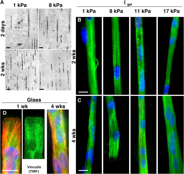
Contractile myocytes provide a test of the hypothesis that cells sense their mechanical as well as molecular microenvironment, altering expression, organization, and/or morphology accordingly. Here, myoblasts were cultured on collagen strips attached to glass or polymer gels of varied elasticity. Subsequent fusion into myotubes occurs independent of substrate flexibility. However, myosin/actin striations emerge later only on gels with stiffness typical of normal muscle (passive Young's modulus, E ∼12 kPa). On glass and much softer or stiffer gels, including gels emulating stiff dystrophic muscle, cells do not striate. In addition, myotubes grown on top of a compliant bottom layer of glass-attached myotubes (but not softer fibroblasts) will striate, whereas the bottom cells will only assemble stress fibers and vinculin-rich adhesions. Unlike sarcomere formation, adhesion strength increases monotonically versus substrate stiffness with strongest adhesion on glass. These findings have major implications for in vivo introduction of stem cells into diseased or damaged striated muscle of altered mechanical composition.
Overview
The claims and statistics of this paper haven’t been checked yet.
Free: it reads the open-access text and checks the first 5 claims that cite a source we can check. Create a free account to check every claim.
Integrity signals
Registry
Cites
Statistics
Figures




Verification
Claims the paper makes
The claims and statistics of this paper haven’t been checked yet.
Free: it reads the open-access text and checks the first 5 claims that cite a source we can check. Create a free account to check every claim.
Claims other papers make about this one
Each is checked against this paper’s own text: does it say what the citing paper says it does?
The source backs part of the claim, not all of it — or backs it for a different population, condition or comparison.
In the citing paper
“Additionally, the stiffness of the cellular environment or niche is an additional mechanical signal that influences for example the differentiation of mesenchymal stem cells into muscle and other cell types (34, 35).”
From The Hippo signal transduction network for exercise physiologists (2016) · cites this paper as [34]
Experimental — accuracy not yet measuredThe cited source says what the claim attributes to it.
In the citing paper
“In studies using biomimetic niches, substrates with an elastic stiffness of young adult skeletal muscle (∼12 kPa Young's modulus, the characteristic elastic rigidity of a material), which is five orders of magnitude softer than traditonal tissue culture plastic), promote MuSC self-renewal29,31 and myoblast differentiation94,95 (Fig. 4).”
From The central role of muscle stem cells in regenerative failure with aging (2015) · cites this paper as [94]
Self-check
Percentages, sums and reported differences re-computed from the paper’s own numbers.
The self-check needs the paper’s full text.
Citations
Cites
Calcium's Role in Mechanotransduction during Muscle Development
Benavides Damm, T., & Egli, M. (2014) · Cellular Physiology and Biochemistry · doi:10.1159/000356667 · Cited by 14,502
Biomechanics. Mechanical Properties of Living Tissues
Fung, Y. C., & Skalak, R. (1982) · Journal of Applied Mechanics · doi:10.1115/1.3162171 · Cited by 6,025
Cell Movement Is Guided by the Rigidity of the Substrate
Lo, C.-M., Wang, H.-B., Dembo, M., & Wang, Y.-L. (2000) · Biophysical Journal · doi:10.1016/s0006-3495(00)76279-5 · Cited by 3,289 · cited 3×
“FAK regulation could also occur by calciumdependent mechanisms (Lo et al., 2000) or stretch-activation (Nakamura et al.”
BackgroundCell locomotion and focal adhesions are regulated by substrate flexibility
Pelham, R. J., & Wang, Y.-L. (1997) · Proceedings of the National Academy of Sciences · doi:10.1073/pnas.94.25.13661 · Cited by 3,027 · cited 7×
“The reason is unclear, but physical aspects of the microenvironment, especially mechanical compliance of matrix (Pelham and Wang, 1997) and perhaps adjacent cells, are becoming increasingly linked to gene expression and protein organization.”
BackgroundInfluentialTaking Cell-Matrix Adhesions to the Third Dimension
Cukierman, E., Pankov, R., Stevens, D. R., & Yamada, K. M. (2001) · Science · doi:10.1126/science.1064829 · Cited by 3,027 · cited 1×
“…adhesion (Choquest et al., 1997; Beningo et al., 2001), posttranslational modification (Pelham and Wang, 1997; Beningo et al., 2001), protein expression (Cukierman et al., 2001), cytoskeletal organization (Cukierman et al., 2001; Engler et al., 2004a), and even cell viability (Wang et al., 2000).”
BackgroundCells lying on a bed of microneedles: An approach to isolate mechanical force
Tan, J. L., Tien, J., Pirone, D. M., Gray, D. S., Bhadriraju, K., & Chen, C. S. (2003) · Proceedings of the National Academy of Sciences · doi:10.1073/pnas.0235407100 · Cited by 1,962 · cited 2×
“The mechanism of coalignment is understood, from theory (Bischofs and Schwartz, 2003), to involve a propagation of cell-induced substrate contractions (Pelham and Wang, 1997; Wang et al., 2001; Tan et al., 2003 ).”
BackgroundSilicone Rubber Substrata: A New Wrinkle in the Study of Cell Locomotion
Harris, A. K., Wild, P., & Stopak, D. (1980) · Science · doi:10.1126/science.6987736 · Cited by 1,348 · cited 1×
“Analogous to the wrinkling of films by cells (Harris et al., 1980; Oliver et al., 1995), cells generate traction forces on collagen-coated polyacrylamide (PA) gel substrates.”
BackgroundExtracellular Matrix Rigidity Causes Strengthening of Integrin–Cytoskeleton Linkages
Choquet, D., Felsenfeld, D. P., & Sheetz, M. P. (1997) · Cell · doi:10.1016/s0092-8674(00)81856-5 · Cited by 1,245 · cited 1×
“Such signals feed back into cell tension (Wang et al., 2002), cell adhesion ( Choquest et al., 1997; Beningo et al., 2001), posttranslational modification (Pelham and Wang, 1997; Beningo et al., 2001), protein expression (Cukierman et al., 2001), cytoskeletal organization (Cukierman et al., 2001; Engler et al., 2004a)…”
BackgroundDetermination of Elastic Moduli of Thin Layers of Soft Material Using the Atomic Force Microscope
Dimitriadis, E. K., Horkay, F., Maresca, J., Kachar, B., & Chadwick, R. S. (2002) · Biophysical Journal · doi:10.1016/s0006-3495(02)75620-8 · Cited by 1,230 · cited 2×
“Mechanics of gels The elastic modulus of various gels was determined by AFM as well as macroscopic methods (Dimitriadis et al., 2002; Engler et al., 2004a).”
MethodSubstrate Compliance versus Ligand Density in Cell on Gel Responses
Engler, A., Bacakova, L., Newman, C., Hategan, A., Griffin, M., & Discher, D. (2004) · Biophysical Journal · doi:10.1016/s0006-3495(04)74140-5 · Cited by 1,141 · cited 16×
“In the absence of stiff ECM resistance, however, assembly and phosphorylation of adhesion proteins are inhibited (Pelham and Wang, 1997), and thus F-actin assembly is reduced (Engler et al., 2004a).”
BackgroundMethodInfluentialSelf-citation
Research works printed but not linked: 1
- 2004, Surf. Sci., In press
Cited by
Matrix elasticity directs stem cell lineage specification
Engler, A. J., Sen, S., Sweeney, H. L., & Discher, D. E. (2006) · Cell · doi:10.1016/j.cell.2006.06.044 · Cited by 13,915
“Such a wide variation in matrix stiffness for differentiated cells is known to influence focal-adhesion structure and the cytoskeleton (Bershadsky et al., 2003; Cukierman et al., 2001; Discher et al., 2005; Engler et al., 2004a; Lo et al., 2000; Pelham and Wang, 1997).”
BackgroundMethodResultInfluentialSelf-citationTissue Cells Feel and Respond to the Stiffness of Their Substrate
Discher, D. E., Janmey, P., & Wang, Y.-L. (2005) · Science · doi:10.1126/science.1116995 · Cited by 6,382
Self-citationTensional homeostasis and the malignant phenotype
Paszek, M. J., Zahir, N., Johnson, K. R., Lakins, J. N., Rozenberg, G. I., Gefen, A., Reinhart-King, C. A., Margulies, S. S., Dembo, M., Boettiger, D., et al. (2005) · Cancer Cell · doi:10.1016/j.ccr.2005.08.010 · Cited by 3,941
“*p % 0.05; **p % 0.01; ***p % 0.001. with the role shown for matrix compliance in smooth muscle cell differentiation (Engler et al., 2004) and extend those findings to incorporate the effect of matrix stiffness on multicellular epithelial tissue morphogenesis.”
BackgroundInfluentialThe control of human mesenchymal cell differentiation using nanoscale symmetry and disorder
Dalby, M. J., Gadegaard, N., Tare, R., Andar, A., Riehle, M. O., Herzyk, P., Wilkinson, C. D. W., & Oreffo, R. O. C. (2007) · Nature Materials · doi:10.1038/nmat2013 · Cited by 2,416
Local force and geometry sensing regulate cell functions
Vogel, V., & Sheetz, M. (2006) · Nature Reviews Molecular Cell Biology · doi:10.1038/nrm1890 · Cited by 2,328
“The response of cells to substrate rigidity in cell culture is cell-specific and seems to be correlated with the rigidity of the native environment of the cell, which can vary significantly between different tissue”
Background
Metadata
- Authors
- Engler, A. J., Griffin, M. A., Sen, S., Bönnemann, C. G., Sweeney, H. L., & Discher, D. E.
- Affiliations
- University of Pennsylvania (US)
- 2Pennsylvania Muscle Institute, University of Pennsylvania, Philadelphia, PA 19104
- Venue
- The Journal of Cell Biology
- Publisher
- Rockefeller University Press
- Published
- 2004-09-13
- Volume
- 166
- Issue
- 6
- Pages
- 877-887
- ISSN
- 0021-9525 (ISSN-L)
- 1540-8140
- PMC
- PMC2172122
- OpenAlex
- W2167243609
- Cited by
- 1,743
- References
- 57
- Citations by year
- 2026: 47 · 2025: 54 · 2024: 63 · 2023: 49 · 2022: 71 · 2021: 84 · 2020: 87 · 2019: 77 · 2018: 86 · 2017: 119 · 2016: 90 · 2015: 88 · 2014: 92 · 2013: 115 · 2012: 128
- FWCI
- 14.63
- Type
- journal article
- Language
- en
- Open access
- green
- www.ncbi.nlm.nih.gov
- Licence
- cc-by-nc-sa
- Locations
- The Journal of Cell Biology (publishedVersion)
- PubMed Central (publishedVersion, cc-by-nc-sa)
- PubMed (publishedVersion)
- ScholarlyCommons (University of Pennsylvania) (submittedVersion)
- eScholarship (California Digital Library) (submittedVersion)
- Topics
- Life Sciences › Biochemistry, Genetics and Molecular Biology › Cell Biology › Cellular Mechanics and Interactions
- Life Sciences › Biochemistry, Genetics and Molecular Biology › Molecular Biology › Muscle Physiology and Disorders
- Health Sciences › Medicine › Surgery › Tissue Engineering and Regenerative Medicine
- Keywords
- myotube differentiation, substrate stiffness, mechanotransduction, myoblast fusion, sarcomere formation, cell adhesion, stress fibers, vinculin, dystrophic muscle, collagen substrate, polymer gels, stem cell transplantation, muscle regeneration
- MeSH terms
- Polyacrylamides, Acrylic Resins, Animals, Cell Adhesion, Cell Differentiation, Cell Line, Cells, Cultured, Elasticity, Fibroblasts, Gels, Mice, Inbred C3H, Mice, Mutant Strains, Models, Biological, Substrate Specificity, Muscle, Skeletal, Muscle Fibers, Skeletal, Microscopy, Atomic Force, Fluorescent Antibody Technique, Direct, Mice
About these checks
Registry, retraction, Cites and statistics results are computed, not judged, with one exception: a printed reference that no index lists is read once by a model before it counts as a citation error. Claims are extracted by a model and kept only when their quote appears verbatim in the text; each cited source is then checked, with a model’s help, for whether it says what the claim relies on. Those verdicts can be wrong — read the claim’s quote and the cited source before relying on one.
Statistics and arithmetic are recomputed, not judged.
Confidence bands are shown only for fields calibrated on our annotated test set. None is calibrated yet, so no band is shown.
What counts as a claim ›How we measure confidence ›How we check this ›
Are you an author, or do you see a problem with this page? Request a review
How to cite
Cite CoChat for the checks on this page, and the paper for its own findings.
- This page
- CoChat. (2026). Citation check: Myotubes differentiate optimally on substrates with tissue-like stiffness. https://app.cochat.ai/papers/10.1083/jcb.200405004
- The paper
- Engler, A. J., Griffin, M. A., Sen, S., Bönnemann, C. G., Sweeney, H. L., & Discher, D. E. (2004). Myotubes differentiate optimally on substrates with tissue-like stiffness. The Journal of Cell Biology. https://doi.org/10.1083/jcb.200405004
Data sources
Bibliographic data: Semantic Scholar, OpenAlex, Crossref.
Contains information from the Semantic Scholar Academic Graph Datasets, made available under the ODC Attribution License (ODC-BY 1.0).
Semantic Scholar’s data is described in: Kinney, R., et al. (2023). The Semantic Scholar Open Data Platform. arXiv:2301.10140.
