

The DOI matches its Crossref record; 1 of the 1 references it cites were found in a registry or an index.
Interactions between heparan sulfate and proteins: the concept of specificity
Abstract
Proteoglycan (PG) coreceptors carry heparan sulfate (HS) chains that mediate interactions with growth factors, morphogens, and receptors. Thus, PGs modulate fundamental processes such as cell survival, division, adhesion, migration, and differentiation. This review summarizes recent biochemical and genetic information that sheds new light on the nature of HS–protein binding. Unexpectedly, many interactions appear to depend more on the overall organization of HS domains than on their fine structure.
Overview
The claims and statistics of this paper haven’t been checked yet.
Run the check — free, no account needed. It reads the open-access text and checks the sources it cites.
Integrity signals
Registry
Cites
Statistics
Figures
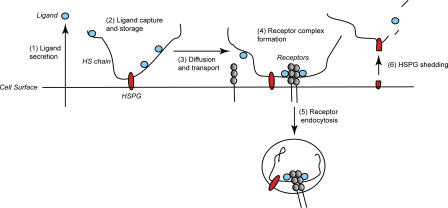
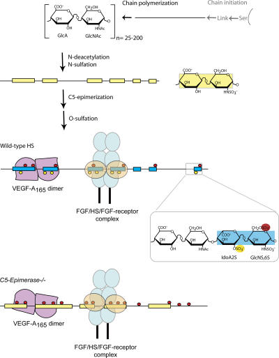
Verification
Claims the paper makes
The claims and statistics of this paper haven’t been checked yet.
Run the check — free, no account needed. It reads the open-access text and checks the sources it cites.
Claims other papers make about this one
Each is checked against this paper’s own text: does it say what the citing paper says it does?
The cited source says what the claim attributes to it.
In the citing paper
“Because heparan sulphate biosynthesis differs depending upon tissue/cell types (Kreuger et al., 2006), the discrepancies in cellular Aβ uptake may result from differences in tissue/cell-specific HSPG structure.”
From Heparan Sulphate Proteoglycan and the Low-Density Lipoprotein Receptor-Related Protein 1 Constitute Major Pathways for Neuronal Amyloid-β Uptake (2011) · cites this paper as Kreuger et al., 2006
Self-check
Percentages, sums and reported differences re-computed from the paper’s own numbers.
The self-check needs the paper’s full text.
Citations
Cites
Mammalian Brain Morphogenesis and Midline Axon Guidance Require Heparan Sulfate
Inatani, M., Irie, F., Plump, A. S., Tessier-Lavigne, M., & Yamaguchi, Y. (2003) · Science · doi:10.1126/science.1090497 · Cited by 431
Research works printed but not linked: 38
- 2003, J. Cell Biol., 162, 341, 10.1083/jcb.200212083 · DOI
- 2003, J. Cell Biol., 163, 637, 10.1083/jcb.200307053 · DOI
- 2004, J. Biol. Chem., 279, 12346, 10.1074/jbc.M313523200 · DOI
- 2005, J. Biol. Chem., 280, 31508, 10.1074/jbc.M414581200 · DOI
- 1992, Annu. Rev. Cell Biol., 8, 365, 10.1146/annurev.cb.08.110192.002053 · DOI
- 1999, Annu. Rev. Biochem., 68, 729, 10.1146/annurev.biochem.68.1.729 · DOI
- 1998, Genes Dev., 12, 1894, 10.1101/gad.12.12.1894 · DOI
- 1996, Nature., 380, 435, 10.1038/380435a0 · DOI
- 2003, Biochem. Soc. Trans., 31, 352, 10.1042/bst0310352 · DOI
- 2001, J. Clin. Invest., 108, 169, 10.1172/JCI200113530 · DOI
- 2001, J. Clin. Invest., 108, 357, 10.1172/JCI13713 · DOI
- 1999, Curr. Biol., 9, 1343, 10.1016/S0960-9822(00)80060-3 · DOI
- 2005, Nat. Rev. Mol. Cell Biol., 6, 530, 10.1038/nrm1681 · DOI
- 2006, Dev. Cell., 10, 625, 10.1016/j.devcel.2006.03.009 · DOI
- 2006, 10.1074/jbc.M600806200 · DOI
- 2002, J. Biol. Chem., 277, 30567, 10.1074/jbc.M203404200 · DOI
- 2002, EMBO J., 21, 6303, 10.1093/emboj/cdf638 · DOI
- 2004, Dev. Cell., 7, 503, 10.1016/j.devcel.2004.08.005 · DOI
- 2005, Biochem. J., 389, 145, 10.1042/BJ20042129 · DOI
- 2004, J. Biol. Chem., 279, 42732, 10.1074/jbc.M405382200 · DOI · Paper page
- 2003, J. Biol. Chem., 278, 28363, 10.1074/jbc.C300219200 · DOI
- 2000, Dev. Biol., 224, 299, 10.1006/dbio.2000.9798 · DOI · Paper page
- 1980, Proc. Natl. Acad. Sci. USA., 77, 6551, 10.1073/pnas.77.11.6551 · DOI · Paper page
- 1995, Biochem. J., 310, 497, 10.1042/bj3100497 · DOI
- 1996, J. Biol. Chem., 271, 17804, 10.1074/jbc.271.30.17804 · DOI
- 2001, J. Biol. Chem., 276, 35429, 10.1074/jbc.M100379200 · DOI
- 2004, J. Biol. Chem., 279, 27239, 10.1074/jbc.M401774200 · DOI
- 2004, Glycobiology., 14, 17R, 10.1093/glycob/cwh051 · DOI
- 1998, J. Biol. Chem., 273, 22936, 10.1074/jbc.273.36.22936 · DOI
- 2000, J. Biol. Chem., 275, 25926, 10.1074/jbc.C000359200 · DOI · Paper page
- 2006, J. Biol. Chem., 281, 1731, 10.1074/jbc.M510760200 · DOI
- 1996, FASEB J., 10, 1270, 10.1096/fasebj.10.11.8836040 · DOI
- 2000, Mol. Cell., 6, 743, 10.1016/S1097-2765(00)00073-3 · DOI · Paper page
- 1999, Cell., 99, 13, 10.1016/S0092-8674(00)80058-6 · DOI
- 1998, J. Biol. Chem., 273, 15487, 10.1074/jbc.273.25.15487 · DOI
- 1997, J. Biol. Chem., 272, 20508, 10.1074/jbc.272.33.20508 · DOI
- 1998, J. Biol. Chem., 273, 12960, 10.1074/jbc.273.21.12960 · DOI
- 2001, J. Clin. Invest., 108, 341, 10.1172/JCI13662 · DOI
Cited by
Sorted by citations
Heparan sulphate proteoglycans fine-tune mammalian physiology
Bishop, J. R., Schuksz, M., & Esko, J. D. (2007) · Nature · doi:10.1038/nature05817 · Cited by 1,631
Franz, S., Rammelt, S., Scharnweber, D., & Simon, J. C. (2011) · Biomaterials · doi:10.1016/j.biomaterials.2011.05.078 · Cited by 1,391
Christianson, H. C., Svensson, K. J., van Kuppevelt, T. H., Li, J.-P., & Belting, M. (2013) · Proceedings of the National Academy of Sciences · doi:10.1073/pnas.1304266110 · Cited by 903
Self-citationDemystifying Heparan Sulfate–Protein Interactions
Xu, D., & Esko, J. D. (2014) · Annual Review of Biochemistry · doi:10.1146/annurev-biochem-060713-035314 · Cited by 805
Effects of Charge on Antibody Tissue Distribution and Pharmacokinetics
Boswell, C. A., Tesar, D. B., Mukhyala, K., Theil, F.-P., Fielder, P. J., & Khawli, L. A. (2010) · Bioconjugate Chemistry · doi:10.1021/bc100261d · Cited by 398
Biological hydrogels as selective diffusion barriers
Lieleg, O., & Ribbeck, K. (2011) · Trends in Cell Biology · doi:10.1016/j.tcb.2011.06.002 · Cited by 387
Kanekiyo, T., Zhang, J., Liu, Q., Liu, C.-C., Zhang, L., & Bu, G. (2011) · The Journal of Neuroscience · doi:10.1523/jneurosci.5491-10.2011 · Cited by 212
Semaphorin 3A Binds to the Perineuronal Nets via Chondroitin Sulfate Type E Motifs in Rodent Brains
Dick, G., Tan, C. L., Alves, J. N., Ehlert, E. M. E., Miller, G. M., Hsieh-Wilson, L. C., Sugahara, K., Oosterhof, A., van Kuppevelt, T. H., Verhaagen, J., et al. (2013) · Journal of Biological Chemistry · doi:10.1074/jbc.m111.310029 · Cited by 160
Malfait, F., Kariminejad, A., Van Damme, T., Gauche, C., Syx, D., Merhi-Soussi, F., Gulberti, S., Symoens, S., Vanhauwaert, S., Willaert, A., et al. (2013) · The American Journal of Human Genetics · doi:10.1016/j.ajhg.2013.04.016 · Cited by 142
Phélippé, M., Gonçalves, O., Thouand, G., Cogne, G., & Laroche, C. (2019) · Algal Research · doi:10.1016/j.algal.2019.101426 · Cited by 84
Blanchard, D. C., Summers, C. H., & Blanchard, R. J. (2013) · Neuroscience & Biobehavioral Reviews · doi:10.1016/j.neubiorev.2013.06.008 · Cited by 60
Auxiliary and Autonomous Proteoglycan Signaling Networks
Elfenbein, A., & Simons, M. (2010) · Methods in Enzymology · doi:10.1016/s0076-6879(10)80001-1 · Cited by 32
Circadian control of heparan sulfate levels times phagocytosis of amyloid beta aggregates
Clark, G. T., Yu, Y., Urban, C. A., Fu, G., Wang, C., Zhang, F., Linhardt, R. J., & Hurley, J. M. (2022) · PLOS Genetics · doi:10.1371/journal.pgen.1009994 · Cited by 31
A personal voyage through the proteoglycan field
Lindahl, U. (2014) · Matrix Biology · doi:10.1016/j.matbio.2014.01.001 · Cited by 28
Self-citationBui, C., Talhaoui, I., Chabel, M., Mulliert, G., Coughtrie, M. W. H., Ouzzine, M., & Fournel-Gigleux, S. (2010) · FEBS Letters · doi:10.1016/j.febslet.2010.08.001 · Cited by 27
Identification of Key Functional Residues in the Active Site of Human β1,4-Galactosyltransferase 7
Talhaoui, I., Bui, C., Oriol, R., Mulliert, G., Gulberti, S., Netter, P., Coughtrie, M. W. H., Ouzzine, M., & Fournel-Gigleux, S. (2010) · Journal of Biological Chemistry · doi:10.1074/jbc.m110.151951 · Cited by 26
Metadata
- Authors
- Kreuger, J., Spillmann, D., Li, J.-P., & Lindahl, U.
- Affiliations
- 1Rudbeck Laboratory, Department of Genetics and Pathology,
- 2Department of Medical Biochemistry and Microbiology, Biomedical Center, Uppsala University, SE-75185 Uppsala, Sweden
- Venue
- The Journal of Cell Biology
- Publisher
- Rockefeller University Press
- Published
- 2006-07-31
- Volume
- 174
- Issue
- 3
- Pages
- 323-327
- ISSN
- 1540-8140
- 0021-9525
- PMC
- PMC2064228
- Cited by
- 449
- References
- 1
- Language
- en
About these checks
Registry, retraction, Cites and statistics results are computed, not judged, with one exception: a printed reference that no index lists is read once by a model before it counts as a citation error. Claims are extracted by a model and kept only when their quote appears verbatim in the text; each cited source is then checked, with a model’s help, for whether it says what the claim relies on. Those verdicts can be wrong — read the claim’s quote and the cited source before relying on one.
Statistics and arithmetic are recomputed, not judged.
Confidence bands are shown only for fields calibrated on our annotated test set. None is calibrated yet, so no band is shown.
What counts as a claim ›How we measure confidence ›How we check this ›
Are you an author, or do you see a problem with this page? Request a review
