

The DOI matches its Crossref record; 45 of the 45 references it cites were found in a registry or an index.
Neurofascin assembles a specialized extracellular matrix at the axon initial segment
Open accessAbstract
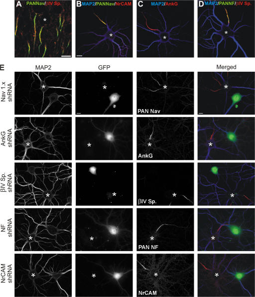
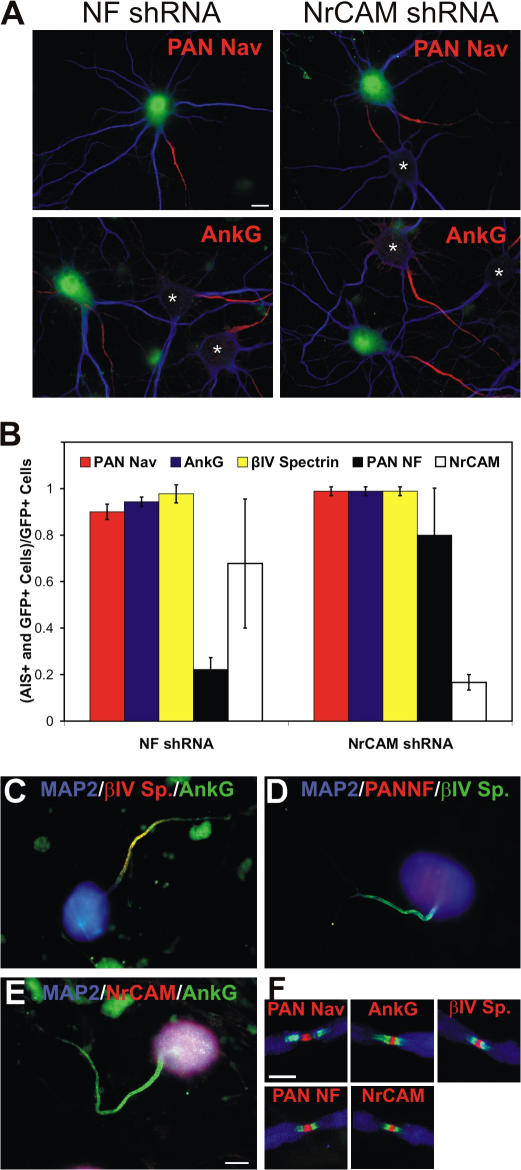
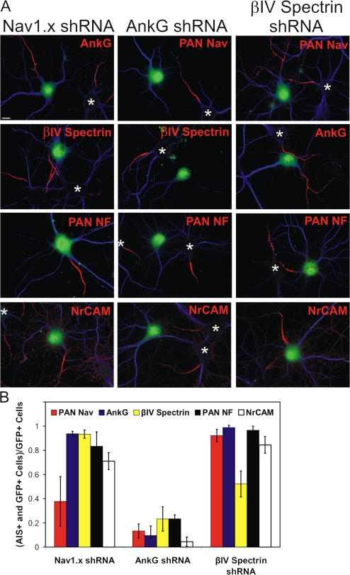
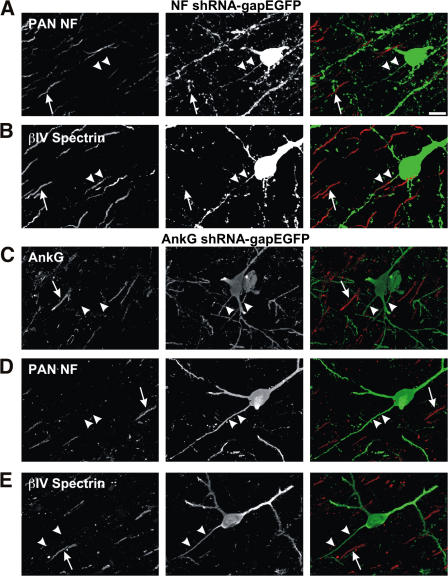
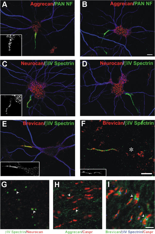
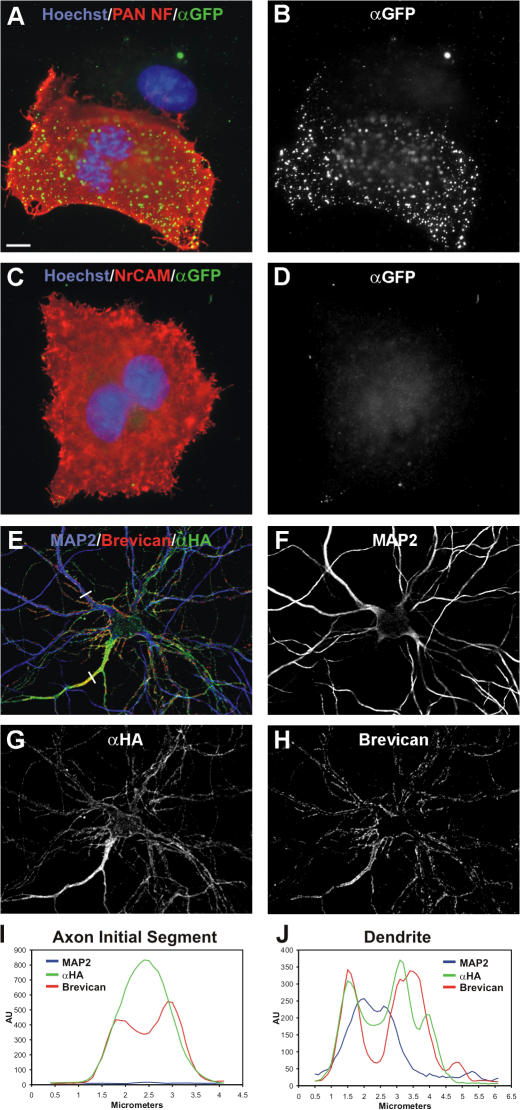
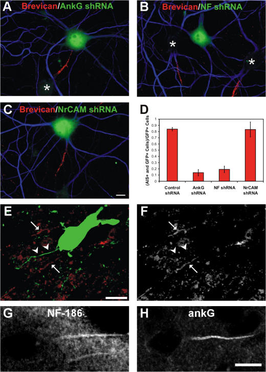
Action potential initiation and propagation requires clustered Na+ (voltage-gated Na+ [Nav]) channels at axon initial segments (AIS) and nodes of Ranvier. In addition to ion channels, these domains are characterized by cell adhesion molecules (CAMs; neurofascin-186 [NF-186] and neuron glia–related CAM [NrCAM]), cytoskeletal proteins (ankyrinG and βIV spectrin), and the extracellular chondroitin-sulfate proteoglycan brevican. Schwann cells initiate peripheral nervous system node formation by clustering NF-186, which then recruits ankyrinG and Nav channels. However, AIS assembly of this protein complex does not require glial contact. To determine the AIS assembly mechanism, we silenced expression of AIS proteins by RNA interference. AnkyrinG knockdown prevented AIS localization of all other AIS proteins. Loss of NF-186, NrCAM, Nav channels, or βIV spectrin did not affect other neuronal AIS proteins. However, loss of NF-186 blocked assembly of the brevican-based AIS extracellular matrix, and NF-186 overexpression caused somatodendritic brevican clustering. Thus, NF-186 assembles and links the specialized brevican-containing AIS extracellular matrix to the intracellular cytoskeleton.
Overview
The claims and statistics of this paper haven’t been checked yet.
Run the check — free, no account needed. It reads the open-access text and checks the sources it cites.
Integrity signals
Registry
Cites
Statistics
Figures
Verification
Claims the paper makes
The claims and statistics of this paper haven’t been checked yet.
Run the check — free, no account needed. It reads the open-access text and checks the sources it cites.
Claims other papers make about this one
Each is checked against this paper’s own text: does it say what the citing paper says it does?
In the citing paper
“Brevican enrichment in the AIS was mediated via direct interaction with NF186, which was diminished by a knockdown of NF186 (John et al., 2006;Hedstrom et al., 2007).”
From The hyaluronan and proteoglycan link proteins: Organizers of the brain extracellular matrix and key molecules for neuronal function and plasticity (2015) · cites this paper as Hedstrom et al., 2007
In the citing paper
“Brevican can interact with NF186 in the AIS as described before (Hedstrom et al., 2007).”
From The hyaluronan and proteoglycan link proteins: Organizers of the brain extracellular matrix and key molecules for neuronal function and plasticity (2015) · cites this paper as Hedstrom et al., 2007
Self-check
Percentages, sums and reported differences re-computed from the paper’s own numbers.
The self-check needs the paper’s full text.
Citations
Cites
Banker, G. A., & Goslin, K. (1998) · Cited by 1,290
Neuronal polarity: from extracellular signals to intracellular mechanisms.
Arimura, N., & Kaibuchi, K. (2007) · Nature Reviews Neuroscience · doi:10.1038/nrn2056 · Cited by 655
Lecticans: organizers of the brain extracellular matrix
Yamaguchi, Y. (2000) · Cellular and Molecular Life Sciences · doi:10.1007/pl00000690 · Cited by 642
Pan, Z., Kao, T., Horvath, Z., Lemos, J. C., Sul, J., Cranstoun, S. D., Bennett, V., Scherer, S. S., & Cooper, E. C. (2006) · Journal of Neuroscience · doi:10.1523/jneurosci.4314-05.2006 · Cited by 586
Zhou, D., Lambert, S., Malen, P. L., Carpenter, S., Boland, L. M., & Bennett, V. (1998) · The Journal of Cell Biology · doi:10.1083/jcb.143.5.1295 · Cited by 568
Perineuronal nets: past and present
Celio, M. R., Spreafico, R., De Biasi, S., & Vitellaro-Zuccarello, L. (1998) · Trends in Neurosciences · doi:10.1016/s0166-2236(98)01298-3 · Cited by 497
Jenkins, S. M., & Bennett, V. (2001) · The Journal of Cell Biology · doi:10.1083/jcb.200109026 · Cited by 446
Unique features of action potential initiation in cortical neurons
Naundorf, B., Wolf, F., & Volgushev, M. (2006) · Nature · doi:10.1038/nature04610 · Cited by 396
In vivo electroporation in the embryonic mouse central nervous system
Saito, T. (2006) · Nature Protocols · doi:10.1038/nprot.2006.276 · Cited by 378
Ango, F., Cristo, G. D., Higashiyama, H., Bennett, V., Wu, P., & Huang, Z. J. (2004) · Cell · doi:10.1016/j.cell.2004.10.004 · Cited by 358
Cited by
Sorted by citations
Items 201–268 of 268
Kinesin Regulation in the Proximal Axon is Essential for Dendrite-selective Transport
Mendoza, C. S., Plowinske, C. R., Montgomery, A. C., Quinones, G. B., Banker, G. A., & Bentley, M. (2024) · Molecular Biology of the Cell · doi:10.1091/mbc.e23-11-0457 · Cited by 5
The axon initial segment as a source of neuropathic pain
Dorrego-Rivas, A., & Grubb, M. S. (2022) · Brain · doi:10.1093/brain/awac156 · Cited by 5
Dong, J., Song, H., Wang, Y., Li, M., Yu, Y., Wang, Y., & Chen, J. (2017) · Frontiers in Neuroscience · doi:10.3389/fnins.2017.00298 · Cited by 5
Chondroitin Sulphate Proteoglycan Axonal Coats in the Human Mediodorsal Thalamic Nucleus
Pantazopoulos, H., Hossain, N. M., Chelini, G., Durning, P. T., Barbas, H., Zikopoulos, B., & Berretta, S. (2022) · Frontiers in Integrative Neuroscience · doi:10.3389/fnint.2022.934764 · Cited by 5
Converging on the Origins of Axonal Ion Channel Clustering
Rasband, M. N. (2009) · PLoS Genetics · doi:10.1371/journal.pgen.1000340 · Cited by 5
Self-citationLive imaging of excitable axonal microdomains in ankyrin-G-GFP micePreprint
Thome, C., Janssen, J. M., Karabulut, S., Acuna, C., D’Este, E., Soyka, S. J., Baum, K., Bock, M., Lehmann, N., Roos, J., et al. (2023) · eLife · doi:10.7554/elife.87078 · Cited by 6
The Nodes of Ranvier: Mechanisms of Assembly and MaintenancePreprint
Rasband, M. N., & Peles, E. (2024) · Cold Spring Harbor Perspectives in Biology · doi:10.1101/cshperspect.a041361 · Cited by 5
Self-citationSingh, J. B., Perelló‐Amorós, B., Schneeberg, J., Mirzapourdelavar, H., Seidenbecher, C. I., Fejtová, A., Dityatev, A. E., & Frischknecht, R. (2025) · EMBO Reports · doi:10.1038/s44319-025-00644-w · Cited by 8
Self-citationHochmeister, S., Pekar, T., Lindner, M., Kitic, M., Haindl, M. T., Storch, M. K., Fazekas, F., & Linington, C. (2014) · PLoS ONE · doi:10.1371/journal.pone.0085393 · Cited by 4
Glucose increases the length and spacing of the lattice structure of the axon initial segment
Wang, Y., Guan, M., Zhang, Y., Zhanghao, K., & Xi, P. (2022) · Microscopy research and technique (Print) · doi:10.1002/jemt.24122 · Cited by 3
Regulation of Axon Initial Segment Diameter by COUP-TFI Fine-tunes Action Potential Generation
Wu, X., Li, H., Huang, J., Xu, M., Xiao, C., & He, S. (2021) · Neuroscience Bulletin · doi:10.1007/s12264-021-00792-8 · Cited by 4
Crystal structure of human Ankyrin G death domain
Liu, Y., Zhang, Y., & Wang, J. (2014) · Proteins Structure Function and Bioinformatics · doi:10.1002/prot.24702 · Cited by 4
Zhu, X., Yu, Y., Jiang, Z., Otani, Y., & Fujitani, M. (2025) · Biomolecules · doi:10.3390/biom15060901 · Cited by 5
Specification and Development of GABAergic Interneurons
Schilling, K. (2019) · Springer eBooks · doi:10.1007/978-3-319-97911-3_11-2 · Cited by 3
Action potential initiation in damaged axon initial segment
Jacques, L., Morris, C. E., Longtin, A., & Joós, B. (2014) · BMC Neuroscience · doi:10.1186/1471-2202-15-s1-p135 · Cited by 3
Specification and Development of GABAergic Interneurons
Schilling, K. (2012) · Springer eBooks · doi:10.1007/978-94-007-1333-8_11 · Cited by 3
Dannemeyer, M. (2016) · doi:10.53846/goediss-5493 · Cited by 3
Axon Initial Segment: Structure, Biological Functions, Diseases, and Therapeutic Targets
Song, D., Yuan, L., Yang, W., Li, W., & Li, J. (2026) · MedComm · doi:10.1002/mco2.70689 · Cited by 2
Nouveautés sur les mécanismes de formation des nœuds de Ranvier
Devaux, J. J. (2014) · Revue Neurologique · doi:10.1016/j.neurol.2014.03.017 · Cited by 2
Location of the axon initial segment assembly can be predicted from neuronal shape
Xu, Z., Angstmann, C. N., Wu, Y., Stefen, H., Parić, E., Fath, T., & Curmi, P. M. G. (2024) · iScience · doi:10.1016/j.isci.2024.109264 · Cited by 2
Structure and Function of Myelinated Axons
Buffington, S. A., & Rasband, M. N. (2013) · Elsevier eBooks · doi:10.1016/b978-0-12-397265-1.00078-2 · Cited by 2
Self-citationThe axon initial segment drives the neuron’s extracellular action potentialPreprint
Bakkum, D. J., Radivojević, M., Obien, M. E. J., Jäckel, D., Frey, U., Takahashi, H., & Hierlemann, A. R. (2018) · bioRxiv (Cold Spring Harbor Laboratory) · doi:10.1101/266734 · Cited by 2
Live imaging of excitable axonal microdomains in ankyrin-G-GFP micePreprint
Thome, C., Janssen, J. M., Karabulut, S., Acuna, C., D’Este, E., Soyka, S. J., Baum, K., Bock, M., Lehmann, N., Hasegawa, M., et al. (2023) · eLife · doi:10.7554/elife.87078.1 · Cited by 2
Darwish, M., Suzuki, S., Ogawa, Y., Takase, A., Tanaka, M., Iijima, Y., Sato, Y., Suzuki, M., Kanegae, Y., Okada, C., et al. (2023) · bioRxiv (Cold Spring Harbor Laboratory) · doi:10.1101/2023.10.29.564567 · Cited by 2
Self-citationLysakowski, A., Govindaraju, A. C., Price, S. D., Gaboyard-Niay, S., Calin‐Jageman, I. E., Chidavaenzi, R. L., Eatock, R. A., Raphael, R. M., & Goldberg, J. M. (2026) · The Journal of Comparative Neurology · doi:10.1002/cne.70127 · Cited by 1
Yamankurt, G. (2013) · Journal of Proteomics & Bioinformatics · doi:10.4172/jpb.1000284 · Cited by 1
Synaptic Origin of Early Sensory-evoked Oscillations in the Immature Thalamus
Sheroziya, M., & Khazipov, R. (2023) · Neuroscience · doi:10.1016/j.neuroscience.2023.09.005 · Cited by 1
Gilloteaux, J., Swert, K. D., Suain, V., & Nicaise, C. (2023) · International Journal of Molecular Sciences · doi:10.3390/ijms242216448 · Cited by 1
Chaotic Neural Circuit Dynamics
Engelken, R. (2018) · eDiss (Georg-August-Universität Göttingen) · doi:10.53846/goediss-6718 · Cited by 2
Sound encoding in mutant mice with disrupted action potential generation
Yamanbaeva, G. (2018) · doi:10.53846/goediss-7005 · Cited by 1
Unveiling the cell biology of hippocampal neurons with dendritic axon originPreprint
Han, Y., Thuenauer, R., Grünewald, K., & Mikhaylova, M. (2023) · bioRxiv (Cold Spring Harbor Laboratory) · doi:10.1101/2023.09.14.557747 · Cited by 1
Baidoe‐Ansah, D., Mirzapourdelavar, H., Aleshin, S. E., Schott, B. H., Seidenbecher, C. I., Kaushik, R., & Dityatev, A. E. (2024) · bioRxiv (Cold Spring Harbor Laboratory) · doi:10.1101/2024.01.26.577401 · Cited by 1
Self-citationDEVELOPMENT OF CORTICAL GABA CIRCUITRY: IDENTIFYING PERIODS OF VULNERABILITY TO SCHIZOPHRENIA
Hoftman, G. D. (2013) · D-Scholarship@Pitt (University of Pittsburgh)
Identification and Characterization of O-mannosylated Proteins
Pacharra, S. (2013) · Kölner Universitäts PublikationsServer (Universität zu Köln)
Peripheral blood biomarkers in Psychiatric Diseases
Castell, M. S. (2012) · TDX (Tesis Doctorals en Xarxa)
Nodal Dynamics afterIn VivoRescue of βIV Spectrin Expression
Bacmeister, C. M., & Thornton, M. A. (2019) · Journal of Neuroscience · doi:10.1523/jneurosci.2149-18.2018
Neuronal polarity: establishing and maintaining the axon initial segment
van Beuningen, S. F. B. (2011) · Utrecht University Repository (Utrecht University)
Ni, G., Hao, X., Cai, X., Qin, J., Zhou, L., Kwan, P., & Chen, Z. (2019) · Acta Epileptologica · doi:10.1186/s42494-019-0004-8
COUP-TFI regulates diameter of the axon initial segment in the mammalian neocortexPreprint
Wu, X., Li, H., Huang, J., Xiao, C., & He, S. (2020) · bioRxiv (Cold Spring Harbor Laboratory) · doi:10.1101/2020.10.07.329284
Pan, X. (2020) · doi:10.33540/99
Biology of Proteoglycans and Associated Glycosaminoglycans
Salinas-Marín, R., Villanueva-Cabello, T. M., & Martínez-Duncker, I. (2021) · Elsevier eBooks · doi:10.1016/b978-0-12-819475-1.00065-1
Macrophage Surveillance and Immunity: Eating, Drinking, and Digesting
Ostrowski, P. P. (2020) · TSpace (University of Toronto)
Specification and Development of GABAergic Interneurons
Schilling, K. (2021) · Springer eBooks · doi:10.1007/978-3-030-23810-0_11
A quantitative analysis of an average cultured neuron
Jähne, S. (2020) · eDiss (Georg-August-Universität Göttingen) · doi:10.53846/goediss-7934
Chan, J. R., & Echeverria, A. (2012) · Faculty Opinions – Post-Publication Peer Review of the Biomedical Literature · doi:10.3410/f.716747900.792102900
AnkG-Neurofascin complex structure reveals binding mechanisms required for integrity of the AISPreprint
He, L., Jiang, W., Li, J., & Wang, C. (2022) · bioRxiv (Cold Spring Harbor Laboratory) · doi:10.1101/2022.04.27.489743
Structure and function of myelinated axons
Buffington, S. A., & Rasband, M. N. (2020) · Elsevier eBooks · doi:10.1016/b978-0-12-814405-3.00040-0
Self-citationActivity-dependent extracellular proteolytic cascade remodels ECM to promote structural plasticityPreprint
Singh, J. B., Perelló‐Amorós, B., Schneeberg, J., Seidenbecher, C. I., Fejtová, A., Dityatev, A. E., & Frischknecht, R. (2024) · bioRxiv (Cold Spring Harbor Laboratory) · doi:10.1101/2024.10.28.620597
Self-citationLive imaging of excitable axonal microdomains in ankyrin-G-GFP micePreprint
Thome, C., Janssen, J. M., Karabulut, S., Acuna, C., D’Este, E., Soyka, S. J., Baum, K., Bock, M., Lehmann, N., Roos, J., et al. (2024) · eLife · doi:10.7554/elife.87078.2
Super‐Resolution Microscopy in Tissue Engineering
Rabiee, N., Akrami‐Hasan‐Kohal, M., & Bencherif, S. A. (2025) · Advanced Materials Interfaces · doi:10.1002/admi.202500437
Telley, L. (2010) · theses.fr (ABES) · doi:10.70675/d6c3af86z9bebz4d64z87d5z290742f0aa95
Pinatel, D. (2015) · theses.fr (ABES) · doi:10.70675/c1997924z1c4bz4f2cz9846z5009e029e1c0
Mechanisms of central nervous system nodes of Ranvier assembly
Freeman, S. A. (2015) · theses.fr (ABES) · doi:10.70675/8a8a095cze5faz4cd3z8fcez185fe0291fb2
Thétiot, M. (2018) · theses.fr (ABES) · doi:10.70675/4fd006d4z6c0ez4fe8zbcd0z0df8b8ef8d85
Haidar, Z. (2019) · theses.fr (ABES) · doi:10.70675/3577ad91z4de1z4642zb506zdfe80f5b9c8a
Revisiting neuronal polarity in the context of PCP
Rivas, A. D. (2021) · theses.fr (ABES) · doi:10.70675/e6c0a489z9417z4ed7z9faaz2b8d89b6869d
Impact of axon initial segment geometry on the electrical properties of neurons
Goethals, S. (2020) · theses.fr (ABES) · doi:10.70675/b3acfdd8z6c50z45e9z9b97z577498c1c5dc
Lanfranchi, M. (2021) · theses.fr (ABES) · doi:10.70675/247335b1zba7fz4005zafbdzdc43c088d75f
Axon initial segment plasticity in neuronal circuit function and memory formation
Gründemann, J. (2026) · Current Opinion in Physiology · doi:10.1016/j.cophys.2026.100981
Live-imaging of endogenous neurofascins reveals glial adhesion shapes developing nodes of RanvierPreprint
Lyster-Binns, P., Marshall-Phelps, K. L., Siems, S. B., Pyrgioti, M., & Almeida, R. (2026) · bioRxiv (Cold Spring Harbor Laboratory) · doi:10.64898/2026.07.24.740328
Intrinsic neuronal polarity: early events in symmetry breakage and neuronal polarization
Sanders, L. (2014) · Utrecht University Repository (Utrecht University)
Metadata
- Authors
- Hedstrom, K. L., Xu, X., Ogawa, Y., Frischknecht, R., Seidenbecher, C. I., Shrager, P., & Rasband, M. N.
- Affiliations
- UConn Health (US)
- University of Rochester Medicine (US)
- Leibniz Institute for Neurobiology (DE)
- Venue
- The Journal of Cell Biology
- Publisher
- Rockefeller University Press
- Published
- 2007-08-20
- Volume
- 178
- Issue
- 5
- Pages
- 875-886
- ISSN
- 0021-9525 (ISSN-L)
- 1540-8140
- PMC
- PMC2064550
- OpenAlex
- W2159315879
- Cited by
- 268
- References
- 45
- Citations by year
- 2026: 4 · 2025: 8 · 2024: 12 · 2023: 10 · 2022: 11 · 2021: 12 · 2020: 15 · 2019: 18 · 2018: 15 · 2017: 15 · 2016: 10 · 2015: 20 · 2014: 17 · 2013: 21 · 2012: 20
- FWCI
- 5.96
- Type
- journal article
- Language
- en
- Open access
- green
- www.ncbi.nlm.nih.gov
- Licence
- cc-by-nc-sa
- Locations
- The Journal of Cell Biology (publishedVersion)
- PubMed Central (publishedVersion, cc-by-nc-sa)
- PubMed (publishedVersion)
- Funders
- National Institute of Neurological Disorders and Stroke (R01NS017965, NS 044916, NS 17965, R01NS044916, R37NS044916)
- Topics
- Life Sciences › Biochemistry, Genetics and Molecular Biology › Molecular Biology › Ion channel regulation and function
- Life Sciences › Neuroscience › Cellular and Molecular Neuroscience › Neuroscience and Neuropharmacology Research
- Life Sciences › Biochemistry, Genetics and Molecular Biology › Molecular Biology › Ion Transport and Channel Regulation
- Keywords
- neurofascin 186, axon initial segment, extracellular matrix assembly, brevican, chondroitin sulfate proteoglycans, ankyrin-G, βIV-spectrin, nodes of Ranvier, Schwann cells, adhesion molecules, cytoskeletal anchoring, action potential generation, RNAi knockdown
- MeSH terms
- Animals, Axons, Cells, Cultured, Cytoskeleton, Extracellular Matrix, Hippocampus, Nerve Growth Factors, Nerve Tissue Proteins, Chondroitin Sulfate Proteoglycans, Ranvier's Nodes, Recombinant Fusion Proteins, RNA, Spectrin, Sodium Channels, Cell Adhesion Molecules, Ankyrins, RNA Interference, Lectins, C-Type, Rats, Brevican
About these checks
Registry, retraction, Cites and statistics results are computed, not judged, with one exception: a printed reference that no index lists is read once by a model before it counts as a citation error. Claims are extracted by a model and kept only when their quote appears verbatim in the text; each cited source is then checked, with a model’s help, for whether it says what the claim relies on. Those verdicts can be wrong — read the claim’s quote and the cited source before relying on one.
Statistics and arithmetic are recomputed, not judged.
Confidence bands are shown only for fields calibrated on our annotated test set. None is calibrated yet, so no band is shown.
What counts as a claim ›How we measure confidence ›How we check this ›
Are you an author, or do you see a problem with this page? Request a review
