

The DOI matches its Crossref record; 45 of the 45 references it cites were found in a registry or an index.
Neurofascin assembles a specialized extracellular matrix at the axon initial segment
Open accessAbstract
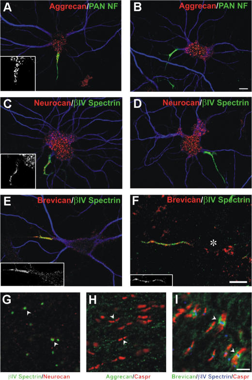
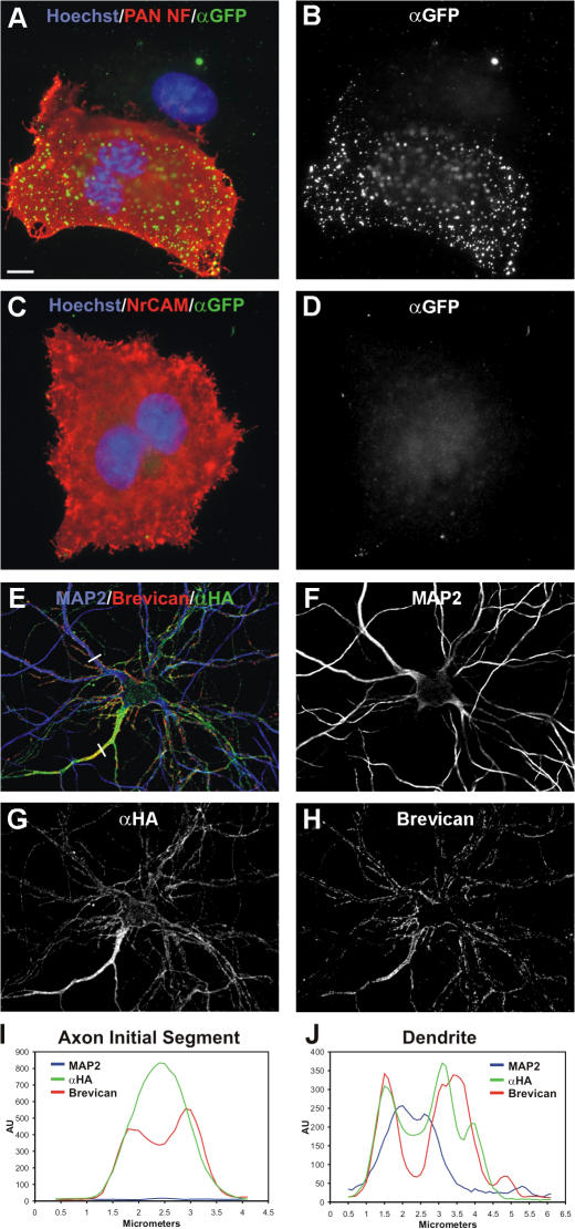
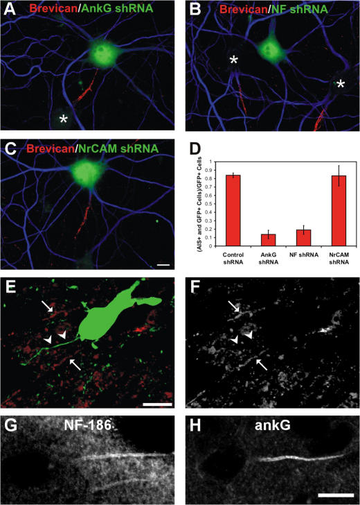
Action potential initiation and propagation requires clustered Na+ (voltage-gated Na+ [Nav]) channels at axon initial segments (AIS) and nodes of Ranvier. In addition to ion channels, these domains are characterized by cell adhesion molecules (CAMs; neurofascin-186 [NF-186] and neuron glia–related CAM [NrCAM]), cytoskeletal proteins (ankyrinG and βIV spectrin), and the extracellular chondroitin-sulfate proteoglycan brevican. Schwann cells initiate peripheral nervous system node formation by clustering NF-186, which then recruits ankyrinG and Nav channels. However, AIS assembly of this protein complex does not require glial contact. To determine the AIS assembly mechanism, we silenced expression of AIS proteins by RNA interference. AnkyrinG knockdown prevented AIS localization of all other AIS proteins. Loss of NF-186, NrCAM, Nav channels, or βIV spectrin did not affect other neuronal AIS proteins. However, loss of NF-186 blocked assembly of the brevican-based AIS extracellular matrix, and NF-186 overexpression caused somatodendritic brevican clustering. Thus, NF-186 assembles and links the specialized brevican-containing AIS extracellular matrix to the intracellular cytoskeleton.
Overview
The claims and statistics of this paper haven’t been checked yet.
Run the check — free, no account needed. It reads the open-access text and checks the sources it cites.
Integrity signals
Registry
Cites
Statistics
Figures
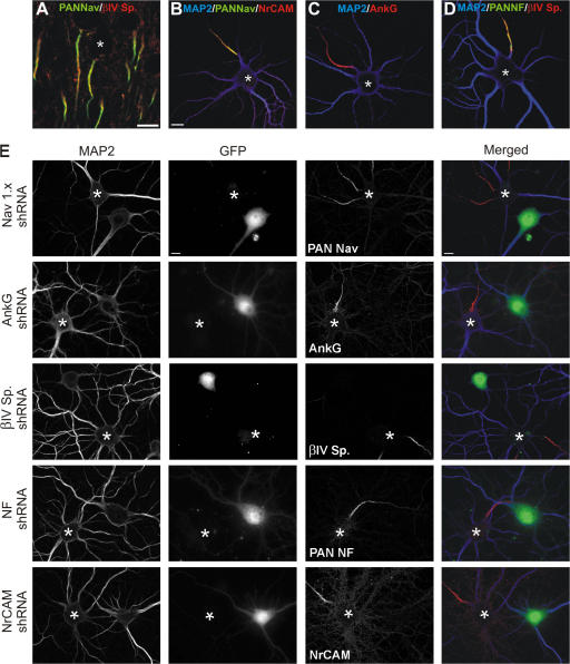
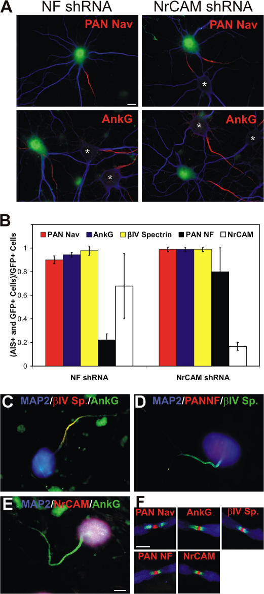
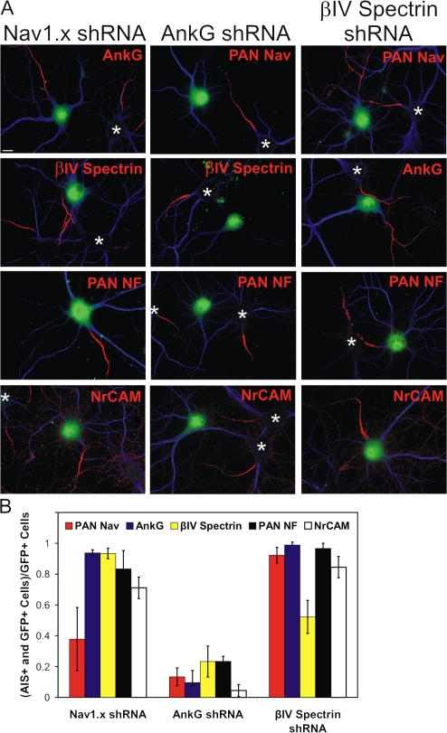
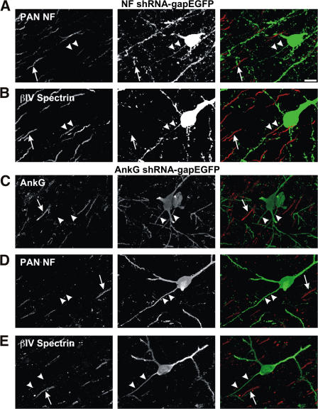
Verification
Claims the paper makes
The claims and statistics of this paper haven’t been checked yet.
Run the check — free, no account needed. It reads the open-access text and checks the sources it cites.
Claims other papers make about this one
Each is checked against this paper’s own text: does it say what the citing paper says it does?
In the citing paper
“Brevican enrichment in the AIS was mediated via direct interaction with NF186, which was diminished by a knockdown of NF186 (John et al., 2006;Hedstrom et al., 2007).”
From The hyaluronan and proteoglycan link proteins: Organizers of the brain extracellular matrix and key molecules for neuronal function and plasticity (2015) · cites this paper as Hedstrom et al., 2007
In the citing paper
“Brevican can interact with NF186 in the AIS as described before (Hedstrom et al., 2007).”
From The hyaluronan and proteoglycan link proteins: Organizers of the brain extracellular matrix and key molecules for neuronal function and plasticity (2015) · cites this paper as Hedstrom et al., 2007
Self-check
Percentages, sums and reported differences re-computed from the paper’s own numbers.
The self-check needs the paper’s full text.
Citations
Cites
Banker, G. A., & Goslin, K. (1998) · Cited by 1,290
Neuronal polarity: from extracellular signals to intracellular mechanisms.
Arimura, N., & Kaibuchi, K. (2007) · Nature Reviews Neuroscience · doi:10.1038/nrn2056 · Cited by 655
Lecticans: organizers of the brain extracellular matrix
Yamaguchi, Y. (2000) · Cellular and Molecular Life Sciences · doi:10.1007/pl00000690 · Cited by 642
Pan, Z., Kao, T., Horvath, Z., Lemos, J. C., Sul, J., Cranstoun, S. D., Bennett, V., Scherer, S. S., & Cooper, E. C. (2006) · Journal of Neuroscience · doi:10.1523/jneurosci.4314-05.2006 · Cited by 586
Zhou, D., Lambert, S., Malen, P. L., Carpenter, S., Boland, L. M., & Bennett, V. (1998) · The Journal of Cell Biology · doi:10.1083/jcb.143.5.1295 · Cited by 568
Perineuronal nets: past and present
Celio, M. R., Spreafico, R., De Biasi, S., & Vitellaro-Zuccarello, L. (1998) · Trends in Neurosciences · doi:10.1016/s0166-2236(98)01298-3 · Cited by 497
Jenkins, S. M., & Bennett, V. (2001) · The Journal of Cell Biology · doi:10.1083/jcb.200109026 · Cited by 446
Unique features of action potential initiation in cortical neurons
Naundorf, B., Wolf, F., & Volgushev, M. (2006) · Nature · doi:10.1038/nature04610 · Cited by 396
In vivo electroporation in the embryonic mouse central nervous system
Saito, T. (2006) · Nature Protocols · doi:10.1038/nprot.2006.276 · Cited by 378
Ango, F., Cristo, G. D., Higashiyama, H., Bennett, V., Wu, P., & Huang, Z. J. (2004) · Cell · doi:10.1016/j.cell.2004.10.004 · Cited by 358
Cited by
Sorted by citations
Items 101–200 of 268
Modulation of semaphorin3A in perineuronal nets during structural plasticity in the adult cerebellum
Carulli, D., Foscarin, S., Faralli, A., Pajaj, E., & Rossi, F. (2013) · Molecular and Cellular Neuroscience · doi:10.1016/j.mcn.2013.08.003 · Cited by 59
Selective filtering defect at the axon initial segment in Alzheimer’s disease mouse models
Sun, X., Wu, Y., Gu, M., Liu, Z., Ma, Y., Li, J., & Zhang, Y. (2014) · Proceedings of the National Academy of Sciences · doi:10.1073/pnas.1411837111 · Cited by 63
Duménieu, M., Oulé, M., Kreutz, M. R., & Lopez‐Rojas, J. (2017) · Frontiers in Cellular Neuroscience · doi:10.3389/fncel.2017.00115 · Cited by 64
SEMA3A Signaling Controls Layer-Specific Interneuron Branching in the Cerebellum
Cioni, J.-M., Telley, L., Saywell, V., Cadilhac, C., Jourdan, C., Huber, A. B., Huang, Z. J., Jahannault-Talignani, C., & Ango, F. (2013) · Current Biology · doi:10.1016/j.cub.2013.04.007 · Cited by 60
Tu, Y.-K., Duman, J. G., & Tolias, K. F. (2018) · Journal of Neuroscience · doi:10.1523/jneurosci.3461-17.2018 · Cited by 63
Rubenstein, E. (2008) · Journal of Neuropathology & Experimental Neurology · doi:10.1097/nen.0b013e31818add4a · Cited by 58
Composition, assembly, and maintenance of excitable membrane domains in myelinated axons
Rasband, M. N. (2010) · Seminars in Cell and Developmental Biology · doi:10.1016/j.semcdb.2010.09.010 · Cited by 62
Self-citationDiversity of Axonal and Dendritic Contributions to Neuronal Output
Goaillard, J., Moubarak, E. M., Tapia, M., & Tell, F. (2020) · Frontiers in Cellular Neuroscience · doi:10.3389/fncel.2019.00570 · Cited by 61
Na+Channel-Dependent Recruitment of Navβ4 to Axon Initial Segments and Nodes of Ranvier
Buffington, S. A., & Rasband, M. N. (2013) · Journal of Neuroscience · doi:10.1523/jneurosci.4051-12.2013 · Cited by 56
Self-citationAnkyrin 3: genetic association with bipolar disorder and relevance to disease pathophysiology
Leussis, M. P., Madison, J. M., & Petryshen, T. L. (2012) · Biology of Mood & Anxiety Disorders · doi:10.1186/2045-5380-2-18 · Cited by 62
Exclusion of Integrins from CNS Axons Is Regulated by Arf6 Activation and the AIS
Franssen, E. H. P., Zhao, R., Koseki, H., Kanamarlapudi, V., Hoogenraad, C. C., Eva, R., & Fawcett, J. W. (2015) · Journal of Neuroscience · doi:10.1523/jneurosci.2850-14.2015 · Cited by 55
Spectrin and Ankyrin-Based Cytoskeletons at Polarized Domains in Myelinated Axons
Susuki, K., & Rasband, M. N. (2008) · Experimental Biology and Medicine · doi:10.3181/0709-mr-243 · Cited by 55
Self-citationStevens, S. R., Longley, C. M., Ogawa, Y., Teliska, L. H., Arumanayagam, A. S., Nair, S., Oses-Prieto, J. A., Burlingame, A. L., Cykowski, M. D., Xue, M., et al. (2021) · eLife · doi:10.7554/elife.66491 · Cited by 53
Self-citationNeural ECM molecules in axonal and synaptic homeostatic plasticity
Frischknecht, R., Chang, K.-J., Rasband, M. N., & Seidenbecher, C. I. (2014) · Progress in brain research · doi:10.1016/b978-0-444-63486-3.00004-9 · Cited by 56
Self-citationMTCL1 plays an essential role in maintaining Purkinje neuron axon initial segment
Satake, T., Yamashita, K., Hayashi, K., Miyatake, S., Tamura‐Nakano, M., Doi, H., Furuta, Y., Shioi, G., Miura, E., Takeo, Y. H., et al. (2017) · The EMBO Journal · doi:10.15252/embj.201695630 · Cited by 55
Cytoskeletal control of axon domain assembly and function
Zhang, C., & Rasband, M. N. (2016) · Current Opinion in Neurobiology · doi:10.1016/j.conb.2016.05.001 · Cited by 52
Self-citationMyelin Structure and Biochemistry
Rasband, M. N., & Macklin, W. B. (2012) · Elsevier eBooks · doi:10.1016/b978-0-12-374947-5.00010-9 · Cited by 54
Self-citationThe making of a node: a co-production of neurons and glia
Eshed‐Eisenbach, Y., & Peles, E. (2013) · Current Opinion in Neurobiology · doi:10.1016/j.conb.2013.06.003 · Cited by 50
Wu, C., Ivanova, E., Cui, J., Lu, Q., & Pan, Z. (2011) · Journal of Neuroscience · doi:10.1523/jneurosci.1861-11.2011 · Cited by 50
Wang, C., Yu, C., Ye, F., Wei, Z., & Zhang, M. (2012) · Proceedings of the National Academy of Sciences · doi:10.1073/pnas.1200613109 · Cited by 47
Chapter 7 Compartmentalizing the Neuronal Plasma Membrane
Lasiecka, Z. M., Yap, C. C., Vakulenko, M., & Winckler, B. (2008) · International review of cytology · doi:10.1016/s1937-6448(08)01607-9 · Cited by 52
Ogawa, Y., Lim, B. C., George, S., Osés-Prieto, J. A., Rasband, J. M., Eshed‐Eisenbach, Y., Hamdan, H., Nair, S., Boato, F., Peles, E., et al. (2023) · Nature Communications · doi:10.1038/s41467-023-42273-8 · Cited by 48
Self-citationDoublecortin (DCX) Mediates Endocytosis of Neurofascin Independently of Microtubule Binding
Yap, C. C., Vakulenko, M., Kruczek, K., Motamedi, B., Digilio, L., Liu, J. S., & Winckler, B. (2012) · The Journal of Neuroscience · doi:10.1523/jneurosci.5318-11.2012 · Cited by 45
Labasque, M., Devaux, J. J., Lévêque, C., & Faivre‐Sarrailh, C. (2011) · Journal of Biological Chemistry · doi:10.1074/jbc.m111.266353 · Cited by 47
Sánchez-Ponce, D., DeFelipe, J., Garrido, J. J., & Muñoz, A. (2012) · PLoS ONE · doi:10.1371/journal.pone.0048557 · Cited by 46
Excitable Domains of Myelinated Nerves
Chang, K.-J., & Rasband, M. N. (2013) · Current topics in membranes · doi:10.1016/b978-0-12-417027-8.00005-2 · Cited by 44
Self-citationGSK3 and β-catenin determines functional expression of sodium channels at the axon initial segment
Tapia, M., del Puerto, A., Puime, A., Sánchez-Ponce, D., Fronzaroli‐Molinières, L., Pallas‐Bazarra, N., Carlier, E., Giraud, P., Debanne, D., Wandosell, F. G., et al. (2012) · Cellular and Molecular Life Sciences · doi:10.1007/s00018-012-1059-5 · Cited by 45
Colombelli, C., Palmisano, M., Eshed‐Eisenbach, Y., Zambroni, D., Pavoni, E., Ferri, C., Saccucci, S., Nicole, S., Soininen, R., McKee, K. K., et al. (2015) · The Journal of Cell Biology · doi:10.1083/jcb.201403111 · Cited by 42
In vivo assembly of the axon initial segment in motor neurons
Bras, B. L., Freal, A., Czarnecki, A., Legendre, P., Bullier, E., Komada, M., Brophy, P., Davenne, M., & Couraud, F. (2013) · Brain Structure and Function · doi:10.1007/s00429-013-0578-7 · Cited by 43
Axon initial segment structure and function in health and disease
Jenkins, P. M., & Bender, K. J. (2024) · Physiological Reviews · doi:10.1152/physrev.00030.2024 · Cited by 44
A Combined Transgenic Proteomic Analysis and Regulated Trafficking of Neuroligin-2
Kang, Y., Ge, Y., Cassidy, R. M., Lam, V., Luo, L., Moon, K.-M., Lewis, R. M., Molday, R. S., Wong, R., Foster, L. J., et al. (2014) · Journal of Biological Chemistry · doi:10.1074/jbc.m114.549279 · Cited by 39
New Insights into ADAMTS Metalloproteases in the Central Nervous System
Mohamedi, Y., Fontanil, T., Cobo, T., Cal, S., & Obaya, Á. J. (2020) · Biomolecules · doi:10.3390/biom10030403 · Cited by 43
Immunoproximity biotinylation reveals the axon initial segment proteome
Zhang, W., Fu, Y., Peng, L., Ogawa, Y., Ding, X., Rasband, A., Zhou, X., Shelly, M., Rasband, M. N., & Zou, P. (2023) · Nature Communications · doi:10.1038/s41467-023-44015-2 · Cited by 40
Self-citationThe actin-spectrin submembrane scaffold restricts endocytosis along proximal axons
Wernert, F., Moparthi, S. B., Pelletier, F., Lainé, J., Simons, E., Moulay, G., Boroni-Rueda, F., Jullien, N., Benkhelifa‐Ziyyat, S., Papandréou, M., et al. (2024) · Science · doi:10.1126/science.ado2032 · Cited by 41
Determinants of voltage-gated sodium channel clustering in neurons
Leterrier, C., Brachet, A., Dargent, B., & Vacher, H. (2010) · Seminars in Cell and Developmental Biology · doi:10.1016/j.semcdb.2010.09.014 · Cited by 39
Levy, C., Brooks, J. M., Chen, J., Su, J., & Fox, M. A. (2014) · The Journal of Comparative Neurology · doi:10.1002/cne.23701 · Cited by 40
Vascak, M., Sun, J., Baer, M. L., Jacobs, K. M., & Povlishock, J. T. (2017) · Frontiers in Cellular Neuroscience · doi:10.3389/fncel.2017.00157 · Cited by 39
NuMA1 promotes axon initial segment assembly through inhibition of endocytosis
Torii, T., Ogawa, Y., Liu, C., Ho, T. S., Hamdan, H., Wang, C.-C., Osés-Prieto, J. A., Burlingame, A. L., & Rasband, M. N. (2019) · The Journal of Cell Biology · doi:10.1083/jcb.201907048 · Cited by 37
Self-citationEnzymatic Degradation of Cortical Perineuronal Nets Reverses GABAergic Interneuron Maturation
Willis, A., Pratt, J. A., & MORRIS, B. J. (2022) · Molecular Neurobiology · doi:10.1007/s12035-022-02772-z · Cited by 38
In vitro maturation of the cisternal organelle in the hippocampal neuron's axon initial segment
Sánchez-Ponce, D., DeFelipe, J., Garrido, J. J., & Muñoz, A. (2011) · Molecular and Cellular Neuroscience · doi:10.1016/j.mcn.2011.06.010 · Cited by 33
Steele-Nicholson, L. J., & Andrews, M. R. (2022) · Cells · doi:10.3390/cells11060937 · Cited by 7
Neurocan Inhibits Semaphorin 3F Induced Dendritic Spine Remodeling Through NrCAM in Cortical Neurons
Mohan, V., Wyatt, E. V., Gotthard, I., Phend, K. D., Diestel, S., Duncan, B. W., Weinberg, R. J., Tripathy, A., & Maness, P. F. (2018) · Frontiers in Cellular Neuroscience · doi:10.3389/fncel.2018.00346 · Cited by 34
Jahan, I., Adachi, R., Egawa, R., Nomura, H., & Kuba, H. (2022) · Journal of Neuroscience · doi:10.1523/jneurosci.0917-22.2022 · Cited by 33
Subcellular Patterning: Axonal Domains with Specialized Structure and Function
Normand, E. A., & Rasband, M. N. (2015) · Developmental Cell · doi:10.1016/j.devcel.2015.01.017 · Cited by 32
Self-citationCK2 accumulation at the axon initial segment depends on sodium channel Nav1
Hien, Y. E., Montersino, A., Castets, F., Leterrier, C., Filhol, O., Vacher, H., & Dargent, B. (2014) · FEBS Letters · doi:10.1016/j.febslet.2014.07.032 · Cited by 32
Duman, J. G., Mulherkar, S. A., Tu, Y.-K., Erikson, K., Tzeng, C. P., Mavratsas, V. C., Ho, T. S., & Tolias, K. F. (2019) · eLife · doi:10.7554/elife.47566 · Cited by 32
Martin, P.-M., Carnaud, M., del Caño, G. G., Irondelle, M., Irinopoulou, T., Girault, J., Dargent, B., & Goutebroze, L. (2008) · Journal of Neuroscience · doi:10.1523/jneurosci.1044-08.2008 · Cited by 32
The dynamic axon initial segment: From neuronal polarity to network homeostasis
Freal, A., & Hoogenraad, C. C. (2025) · Neuron · doi:10.1016/j.neuron.2025.01.004 · Cited by 34
Alshammari, M. A., Khan, M. R., Alasmari, F. F., Alshehri, A. O., Ali, R., Boudjelal, M., Al-Hosaini, K. A., Niazy, A. A., & Alshammari, T. K. (2019) · Neural Plasticity · doi:10.1155/2019/4893103 · Cited by 30
Maintenance of neuronal polarity
Ho, T. S., & Rasband, M. N. (2010) · Developmental Neurobiology · doi:10.1002/dneu.20843 · Cited by 30
Self-citationAxon glycoprotein routing in nerve polarity, function, and repair
Abad‐Rodríguez, J., & Díez‐Revuelta, N. (2015) · Trends in Biochemical Sciences · doi:10.1016/j.tibs.2015.03.015 · Cited by 27
Oligodendrocyte Myelin Glycoprotein Does Not Influence Node of Ranvier Structure or Assembly
Chang, K.-J., Susuki, K., Dours-Zimmermann, M. T., Zimmermann, D. R., & Rasband, M. N. (2010) · Journal of Neuroscience · doi:10.1523/jneurosci.1698-10.2010 · Cited by 28
Self-citationLocally Activating TrkB Receptor Generates Actin Waves and Specifies Axonal Fate
Woo, D., Seo, Y., Jung, H., Kim, S., Kim, N., Park, S., Lee, H., Lee, S., Cho, K., & Heo, W. D. (2019) · Cell chemical biology · doi:10.1016/j.chembiol.2019.10.006 · Cited by 27
Steinecke, A., Hozhabri, E., Tapanes, S. A., Ishino, Y., Zeng, H., Kamasawa, N., & Taniguchi, H. (2017) · eNeuro · doi:10.1523/eneuro.0057-17.2017 · Cited by 29
Ultrastructure of dorsal root ganglia
Haberberger, R. V., Kuramatilake, J., Barry, C., & Matusica, D. (2023) · Cell and Tissue Research · doi:10.1007/s00441-023-03770-w · Cited by 27
Shabani, Z., Ghadiri, T., Karimipour, M., Sadigh‐Eteghad, S., Mahmoudi, J., Mehrad, H., & Farhoudi, M. (2021) · International Journal of Biological Macromolecules · doi:10.1016/j.ijbiomac.2021.01.006 · Cited by 30
IκBα is not required for axon initial segment assembly
Buffington, S. A., Sobotzik, J.-M., Schultz, C., & Rasband, M. N. (2012) · Molecular and Cellular Neuroscience · doi:10.1016/j.mcn.2012.03.003 · Cited by 27
Self-citationSiddiqui, S., Horvat‐Broecker, A., & Faissner, A. (2009) · Glia · doi:10.1002/glia.20860 · Cited by 26
Effects of Amyloid-β Plaque Proximity on the Axon Initial Segment of Pyramidal Cells
León‐Espinosa, G., DeFelipe, J., & Muñoz, A. (2012) · Journal of Alzheimer s Disease · doi:10.3233/jad-2012-112036 · Cited by 25
The Ins and Outs of Polarized Axonal Domains
Zollinger, D. R., Baalman, K. L., & Rasband, M. N. (2015) · Annual Review of Cell and Developmental Biology · doi:10.1146/annurev-cellbio-100913-013107 · Cited by 24
Self-citationPathophysiological Roles of Abnormal Axon Initial Segments in Neurodevelopmental Disorders
Fujitani, M., Otani, Y., & Miyajima, H. (2021) · Cells · doi:10.3390/cells10082110 · Cited by 20
Intrinsic Mechanisms Regulating Axon Regeneration
Eva, R., Andrews, M. R., Franssen, E. H. P., & Fawcett, J. W. (2012) · International review of neurobiology · doi:10.1016/b978-0-12-407178-0.00004-1 · Cited by 23
Niekisch, H., Steinhardt, J., Berghäuser, J., Bertazzoni, S., Kaschinski, E., Kasper, J., Kisse, M., Mitlöhner, J., Singh, J. B., Weber, J., et al. (2019) · The Journal of Neuroscience · doi:10.1523/jneurosci.2499-18.2019 · Cited by 22
Self-citationWójtowicz, T., Brzdąk, P., & Mozrzymas, J. W. (2015) · Frontiers in Cellular Neuroscience · doi:10.3389/fncel.2015.00313 · Cited by 20
Kuehn, C., & Duch, C. (2012) · European Journal of Neuroscience · doi:10.1111/ejn.12104 · Cited by 24
Formation and disruption of functional domains in myelinated CNS axons
Griggs, R. B., Yermakov, L. M., & Susuki, K. (2016) · Neuroscience Research · doi:10.1016/j.neures.2016.09.010 · Cited by 22
Dorrego-Rivas, A., Ezan, J., Moreau, M. M., Poirault‐Chassac, S., Aubailly, N., Neve, J. D., Blanchard, C., Castets, F., Freal, A., Battefeld, A., et al. (2022) · Science Advances · doi:10.1126/sciadv.abo6333 · Cited by 22
Myelination defects in the medial prefrontal cortex of Fkbp5 knockout mice
Choi, K., Lee, J. L., & Kang, H. J. (2021) · The FASEB Journal · doi:10.1096/fj.202001883r · Cited by 19
Sculpting excitable membranes: voltage-gated ion channel delivery and distribution
Tyagi, S., Higerd‐Rusli, G. P., Akin, E. J., Waxman, S. G., & Dib‐Hajj, S. D. (2025) · Nature reviews. Neuroscience · doi:10.1038/s41583-025-00917-2 · Cited by 29
Papandréou, M., Vacher, H., Fache, M., Klingler, E., Boroni-Rueda, F., Ferracci, G., Debarnot, C., Pipéroglou, C., del Caño, G. G., Goutebroze, L., et al. (2015) · Journal of Neurochemistry · doi:10.1111/jnc.13158 · Cited by 18
Ecm29-mediated proteasomal distribution modulates excitatory GABA responses in the developing brain
Lee, M., Liu, Y., Chen, C., Lu, C.-H., Lu, S.-T., Huang, T., Hsu, M.-T., Hsueh, Y., & Cheng, P. (2020) · The Journal of Cell Biology · doi:10.1083/jcb.201903033 · Cited by 18
Structural basis for the membrane association of ankyrinG via palmitoylation
Fujiwara, Y., KONDO, H. X., Shirota, M., Kobayashi, M. S., Takeshita, K., Nakagawa, A., Okamura, Y., & Kinoshita, K. (2016) · Scientific Reports · doi:10.1038/srep23981 · Cited by 17
The role of the membrane-associated periodic skeleton in axons
Costa, A. R., & Sousa, M. M. (2021) · Cellular and Molecular Life Sciences · doi:10.1007/s00018-021-03867-x · Cited by 16
Organization of the axon initial segment: Actin like a fence
Huang, Y.-M., & Rasband, M. N. (2016) · The Journal of Cell Biology · doi:10.1083/jcb.201609084 · Cited by 16
Self-citationExtracellular Tau Oligomers Damage the Axon Initial Segment
Best, M. N., Lim, Y., Ferenc, N. N., Kim, N., Min, L. Y., Wang, D. B., Sharifi, K., Wasserman, A. E., McTavish, S. A., Siller, K. H., et al. (2023) · Journal of Alzheimer s Disease · doi:10.3233/jad-221284 · Cited by 15
Live imaging of excitable axonal microdomains in ankyrin-G-GFP mice
Thome, C., Janssen, J. M., Karabulut, S., Acuna, C., D’Este, E., Soyka, S. J., Baum, K., Bock, M., Lehmann, N., Roos, J., et al. (2025) · eLife · doi:10.7554/elife.87078.3 · Cited by 14
Balaraman, Y., Lahiri, D. K., & Nürnberger, J. I. (2015) · Complex Psychiatry · doi:10.1159/000371886 · Cited by 15
He, L., Jiang, W., Li, J., & Wang, C. (2022) · Journal of Biological Chemistry · doi:10.1016/j.jbc.2022.102272 · Cited by 12
The node of Ranvier influences the in vivo axonal transport of mitochondria and signaling endosomes
Tosolini, A. P., Abatecola, F., Negro, S., Sleigh, J. N., & Schiavo, G. (2024) · iScience · doi:10.1016/j.isci.2024.111158 · Cited by 14
Neurocan regulates axon initial segment organization and neuronal activity
Baidoe‐Ansah, D., Mirzapourdelavar, H., Aleshin, S. E., Schott, B. H., Seidenbecher, C. I., Kaushik, R., & Dityatev, A. E. (2025) · Matrix Biology · doi:10.1016/j.matbio.2025.01.001 · Cited by 14
Self-citationEbel, J., Beuter, S., Wuchter, J., Kriebel, M., & Volkmer, H. (2013) · Advances in neurobiology · doi:10.1007/978-1-4614-8090-7_10 · Cited by 14
Teliska, L. H., Costa, I. D., Sert, O., Twiss, J. L., & Rasband, M. N. (2022) · Journal of Neuroscience · doi:10.1523/jneurosci.1261-22.2022 · Cited by 14
Self-citationBolós, M., Terreros-Roncal, J., Perea, Jr., Pallas-Bazarra, N., Ávila, J., & Llorens-Martín, M. (2019) · The Journal of Neuroscience · doi:10.1523/jneurosci.2253-18.2019 · Cited by 14
Na+ channels get anchored…with a little help
Rasband, M. N. (2008) · The Journal of Cell Biology · doi:10.1083/jcb.200811086 · Cited by 13
Self-citationWang, H., Foquet, B., Dewell, R. B., Song, H., Dierick, H. A., & Gabbiani, F. (2020) · Journal of Comparative Physiology A · doi:10.1007/s00359-019-01396-4 · Cited by 12
Malavasi, E. L. V., Ghosh, A., Booth, D. G., Zagnoni, M., Sherman, D. L., & Brophy, P. (2021) · eLife · doi:10.7554/elife.68089 · Cited by 13
Calcineurin Signaling Mediates Disruption of the Axon Initial Segment Cytoskeleton after Injury
Zhao, Y., Wu, X., Chen, X., Li, J., Tian, C., Chen, J., Xiao, C., Zhong, G., & He, S. (2020) · iScience · doi:10.1016/j.isci.2020.100880 · Cited by 12
Potassium channel clustering: mechanisms shaping axonal excitability
Escobedo, G., & Rasband, M. N. (2025) · Frontiers in Cellular Neuroscience · doi:10.3389/fncel.2025.1627517 · Cited by 11
Self-citationEndogenously expressed Ranbp2 is not at the axon initial segment
Ogawa, Y., & Rasband, M. N. (2021) · Journal of Cell Science · doi:10.1242/jcs.256180 · Cited by 11
Self-citationKneynsberg, A., & Kanaan, N. M. (2017) · eNeuro · doi:10.1523/eneuro.0043-17.2017 · Cited by 11
Unveiling the cell biology of hippocampal neurons with dendritic axon origin
Han, Y., Hacker, D., Donders, B. C., Parperis, C., Thuenauer, R., Leterrier, C., Grünewald, K., & Mikhaylova, M. (2024) · The Journal of Cell Biology · doi:10.1083/jcb.202403141 · Cited by 8
Ankyrin-G induces nucleoporin Nup358 to associate with the axon initial segment of neurons
Khalaf, B., Roncador, A., Pischedda, F., Casini, A., Thomas, S., Piccoli, G., Kiebler, M. A., & Macchi, P. (2019) · Journal of Cell Science · doi:10.1242/jcs.222802 · Cited by 10
Bekku, Y., & Oohashi, T. (2019) · Advances in experimental medicine and biology · doi:10.1007/978-981-32-9636-7_8 · Cited by 8
Axon glycoprotein routing in nerve polarity, function, and repair
ez-Revuelta, N. D. ́. (2015) · Cited by 7
P2Y1 Purinergic Receptor Modulate Axon Initial Segment Initial Development
Zhang, W., Bonadiman, A., Ciorraga, M., Benítez, M. J., & Garrido, J. J. (2019) · Frontiers in Cellular Neuroscience · doi:10.3389/fncel.2019.00152 · Cited by 7
Pletto, D. R., Capra, S., Finardi, A., Colciaghi, F., Nobili, P., Battaglia, G. S., Locatelli, D., & Cagnoli, C. M. (2018) · PLoS ONE · doi:10.1371/journal.pone.0199105 · Cited by 7
AP2A2 mutation and defective endocytosis in a Malian family with hereditary spastic paraplegia
Diarra, S., Ghosh, S., Cissé, L., Coulibaly, T., Yalcouyé, A., Harmison, G. G., Diallo, S., Diallo, S. H., Coulibaly, O., Schindler, A. B., et al. (2024) · Neurobiology of Disease · doi:10.1016/j.nbd.2024.106537 · Cited by 6
Astrocyte‐derived factors regulate CNS myelination
Seiler, S., Rudolf, F., Gomes, F. R., Pavlovic, A., Nebel, J., Seidenbecher, C. I., & Foo, L. C. (2024) · Glia · doi:10.1002/glia.24596 · Cited by 6
Self-citationDoyle, G. A. R., Lai, A. T., Chou, A. D., Wang, M., Gai, X., Rappaport, E. F., & Berrettini, W. H. (2012) · Bipolar Disorders · doi:10.1111/bdi.12002 · Cited by 6
Nicholson, L., Gervasi, N., Falières, T., Leroy, A., Miremont, D., Zala, D., & Hanus, C. (2020) · Frontiers in Cellular Neuroscience · doi:10.3389/fncel.2020.00180 · Cited by 6
Metadata
- Authors
- Hedstrom, K. L., Xu, X., Ogawa, Y., Frischknecht, R., Seidenbecher, C. I., Shrager, P., & Rasband, M. N.
- Affiliations
- UConn Health (US)
- University of Rochester Medicine (US)
- Leibniz Institute for Neurobiology (DE)
- Venue
- The Journal of Cell Biology
- Publisher
- Rockefeller University Press
- Published
- 2007-08-20
- Volume
- 178
- Issue
- 5
- Pages
- 875-886
- ISSN
- 0021-9525 (ISSN-L)
- 1540-8140
- PMC
- PMC2064550
- OpenAlex
- W2159315879
- Cited by
- 268
- References
- 45
- Citations by year
- 2026: 4 · 2025: 8 · 2024: 12 · 2023: 10 · 2022: 11 · 2021: 12 · 2020: 15 · 2019: 18 · 2018: 15 · 2017: 15 · 2016: 10 · 2015: 20 · 2014: 17 · 2013: 21 · 2012: 20
- FWCI
- 5.96
- Type
- journal article
- Language
- en
- Open access
- green
- www.ncbi.nlm.nih.gov
- Licence
- cc-by-nc-sa
- Locations
- The Journal of Cell Biology (publishedVersion)
- PubMed Central (publishedVersion, cc-by-nc-sa)
- PubMed (publishedVersion)
- Funders
- National Institute of Neurological Disorders and Stroke (R01NS017965, NS 044916, NS 17965, R01NS044916, R37NS044916)
- Topics
- Life Sciences › Biochemistry, Genetics and Molecular Biology › Molecular Biology › Ion channel regulation and function
- Life Sciences › Neuroscience › Cellular and Molecular Neuroscience › Neuroscience and Neuropharmacology Research
- Life Sciences › Biochemistry, Genetics and Molecular Biology › Molecular Biology › Ion Transport and Channel Regulation
- Keywords
- neurofascin 186, axon initial segment, extracellular matrix assembly, brevican, chondroitin sulfate proteoglycans, ankyrin-G, βIV-spectrin, nodes of Ranvier, Schwann cells, adhesion molecules, cytoskeletal anchoring, action potential generation, RNAi knockdown
- MeSH terms
- Animals, Axons, Cells, Cultured, Cytoskeleton, Extracellular Matrix, Hippocampus, Nerve Growth Factors, Nerve Tissue Proteins, Chondroitin Sulfate Proteoglycans, Ranvier's Nodes, Recombinant Fusion Proteins, RNA, Spectrin, Sodium Channels, Cell Adhesion Molecules, Ankyrins, RNA Interference, Lectins, C-Type, Rats, Brevican
About these checks
Registry, retraction, Cites and statistics results are computed, not judged, with one exception: a printed reference that no index lists is read once by a model before it counts as a citation error. Claims are extracted by a model and kept only when their quote appears verbatim in the text; each cited source is then checked, with a model’s help, for whether it says what the claim relies on. Those verdicts can be wrong — read the claim’s quote and the cited source before relying on one.
Statistics and arithmetic are recomputed, not judged.
Confidence bands are shown only for fields calibrated on our annotated test set. None is calibrated yet, so no band is shown.
What counts as a claim ›How we measure confidence ›How we check this ›
Are you an author, or do you see a problem with this page? Request a review
