

The DOI matches its Crossref record; 45 of the 45 references it cites were found in a registry or an index.
Neurofascin assembles a specialized extracellular matrix at the axon initial segment
Open accessAbstract
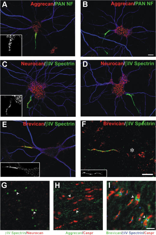
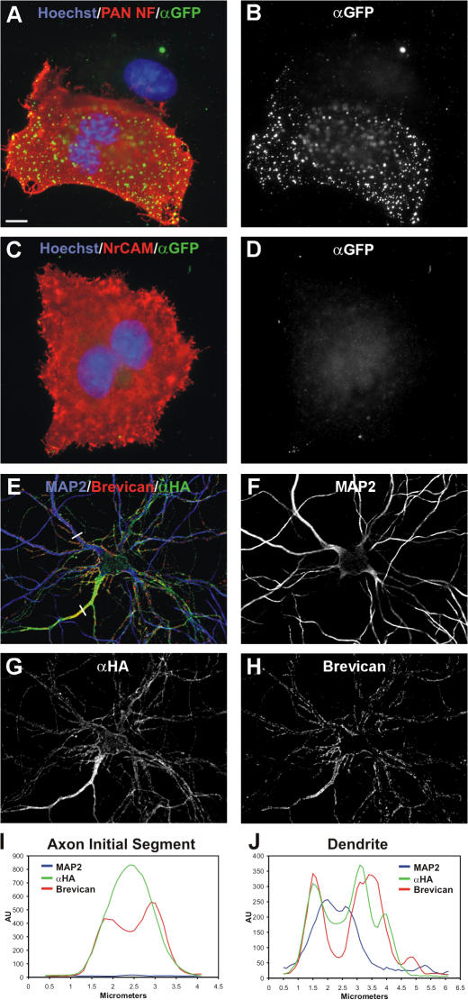
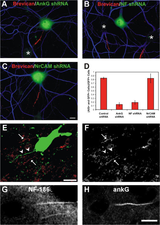
Action potential initiation and propagation requires clustered Na+ (voltage-gated Na+ [Nav]) channels at axon initial segments (AIS) and nodes of Ranvier. In addition to ion channels, these domains are characterized by cell adhesion molecules (CAMs; neurofascin-186 [NF-186] and neuron glia–related CAM [NrCAM]), cytoskeletal proteins (ankyrinG and βIV spectrin), and the extracellular chondroitin-sulfate proteoglycan brevican. Schwann cells initiate peripheral nervous system node formation by clustering NF-186, which then recruits ankyrinG and Nav channels. However, AIS assembly of this protein complex does not require glial contact. To determine the AIS assembly mechanism, we silenced expression of AIS proteins by RNA interference. AnkyrinG knockdown prevented AIS localization of all other AIS proteins. Loss of NF-186, NrCAM, Nav channels, or βIV spectrin did not affect other neuronal AIS proteins. However, loss of NF-186 blocked assembly of the brevican-based AIS extracellular matrix, and NF-186 overexpression caused somatodendritic brevican clustering. Thus, NF-186 assembles and links the specialized brevican-containing AIS extracellular matrix to the intracellular cytoskeleton.
Overview
The claims and statistics of this paper haven’t been checked yet.
Run the check — free, no account needed. It reads the open-access text and checks the sources it cites.
Integrity signals
Registry
Cites
Statistics
Figures
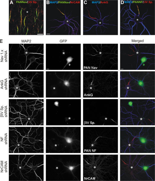
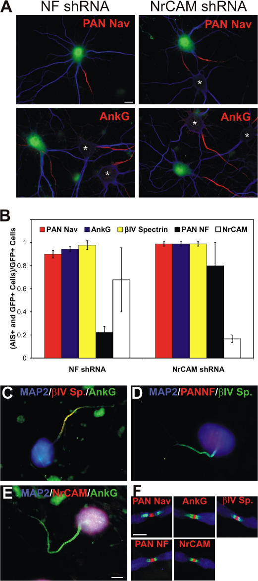
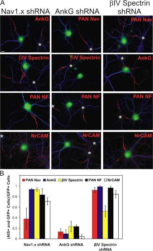
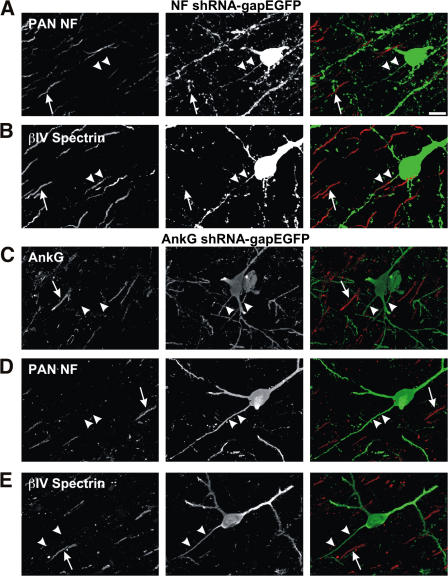
Verification
Claims the paper makes
The claims and statistics of this paper haven’t been checked yet.
Run the check — free, no account needed. It reads the open-access text and checks the sources it cites.
Claims other papers make about this one
Each is checked against this paper’s own text: does it say what the citing paper says it does?
In the citing paper
“Brevican enrichment in the AIS was mediated via direct interaction with NF186, which was diminished by a knockdown of NF186 (John et al., 2006;Hedstrom et al., 2007).”
From The hyaluronan and proteoglycan link proteins: Organizers of the brain extracellular matrix and key molecules for neuronal function and plasticity (2015) · cites this paper as Hedstrom et al., 2007
In the citing paper
“Brevican can interact with NF186 in the AIS as described before (Hedstrom et al., 2007).”
From The hyaluronan and proteoglycan link proteins: Organizers of the brain extracellular matrix and key molecules for neuronal function and plasticity (2015) · cites this paper as Hedstrom et al., 2007
Self-check
Percentages, sums and reported differences re-computed from the paper’s own numbers.
The self-check needs the paper’s full text.
Citations
Cites
Banker, G. A., & Goslin, K. (1998) · Cited by 1,290
Neuronal polarity: from extracellular signals to intracellular mechanisms.
Arimura, N., & Kaibuchi, K. (2007) · Nature Reviews Neuroscience · doi:10.1038/nrn2056 · Cited by 655
Lecticans: organizers of the brain extracellular matrix
Yamaguchi, Y. (2000) · Cellular and Molecular Life Sciences · doi:10.1007/pl00000690 · Cited by 642
Pan, Z., Kao, T., Horvath, Z., Lemos, J. C., Sul, J., Cranstoun, S. D., Bennett, V., Scherer, S. S., & Cooper, E. C. (2006) · Journal of Neuroscience · doi:10.1523/jneurosci.4314-05.2006 · Cited by 586
Zhou, D., Lambert, S., Malen, P. L., Carpenter, S., Boland, L. M., & Bennett, V. (1998) · The Journal of Cell Biology · doi:10.1083/jcb.143.5.1295 · Cited by 568
Perineuronal nets: past and present
Celio, M. R., Spreafico, R., De Biasi, S., & Vitellaro-Zuccarello, L. (1998) · Trends in Neurosciences · doi:10.1016/s0166-2236(98)01298-3 · Cited by 497
Jenkins, S. M., & Bennett, V. (2001) · The Journal of Cell Biology · doi:10.1083/jcb.200109026 · Cited by 446
Unique features of action potential initiation in cortical neurons
Naundorf, B., Wolf, F., & Volgushev, M. (2006) · Nature · doi:10.1038/nature04610 · Cited by 396
In vivo electroporation in the embryonic mouse central nervous system
Saito, T. (2006) · Nature Protocols · doi:10.1038/nprot.2006.276 · Cited by 378
Ango, F., Cristo, G. D., Higashiyama, H., Bennett, V., Wu, P., & Huang, Z. J. (2004) · Cell · doi:10.1016/j.cell.2004.10.004 · Cited by 358
Cited by
Sorted by citations
The roles of perineuronal nets and the perinodal extracellular matrix in neuronal function
Fawcett, J. W., Oohashi, T., & Pizzorusso, T. (2019) · Nature Reviews Neuroscience · doi:10.1038/s41583-019-0196-3 · Cited by 589
Debanne, D., Campanac, E., Bialowas, A., Carlier, E., & Alcaraz, G. (2011) · Physiological Reviews · doi:10.1152/physrev.00048.2009 · Cited by 633
Brain extracellular matrix affects AMPA receptor lateral mobility and short-term synaptic plasticity
Frischknecht, R., Heine, M., Perrais, D., Seidenbecher, C. I., Choquet, D., & Gundelfinger, E. D. (2009) · Nature Neuroscience · doi:10.1038/nn.2338 · Cited by 554
Self-citationEstablishment of axon-dendrite polarity in developing neurons.
Barnes, A. P., & Polleux, F. (2009) · Annual Review of Neuroscience · doi:10.1146/annurev.neuro.31.060407.125536 · Cited by 523
Extracellular matrix of the central nervous system: from neglect to challenge
Zimmermann, D. R., & Dours-Zimmermann, M. T. (2008) · Histochemistry and Cell Biology · doi:10.1007/s00418-008-0485-9 · Cited by 465
The axon initial segment and the maintenance of neuronal polarity
Rasband, M. N. (2010) · Nature Reviews Neuroscience · doi:10.1038/nrn2852 · Cited by 442
Self-citationCell-type-dependent molecular composition of the axon initial segment
Lorincz, A., & Nusser, Z. (2008) · The Journal of Neuroscience · doi:10.1523/jneurosci.4833-08.2008 · Cited by 382
AnkyrinG is required for maintenance of the axon initial segment and neuronal polarity
Hedstrom, K. L., Ogawa, Y., & Rasband, M. N. (2008) · The Journal of Cell Biology · doi:10.1083/jcb.200806112 · Cited by 371
Self-citationThe Axon Initial Segment: An Updated Viewpoint
Leterrier, C. (2018) · Journal of Neuroscience · doi:10.1523/jneurosci.1922-17.2018 · Cited by 317
Crosstalk between glia, extracellular matrix and neurons
Song, I., & Dityatev, A. E. (2017) · Brain Research Bulletin · doi:10.1016/j.brainresbull.2017.03.003 · Cited by 270
Nanoscale Architecture of the Axon Initial Segment Reveals an Organized and Robust Scaffold
Leterrier, C., Potier, J., Caillol, G., Debarnot, C., Boroni-Rueda, F., & Dargent, B. (2015) · Cell Reports · doi:10.1016/j.celrep.2015.11.051 · Cited by 249
Galiano, M. R., Jha, S., Ho, T. S., Zhang, C., Ogawa, Y., Chang, K.-J., Stankewich, M. C., Mohler, P. J., & Rasband, M. N. (2012) · Cell · doi:10.1016/j.cell.2012.03.039 · Cited by 245
Self-citationDityatev, A. E., Seidenbecher, C. I., & Schachner, M. (2010) · Trends in Neurosciences · doi:10.1016/j.tins.2010.08.003 · Cited by 235
Self-citationKochlamazashvili, G., Henneberger, C., Bukalo, O., Dvoretskova, E., Senkov, O., Lievens, P. M.-J., Westenbroek, R., Engel, A. K., Catterall, W. A., Rusakov, D. A., et al. (2010) · Neuron · doi:10.1016/j.neuron.2010.05.030 · Cited by 215
Disruption of the Axon Initial Segment Cytoskeleton Is a New Mechanism for Neuronal Injury
Schafer, D. P., Jha, S., Liu, F., Akella, T., McCullough, L. D., & Rasband, M. N. (2009) · Journal of Neuroscience · doi:10.1523/jneurosci.3376-09.2009 · Cited by 220
Self-citationAnimal models of multiple sclerosis—Potentials and limitations
Mix, E., Meyer-Rienecker, H., Hartung, H., & Zettl, U. K. (2010) · Progress in Neurobiology · doi:10.1016/j.pneurobio.2010.06.005 · Cited by 224
Gumy, L. F., Katrukha, E. A., Grigoriev, I., Jaarsma, D., Kapitein, L. C., Akhmanova, A., & Hoogenraad, C. C. (2017) · Neuron · doi:10.1016/j.neuron.2017.03.046 · Cited by 186
Three mechanisms assemble central nervous system nodes of Ranvier
Susuki, K., Chang, K.-J., Zollinger, D. R., Liu, Y., Ogawa, Y., Eshed-Eisenbach, Y., Dours-Zimmermann, M. T., Oses-Prieto, J. A., Burlingame, A. L., Seidenbecher, C. I., et al. (2013) · Neuron · doi:10.1016/j.neuron.2013.03.005 · Cited by 186
“In support of this notion, Bcan was shown to bind NF186 (Hedstrom et al., 2007).”
BackgroundMethodResultInfluentialSelf-citationAxon initial segments: structure, function, and disease
Huang, C. Y., & Rasband, M. N. (2018) · Annals of the New York Academy of Sciences · doi:10.1111/nyas.13718 · Cited by 204
Self-citationNav1.1 is predominantly expressed in nodes of Ranvier and axon initial segments
Duflocq, A., Bras, B. L., Bullier, E., Couraud, F., & Davenne, M. (2008) · Molecular and Cellular Neuroscience · doi:10.1016/j.mcn.2008.06.008 · Cited by 188
Membrane Domains Based on Ankyrin and Spectrin Associated with Cell-Cell InteractionsPreprint
Bennett, V., & Healy, J. (2009) · Cold Spring Harbor Perspectives in Biology · doi:10.1101/cshperspect.a003012 · Cited by 191
The Nodes of Ranvier: Molecular Assembly and Maintenance
Rasband, M. N., & Peles, E. (2016) · Cold Spring Harbor Perspectives in Biology · doi:10.1101/cshperspect.a020495 · Cited by 190
Self-citationAxon Initial Segment Cytoskeleton: Architecture, Development, and Role in Neuron Polarity
Jones, S. L., & Svitkina, T. (2016) · Neural Plasticity · doi:10.1155/2016/6808293 · Cited by 191
The axonal cytoskeleton: from organization to function
Kevenaar, J. T., & Hoogenraad, C. C. (2015) · Frontiers in Molecular Neuroscience · doi:10.3389/fnmol.2015.00044 · Cited by 200
Bréchet, A., Fache, M., Brachet, A., Ferracci, G., Baude, A., Irondelle, M., Pereira, S., Leterrier, C., & Dargent, B. (2008) · The Journal of Cell Biology · doi:10.1083/jcb.200805169 · Cited by 177
The nano-architecture of the axonal cytoskeleton
Leterrier, C., Dubey, P., & Roy, S. (2017) · Nature reviews. Neuroscience · doi:10.1038/nrn.2017.129 · Cited by 175
Mechanisms of node of Ranvier assembly
Rasband, M. N., & Peles, E. (2020) · Nature reviews. Neuroscience · doi:10.1038/s41583-020-00406-8 · Cited by 180
Self-citationEnd-binding proteins EB3 and EB1 link microtubules to ankyrin G in the axon initial segment
Leterrier, C., Vacher, H., Fache, M., d'Ortoli, S. A., Castets, F., Autillo‐Touati, A., & Dargent, B. (2011) · Proceedings of the National Academy of Sciences · doi:10.1073/pnas.1018671108 · Cited by 171
Spectrin- and Ankyrin-Based Membrane Domains and the Evolution of Vertebrates
Bennett, V., & Lorenzo, D. N. (2013) · Current topics in membranes · doi:10.1016/b978-0-12-417027-8.00001-5 · Cited by 159
Zonta, B., Desmazières, A., Rinaldi, A., Tait, S., Sherman, D. L., Nolan, M. F., & Brophy, P. (2011) · Neuron · doi:10.1016/j.neuron.2011.02.021 · Cited by 162
Short- and long-term plasticity at the axon initial segment
Grubb, M., Shu, Y.-S., Kuba, H., Rasband, M., Wimmer, V., & Bender, K. J. (2011) · Journal of Neuroscience · doi:10.1523/jneurosci.4064-11.2011 · Cited by 157
Self-citationCell-Permeant Large Stokes Shift Dyes for Transfection-Free Multicolor Nanoscopy
Butkevich, A. N., Lukinavičius, G., D’Este, E., & Hell, S. W. (2017) · Journal of the American Chemical Society · doi:10.1021/jacs.7b06412 · Cited by 151
Molecular Microdomains in a Sensory Terminal, the Vestibular Calyx Ending
Lysakowski, A., Gaboyard-Niay, S., Calin‐Jageman, I. E., Chatlani, S., Price, S. D., & Eatock, R. A. (2011) · Journal of Neuroscience · doi:10.1523/jneurosci.0521-11.2011 · Cited by 150
Ultrastructure of the axonal periodic scaffold reveals a braid-like organization of actin rings
Vassilopoulos, S., Gibaud, S., Jimenez, A., Caillol, G., & Leterrier, C. (2019) · Nature Communications · doi:10.1038/s41467-019-13835-6 · Cited by 143
The intriguing nature of dorsal root ganglion neurons: Linking structure with polarity and function
Nascimento, A. I., Mar, F. M., & Sousa, M. M. (2018) · Progress in Neurobiology · doi:10.1016/j.pneurobio.2018.05.002 · Cited by 149
Geissler, M., Gottschling, C., Aguado, A., Rauch, U., Wetzel, C. H., Hatt, H., & Faissner, A. (2013) · The Journal of Neuroscience · doi:10.1523/jneurosci.3275-12.2013 · Cited by 140
The role of NrCAM in neural development and disorders—Beyond a simple glue in the brain
Sakurai, T. (2011) · Molecular and Cellular Neuroscience · doi:10.1016/j.mcn.2011.12.002 · Cited by 150
Zempel, H., Dennissen, F. J. A., Kumar, Y., Luedtke, J., Biernat, J., Mandelkow, E., & Mandelkow�, E. (2017) · Journal of Biological Chemistry · doi:10.1074/jbc.m117.784702 · Cited by 147
Postsynaptic Density-93 Clusters Kv1 Channels at Axon Initial Segments Independently of Caspr2
Ogawa, Y., Horresh, I., Trimmer, J. S., Bredt, D. S., Peles, E., & Rasband, M. N. (2008) · Journal of Neuroscience · doi:10.1523/jneurosci.4431-07.2008 · Cited by 141
Self-citationBrackenbury, W. J., Calhoun, J. D., Chen, C., Miyazaki, H., Nukina, N., Oyama, F., Ranscht, B., & Isom, L. L. (2010) · Proceedings of the National Academy of Sciences · doi:10.1073/pnas.0909434107 · Cited by 139
Axon Initial Segment–Associated Microglia
Baalman, K., Marin, M. A., Ho, T., Godoy, M. I., Cherian, L., Robertson, C., & Rasband, M. (2015) · Journal of Neuroscience · doi:10.1523/jneurosci.3751-14.2015 · Cited by 129
Self-citationConverting juvenile into adult plasticity: a role for the brain's extracellular matrix
Gundelfinger, E. D., Frischknecht, R., Choquet, D., & Heine, M. (2010) · European Journal of Neuroscience · doi:10.1111/j.1460-9568.2010.07253.x · Cited by 136
Self-citationPanzanelli, P., Gunn, B. G., Schlatter, M. C., Benke, D., Tyagarajan, S. K., Scheiffele, P., Belelli, D., Lambert, J. J., Rudolph, U., & Fritschy, J. (2011) · The Journal of Physiology · doi:10.1113/jphysiol.2011.216028 · Cited by 140
Molecular mechanisms of node of Ranvier formation
Susuki, K., & Rasband, M. N. (2008) · Current Opinion in Cell Biology · doi:10.1016/j.ceb.2008.09.007 · Cited by 138
Self-citationOohashi, T., Edamatsu, M., Bekku, Y., & Carulli, D. (2015) · Experimental Neurology · doi:10.1016/j.expneurol.2015.09.010 · Cited by 131
Duman, J. G., Tzeng, C. P., Tu, Y.-K., Munjal, T., Schwechter, B., Ho, T. S., & Tolias, K. F. (2013) · Journal of Neuroscience · doi:10.1523/jneurosci.3978-12.2013 · Cited by 135
Gasser, A. S., Ho, T. S., Cheng, X., Chang, K.-J., Waxman, S. G., Rasband, M. N., & Dib‐Hajj, S. D. (2012) · Journal of Neuroscience · doi:10.1523/jneurosci.5434-11.2012 · Cited by 130
Self-citationSorting of Dendritic and Axonal Vesicles at the Pre-axonal Exclusion Zone
Farı́as, G. G., Guardia, C. M. A., Britt, D. J., Guo, X., & Bonifacino, J. S. (2015) · Cell Reports · doi:10.1016/j.celrep.2015.09.074 · Cited by 128
Rbfox Splicing Factors Promote Neuronal Maturation and Axon Initial Segment Assembly
Jacko, M., Weyn‐Vanhentenryck, S. M., Smerdon, J. W., Yan, R., Feng, H., Williams, D. J., Pai, J. A., Xu, K., Wichterle, H., & Zhang, C. (2018) · Neuron · doi:10.1016/j.neuron.2018.01.020 · Cited by 131
Ankyrin protein networks in membrane formation and stabilization
Cunha, S. R., & Mohler, P. J. (2009) · Journal of Cellular and Molecular Medicine · doi:10.1111/j.1582-4934.2009.00943.x · Cited by 125
Versican V2 Assembles the Extracellular Matrix Surrounding the Nodes of Ranvier in the CNS
Dours-Zimmermann, M. T., Maurer, K., Rauch, U., Stoffel, W., Fassler, R., & Zimmermann, D. R. (2009) · Journal of Neuroscience · doi:10.1523/jneurosci.4158-08.2009 · Cited by 123
Bral1: its role in diffusion barrier formation and conduction velocity in the CNS
Bekku, Y., Vargová, L., Goto, Y., Vorísek, I., Dmytrenko, L., Narasaki, M., Ohtsuka, A., Fässler, R., Ninomiya, Y., Syková, E., et al. (2010) · The Journal of Neuroscience · doi:10.1523/jneurosci.5598-09.2010 · Cited by 123
Calcineurin Signaling Mediates Activity-Dependent Relocation of the Axon Initial Segment
Evans, M. D., Sammons, R. P., Lebron, S., Dumitrescu, A. S., Watkins, T. B. K., Uebele, V. N., Renger, J. J., & Grubb, M. S. (2013) · Journal of Neuroscience · doi:10.1523/jneurosci.0277-13.2013 · Cited by 122
Horresh, I., Poliak, S., Grant, S., Bredt, D., Rasband, M., & Peles, E. (2008) · Journal of Neuroscience · doi:10.1523/jneurosci.3398-08.2008 · Cited by 116
Self-citationBaines, A. J. (2010) · PROTOPLASMA · doi:10.1007/s00709-010-0181-1 · Cited by 125
A hierarchy of ankyrin-spectrin complexes clusters sodium channels at nodes of Ranvier
Ho, T. S., Zollinger, D. R., Chang, K.-J., Xu, M., Cooper, E. C., Stankewich, M. C., Bennett, V., & Rasband, M. N. (2014) · Nature Neuroscience · doi:10.1038/nn.3859 · Cited by 116
Self-citationThe functional organization and assembly of the axon initial segment
Ogawa, Y., & Rasband, M. N. (2008) · Current Opinion in Neurobiology · doi:10.1016/j.conb.2008.08.008 · Cited by 116
Self-citationBrevican: A key proteoglycan in the perisynaptic extracellular matrix of the brain
Frischknecht, R., & Seidenbecher, C. I. (2012) · The International Journal of Biochemistry & Cell Biology · doi:10.1016/j.biocel.2012.03.022 · Cited by 112
Self-citationJones, S. L., Korobova, F., & Svitkina, T. (2014) · Journal of Cell Biology · doi:10.1083/jcb.201401045 · Cited by 111
The axon initial segment in nervous system disease and injury
Buffington, S. A., & Rasband, M. N. (2011) · European Journal of Neuroscience · doi:10.1111/j.1460-9568.2011.07875.x · Cited by 114
Self-citationMapping axon initial segment structure and function by multiplexed proximity biotinylation
Hamdan, H., Lim, B. C., Torii, T., Joshi, A. R., Koenning, M., Smith, C. L., Palmer, D. J., Ng, P., Leterrier, C., Osés-Prieto, J. A., et al. (2020) · Nature Communications · doi:10.1038/s41467-019-13658-5 · Cited by 110
Self-citationBuilding and maintaining the axon initial segment
Grubb, M. S., & Burrone, J. (2010) · Current Opinion in Neurobiology · doi:10.1016/j.conb.2010.04.012 · Cited by 106
Bekku, Y., Rauch, U., Ninomiya, Y., & Oohashi, T. (2009) · Journal of Neurochemistry · doi:10.1111/j.1471-4159.2009.05873.x · Cited by 104
Moore, A. R., Filipovic, R., Mo, Z., Rasband, M. N., Zečević, N., & Antic, S. D. (2008) · Cerebral Cortex · doi:10.1093/cercor/bhn206 · Cited by 103
Self-citationLocalized Myosin II Activity Regulates Assembly and Plasticity of the Axon Initial Segment
Berger, S. L., Leo‐Macías, A., Yuen, S. K., Khatri, L., Pfennig, S., Zhang, Y., Agulló-Pascual, E., Caillol, G., Zhu, M., Rothenberg, E., et al. (2018) · Neuron · doi:10.1016/j.neuron.2017.12.039 · Cited by 100
Brachet, A., Leterrier, C., Irondelle, M., Fache, M., Racine, V., Sibarita, J., Choquet, D., & Dargent, B. (2010) · The Journal of Cell Biology · doi:10.1083/jcb.201003042 · Cited by 97
Ankyrin-G Directly Binds to Kinesin-1 to Transport Voltage-Gated Na+ Channels into Axons
Barry, J., Gu, Y., Jukkola, P. I., O’Neill, B., Gu, H. H., Mohler, P. J., Rajamani, K. T., & Gu, C. (2014) · Developmental Cell · doi:10.1016/j.devcel.2013.11.023 · Cited by 97
Petersen, J. D., Kaech, S., & Banker, G. A. (2014) · Journal of Neuroscience · doi:10.1523/jneurosci.3779-13.2014 · Cited by 96
Leterrier, C., & Dargent, B. (2013) · Seminars in Cell and Developmental Biology · doi:10.1016/j.semcdb.2013.11.001 · Cited by 100
Huang, C. Y., Zhang, C., Ho, T. S., Osés-Prieto, J. A., Burlingame, A. L., Lalonde, J. W., Noebels, J. L., Leterrier, C., & Rasband, M. N. (2017) · Journal of Neuroscience · doi:10.1523/jneurosci.2112-17.2017 · Cited by 95
Self-citationSarmiere, P. D., Weigle, C. M., & Tamkun, M. M. (2008) · BMC Neuroscience · doi:10.1186/1471-2202-9-112 · Cited by 94
Glia unglued: How signals from the extracellular matrix regulate the development of myelinating glia
Colognato, H., & Tzvetanova, I. D. (2011) · Developmental Neurobiology · doi:10.1002/dneu.20966 · Cited by 92
Axon initial segment dysfunction in epilepsy
Wimmer, V. C., Reid, C. A., So, E., Berkovic, S. F., & Petrou, S. (2010) · The Journal of Physiology · doi:10.1113/jphysiol.2010.188417 · Cited by 93
Organization and maintenance of molecular domains in myelinated axons
Buttermore, E. D., Thaxton, C., & Bhat, M. A. (2013) · Journal of Neuroscience Research · doi:10.1002/jnr.23197 · Cited by 96
Nodes of Ranvier during development and repair in the CNS
Lubetzki, C., Sol‐Foulon, N., & Desmazières, A. (2020) · Nature Reviews Neurology · doi:10.1038/s41582-020-0375-x · Cited by 95
Neuro-glial interactions at the nodes of Ranvier: implication in health and diseases
Faivre‐Sarrailh, C., & Devaux, J. J. (2013) · Frontiers in Cellular Neuroscience · doi:10.3389/fncel.2013.00196 · Cited by 90
Neurofascin: A switch between neuronal plasticity and stability
Kriebel, M., Wuchter, J., Trinks, S., & Volkmer, H. (2012) · The International Journal of Biochemistry & Cell Biology · doi:10.1016/j.biocel.2012.01.012 · Cited by 86
The crosstalk of hyaluronan-based extracellular matrix and synapses
Frischknecht, R., & Seidenbecher, C. I. (2008) · Neuron Glia Biology · doi:10.1017/s1740925x09990226 · Cited by 85
Self-citationTai, Y., Gallo, N. B., Wang, M., Yu, J.-R., & Aelst, L. V. (2019) · Neuron · doi:10.1016/j.neuron.2019.02.009 · Cited by 86
GoFDR: A sequence alignment based method for predicting protein functions
Gong, Q., Ning, W., & Tian, W. (2015) · Methods · doi:10.1016/j.ymeth.2015.08.009 · Cited by 84
Structural basis of diverse membrane target recognitions by ankyrins
Wang, C., Wei, Z., Chen, K., Ye, F., Yu, C., Bennett, V., & Zhang, M. (2014) · eLife · doi:10.7554/elife.04353 · Cited by 84
Acceleration of conduction velocity linked to clustering of nodal components precedes myelination
Freeman, S. A., Desmazières, A., Simonnet, J., Gatta, M., Pfeiffer, F., Aigrot, M. S., Rappeneau, Q., Guerreiro, S., Michel, P. P., Yanagawa, Y., et al. (2015) · Proceedings of the National Academy of Sciences · doi:10.1073/pnas.1419099112 · Cited by 80
The Axon Initial Segment, 50 Years Later
Leterrier, C. (2015) · Current topics in membranes · doi:10.1016/bs.ctm.2015.10.005 · Cited by 87
He, M., Jenkins, P. M., & Bennett, V. (2012) · Journal of Biological Chemistry · doi:10.1074/jbc.m112.417501 · Cited by 78
Cooperative Interactions between 480 kDa Ankyrin-G and EB Proteins Assemble the Axon Initial Segment
Freal, A., Fassier, C., Bras, B. L., Bullier, E., Gois, S. D., Hazan, J., Hoogenraad, C. C., & Couraud, F. (2016) · Journal of Neuroscience · doi:10.1523/jneurosci.3219-15.2016 · Cited by 79
Voltage-gated sodium channel organization in neurons: Protein interactions and trafficking pathways
Leterrier, C., Brachet, A., Fache, M., & Dargent, B. (2010) · Neuroscience Letters · doi:10.1016/j.neulet.2010.08.079 · Cited by 75
Feedback-Driven Assembly of the Axon Initial Segment
Freal, A., Rai, D., Tas, R. P., Pan, X., Katrukha, E. A., van de Willige, D., Stucchi, R., Aher, A., Yang, C., Altelaar, M. A. F., et al. (2019) · Neuron · doi:10.1016/j.neuron.2019.07.029 · Cited by 75
Activity-Dependent and Activity-Independent Development of the Axon Initial Segment
Kuba, H., Adachi, R., & Ohmori, H. (2014) · Journal of Neuroscience · doi:10.1523/jneurosci.4357-13.2014 · Cited by 73
Alpizar, S. A., Baker, A. L., Gulledge, A. T., & Hoppa, M. B. (2019) · Frontiers in Cellular Neuroscience · doi:10.3389/fncel.2019.00001 · Cited by 73
Zhu, S., Cordner, Z. A., Xiong, J., Chiu, C.-T., Artola, A., Zuo, Y., Nelson, A. D., Kim, T.-Y., Zaika, N., Woolums, B. M., et al. (2017) · Proceedings of the National Academy of Sciences · doi:10.1073/pnas.1700689114 · Cited by 71
Kriebel, M., Metzger, J., Trinks, S., Chugh, D., Harvey, R. J., Harvey, K., & Volkmer, H. (2011) · Journal of Biological Chemistry · doi:10.1074/jbc.m110.212191 · Cited by 70
Buttermore, E. D., Piochon, C., Wallace, M. L., Philpot, B. D., Hansel, C., & Bhat, M. A. (2012) · Journal of Neuroscience · doi:10.1523/jneurosci.5602-11.2012 · Cited by 68
Diffusion Barriers, Mechanical Forces, and the Biophysics of Phagocytosis
Ostrowski, P. P., Grinstein, S., & Freeman, S. A. (2016) · Developmental Cell · doi:10.1016/j.devcel.2016.06.023 · Cited by 75
Pablo, J. L., Wang, C., Presby, M. M., & Pitt, G. S. (2016) · Proceedings of the National Academy of Sciences · doi:10.1073/pnas.1521194113 · Cited by 67
Ankyrin G Membrane Partners Drive the Establishment and Maintenance of the Axon Initial Segment
Leterrier, C., Clerc, N., Boroni-Rueda, F., Montersino, A., Dargent, B., & Castets, F. (2017) · Frontiers in Cellular Neuroscience · doi:10.3389/fncel.2017.00006 · Cited by 65
FGF14 localization and organization of the axon initial segment
Xiao, M., Bosch, M. K., Nerbonne, J. M., & Ornitz, D. M. (2013) · Molecular and Cellular Neuroscience · doi:10.1016/j.mcn.2013.07.008 · Cited by 63
The role of Gpi-anchored axonal glycoproteins in neural development and neurological disorders.
Gennarini, G., Bizzoca, A., Picocci, S., Puzzo, D., Corsi, P., & Furley, A. (2017) · Molecular and cellular neurosciences · doi:10.1016/j.mcn.2016.11.006 · Cited by 59
Appeltshauser, L., Junghof, H., Messinger, J., Linke, J., Haarmann, A., Ayzenberg, I., Baka, P., Dorst, J., Fisse, A. L., Grüter, T., et al. (2022) · Brain · doi:10.1093/brain/awac418 · Cited by 67
Wang, C.-C., Ortiz-Gonzalez, X. R., Yum, S. W., Gill, S. M., White, A. L., Kelter, E., Seaver, L. H., Lee, S., Wiley, G. B., Gaffney, P. M., et al. (2018) · The American Journal of Human Genetics · doi:10.1016/j.ajhg.2018.04.012 · Cited by 63
Self-citationBakkum, D. J., Obien, M. E. J., Radivojević, M., Jäckel, D., Frey, U., Takahashi, H., & Hierlemann, A. R. (2018) · Advanced Biosystems · doi:10.1002/adbi.201800308 · Cited by 73
Metadata
- Authors
- Hedstrom, K. L., Xu, X., Ogawa, Y., Frischknecht, R., Seidenbecher, C. I., Shrager, P., & Rasband, M. N.
- Affiliations
- UConn Health (US)
- University of Rochester Medicine (US)
- Leibniz Institute for Neurobiology (DE)
- Venue
- The Journal of Cell Biology
- Publisher
- Rockefeller University Press
- Published
- 2007-08-20
- Volume
- 178
- Issue
- 5
- Pages
- 875-886
- ISSN
- 0021-9525 (ISSN-L)
- 1540-8140
- PMC
- PMC2064550
- OpenAlex
- W2159315879
- Cited by
- 268
- References
- 45
- Citations by year
- 2026: 4 · 2025: 8 · 2024: 12 · 2023: 10 · 2022: 11 · 2021: 12 · 2020: 15 · 2019: 18 · 2018: 15 · 2017: 15 · 2016: 10 · 2015: 20 · 2014: 17 · 2013: 21 · 2012: 20
- FWCI
- 5.96
- Type
- journal article
- Language
- en
- Open access
- green
- www.ncbi.nlm.nih.gov
- Licence
- cc-by-nc-sa
- Locations
- The Journal of Cell Biology (publishedVersion)
- PubMed Central (publishedVersion, cc-by-nc-sa)
- PubMed (publishedVersion)
- Funders
- National Institute of Neurological Disorders and Stroke (R01NS017965, NS 044916, NS 17965, R01NS044916, R37NS044916)
- Topics
- Life Sciences › Biochemistry, Genetics and Molecular Biology › Molecular Biology › Ion channel regulation and function
- Life Sciences › Neuroscience › Cellular and Molecular Neuroscience › Neuroscience and Neuropharmacology Research
- Life Sciences › Biochemistry, Genetics and Molecular Biology › Molecular Biology › Ion Transport and Channel Regulation
- Keywords
- neurofascin 186, axon initial segment, extracellular matrix assembly, brevican, chondroitin sulfate proteoglycans, ankyrin-G, βIV-spectrin, nodes of Ranvier, Schwann cells, adhesion molecules, cytoskeletal anchoring, action potential generation, RNAi knockdown
- MeSH terms
- Animals, Axons, Cells, Cultured, Cytoskeleton, Extracellular Matrix, Hippocampus, Nerve Growth Factors, Nerve Tissue Proteins, Chondroitin Sulfate Proteoglycans, Ranvier's Nodes, Recombinant Fusion Proteins, RNA, Spectrin, Sodium Channels, Cell Adhesion Molecules, Ankyrins, RNA Interference, Lectins, C-Type, Rats, Brevican
About these checks
Registry, retraction, Cites and statistics results are computed, not judged, with one exception: a printed reference that no index lists is read once by a model before it counts as a citation error. Claims are extracted by a model and kept only when their quote appears verbatim in the text; each cited source is then checked, with a model’s help, for whether it says what the claim relies on. Those verdicts can be wrong — read the claim’s quote and the cited source before relying on one.
Statistics and arithmetic are recomputed, not judged.
Confidence bands are shown only for fields calibrated on our annotated test set. None is calibrated yet, so no band is shown.
What counts as a claim ›How we measure confidence ›How we check this ›
Are you an author, or do you see a problem with this page? Request a review
