

The DOI matches its Crossref record; 45 of the 45 references it cites were found in a registry or an index.
Neurofascin assembles a specialized extracellular matrix at the axon initial segment
Open accessAbstract
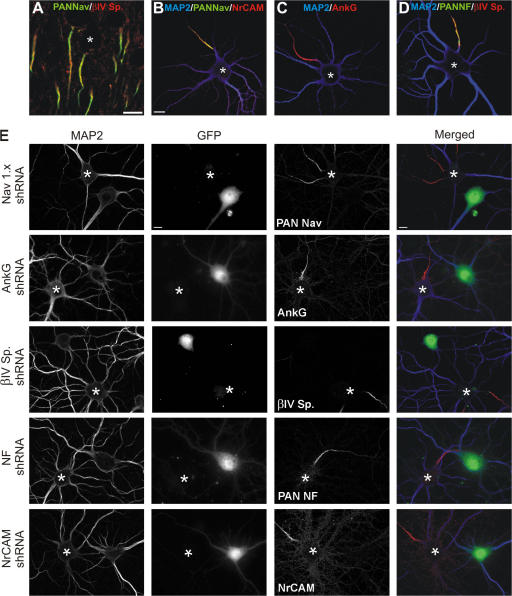
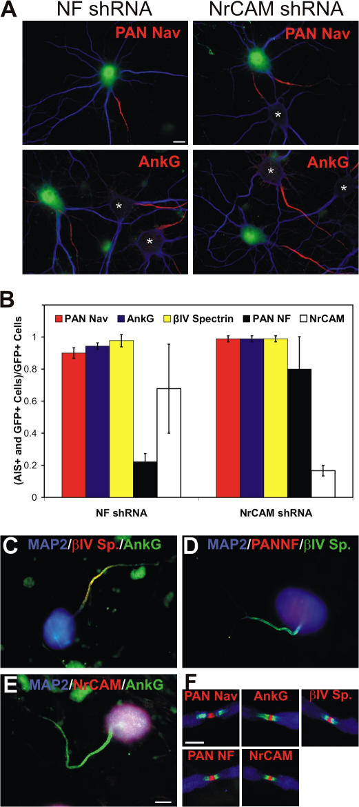
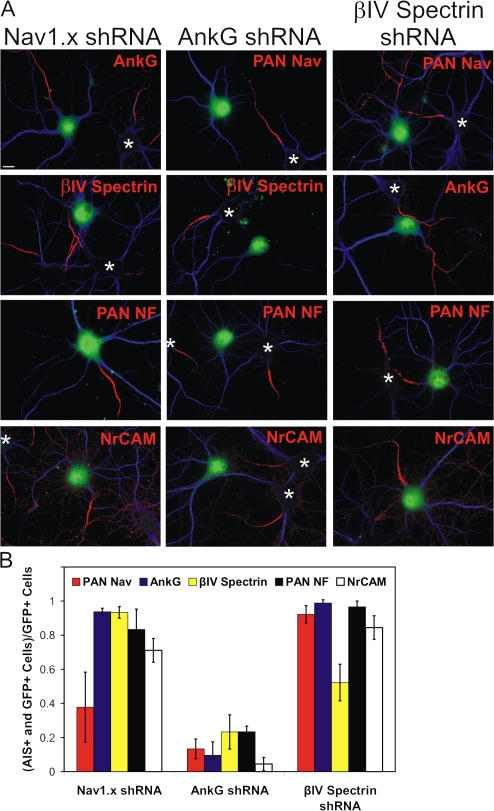
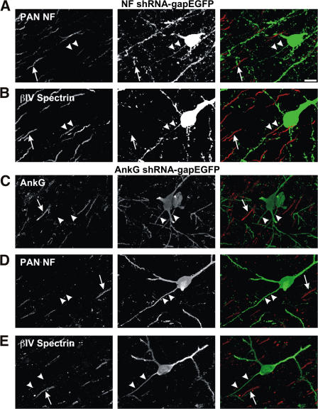
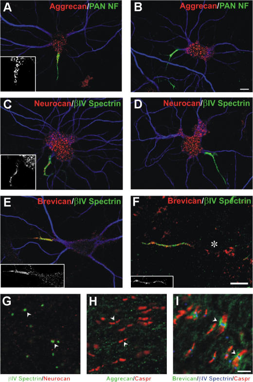
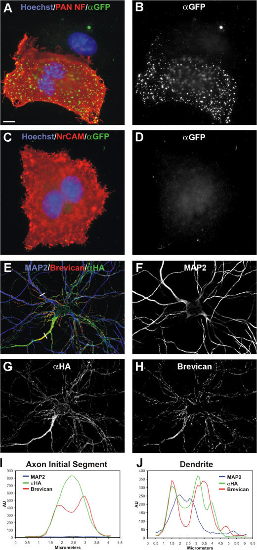
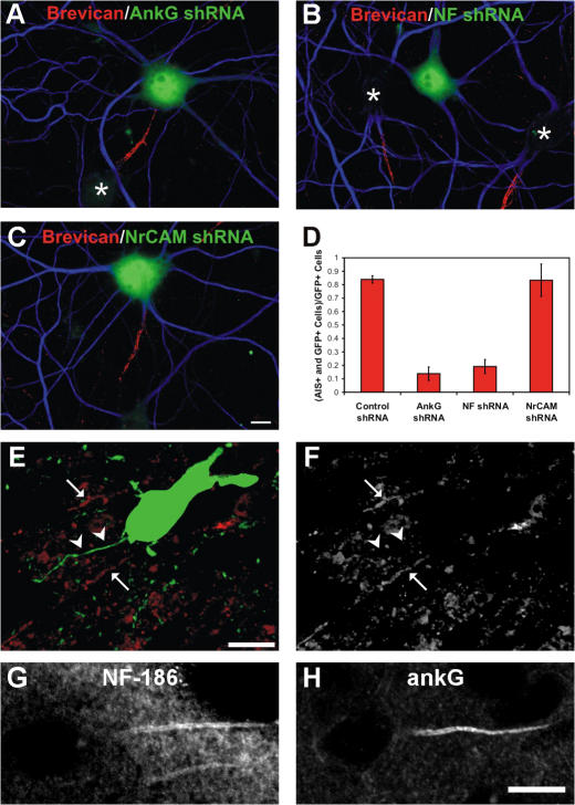
Action potential initiation and propagation requires clustered Na+ (voltage-gated Na+ [Nav]) channels at axon initial segments (AIS) and nodes of Ranvier. In addition to ion channels, these domains are characterized by cell adhesion molecules (CAMs; neurofascin-186 [NF-186] and neuron glia–related CAM [NrCAM]), cytoskeletal proteins (ankyrinG and βIV spectrin), and the extracellular chondroitin-sulfate proteoglycan brevican. Schwann cells initiate peripheral nervous system node formation by clustering NF-186, which then recruits ankyrinG and Nav channels. However, AIS assembly of this protein complex does not require glial contact. To determine the AIS assembly mechanism, we silenced expression of AIS proteins by RNA interference. AnkyrinG knockdown prevented AIS localization of all other AIS proteins. Loss of NF-186, NrCAM, Nav channels, or βIV spectrin did not affect other neuronal AIS proteins. However, loss of NF-186 blocked assembly of the brevican-based AIS extracellular matrix, and NF-186 overexpression caused somatodendritic brevican clustering. Thus, NF-186 assembles and links the specialized brevican-containing AIS extracellular matrix to the intracellular cytoskeleton.
Overview
The claims and statistics of this paper haven’t been checked yet.
Run the check — free, no account needed. It reads the open-access text and checks the sources it cites.
Integrity signals
Registry
Cites
Statistics
Figures
Verification
Claims the paper makes
The claims and statistics of this paper haven’t been checked yet.
Run the check — free, no account needed. It reads the open-access text and checks the sources it cites.
Claims other papers make about this one
Each is checked against this paper’s own text: does it say what the citing paper says it does?
In the citing paper
“Brevican enrichment in the AIS was mediated via direct interaction with NF186, which was diminished by a knockdown of NF186 (John et al., 2006;Hedstrom et al., 2007).”
From The hyaluronan and proteoglycan link proteins: Organizers of the brain extracellular matrix and key molecules for neuronal function and plasticity (2015) · cites this paper as Hedstrom et al., 2007
In the citing paper
“Brevican can interact with NF186 in the AIS as described before (Hedstrom et al., 2007).”
From The hyaluronan and proteoglycan link proteins: Organizers of the brain extracellular matrix and key molecules for neuronal function and plasticity (2015) · cites this paper as Hedstrom et al., 2007
Self-check
Percentages, sums and reported differences re-computed from the paper’s own numbers.
The self-check needs the paper’s full text.
Citations
Cites
Banker, G. A., & Goslin, K. (1998) · Cited by 1,290
Neuronal polarity: from extracellular signals to intracellular mechanisms.
Arimura, N., & Kaibuchi, K. (2007) · Nature Reviews Neuroscience · doi:10.1038/nrn2056 · Cited by 655
Lecticans: organizers of the brain extracellular matrix
Yamaguchi, Y. (2000) · Cellular and Molecular Life Sciences · doi:10.1007/pl00000690 · Cited by 642
Pan, Z., Kao, T., Horvath, Z., Lemos, J. C., Sul, J., Cranstoun, S. D., Bennett, V., Scherer, S. S., & Cooper, E. C. (2006) · Journal of Neuroscience · doi:10.1523/jneurosci.4314-05.2006 · Cited by 586
Zhou, D., Lambert, S., Malen, P. L., Carpenter, S., Boland, L. M., & Bennett, V. (1998) · The Journal of Cell Biology · doi:10.1083/jcb.143.5.1295 · Cited by 568
Perineuronal nets: past and present
Celio, M. R., Spreafico, R., De Biasi, S., & Vitellaro-Zuccarello, L. (1998) · Trends in Neurosciences · doi:10.1016/s0166-2236(98)01298-3 · Cited by 497
Jenkins, S. M., & Bennett, V. (2001) · The Journal of Cell Biology · doi:10.1083/jcb.200109026 · Cited by 446
Unique features of action potential initiation in cortical neurons
Naundorf, B., Wolf, F., & Volgushev, M. (2006) · Nature · doi:10.1038/nature04610 · Cited by 396
In vivo electroporation in the embryonic mouse central nervous system
Saito, T. (2006) · Nature Protocols · doi:10.1038/nprot.2006.276 · Cited by 378
Ango, F., Cristo, G. D., Higashiyama, H., Bennett, V., Wu, P., & Huang, Z. J. (2004) · Cell · doi:10.1016/j.cell.2004.10.004 · Cited by 358
A Targeting Motif Involved in Sodium Channel Clustering at the Axonal Initial Segment
Garrido, J. J., Giraud, P., Carlier, E., Fernandes, F., Moussif, A., Fache, M., Debanne, D., & Dargent, B. (2003) · Science · doi:10.1126/science.1085167 · Cited by 358
Neurofascins Are Required to Establish Axonal Domains for Saltatory Conduction
Sherman, D. L., Tait, S., Melrose, S., Johnson, R., Zonta, B., Court, F. A., Macklin, W. B., Meek, S. E., Smith, A., Cottrell, D. F., et al. (2005) · Neuron · doi:10.1016/j.neuron.2005.10.019 · Cited by 342
Site of Action Potential Initiation in Layer 5 Pyramidal Neurons
Palmer, L. M., & Stuart, G. J. (2006) · Journal of Neuroscience · doi:10.1523/jneurosci.4812-05.2006 · Cited by 336
Induction of sodium channel clustering by oligodendrocytes
Kaplan, M. R., Meyer‐Franke, A., Lambert, S., Bennett, V., Duncan, I. D., Levinson, S. R., & Barres, B. A. (1997) · Nature · doi:10.1038/386724a0 · Cited by 332
Gliomedin mediates Schwann cell-axon interaction and the molecular assembly of the nodes of Ranvier
Eshed, Y., Feinberg, K., Poliak, S., Sabanay, H., Sarig-Nadir, O., Spiegel, I., Bermingham, J. R., & Peles, E. (2005) · Neuron · doi:10.1016/j.neuron.2005.06.026 · Cited by 321
Komada, M., & Soriano, P. M. (2002) · The Journal of Cell Biology · doi:10.1083/jcb.200110003 · Cited by 291
A Novel Targeting Signal for Proximal Clustering of the Kv2.1 K+ Channel in Hippocampal Neurons
Seung Taik, L. I. M., Antonucci-Durgan, D. E., Scannevin, R. H., & Trimmer, J. S. (2000) · Neuron · doi:10.1016/s0896-6273(00)80902-2 · Cited by 288
Brakebusch, C., Seidenbecher, C. I., Asztely, F., Rauch, U., Matthies, H., Meyer, H., Krug, M., Böckers, T. M., Zhou, X., Kreutz, M. R., et al. (2002) · Molecular and Cellular Biology · doi:10.1128/mcb.22.21.7417-7427.2002 · Cited by 264
Self-citationWeber, P., Bartsch, U., Rasband, M. N., Czaniera, R., Lang, Y., Bluethmann, H., Margolis, R. U., Levinson, S. R., Shrager, P., Montag, D., et al. (1999) · The Journal of Neuroscience · doi:10.1523/jneurosci.19-11-04245.1999 · Cited by 247
Self-citationIdentification of a Conserved Ankyrin-binding Motif in the Family of Sodium Channel α Subunits
Lemaillet, G., Walker, B. J., & Lambert, S. (2003) · Journal of Biological Chemistry · doi:10.1074/jbc.m303327200 · Cited by 243
Garver, T. D., Ren, Q., Tuvia, S., & Bennett, V. (1997) · The Journal of Cell Biology · doi:10.1083/jcb.137.3.703 · Cited by 243
Shu, Y., Duque, A., Yu, Y., Haider, B., & McCormick, D. A. (2006) · Journal of Neurophysiology · doi:10.1152/jn.00922.2006 · Cited by 229
Dzhashiashvili, Y., Zhang, Y., Galinska, J., Lam, I., Grumet, M., & Salzer, J. L. (2007) · The Journal of Cell Biology · doi:10.1083/jcb.200612012 · Cited by 206
βIV spectrin is recruited to axon initial segments and nodes of Ranvier by ankyrinG
Yang, Y., Ogawa, Y., Hedstrom, K. L., & Rasband, M. N. (2007) · The Journal of Cell Biology · doi:10.1083/jcb.200610128 · Cited by 198
Self-citationInda, M. C., DeFelipe, J., & Muñoz, A. (2006) · Proceedings of the National Academy of Sciences · doi:10.1073/pnas.0511197103 · Cited by 192
Davis, J. Q., McLaughlin, T. M., & Bennett, V. (1993) · The Journal of Cell Biology · doi:10.1083/jcb.121.1.121 · Cited by 170
Uncovering multiple axonal targeting pathways in hippocampal neurons
Wisco, D., Anderson, E. D., Chang, M. C., Norden, C., Boiko, T., Fölsch, H., & Winckler, B. (2003) · The Journal of Cell Biology · doi:10.1083/jcb.200307069 · Cited by 163
Spectrins and AnkyrinB Constitute a Specialized Paranodal Cytoskeleton
Ogawa, Y., Schafer, D. P., Horresh, I., Bar, V., Hales, K., Yang, Y., Susuki, K., Peles, E., Stankewich, M. C., & Rasband, M. N. (2006) · Journal of Neuroscience · doi:10.1523/jneurosci.0425-06.2006 · Cited by 162
Self-citationGlial Membranes at the Node of Ranvier Prevent Neurite Outgrowth
Huang, J. K., Phillips, G. R., Roth, A. D., Pedraza, L., Shan, W., Belkaïd, W., Mi, S., Svenningsen, Å. F., Florens, L. A., Yates, J. R., et al. (2005) · Science · doi:10.1126/science.1118313 · Cited by 160
Khaliq, Z. M., & Raman, I. M. (2006) · Journal of Neuroscience · doi:10.1523/jneurosci.4664-05.2006 · Cited by 157
Yang, Y., Lacas-Gervais, S., Morest, D., Solimena, M., & Rasband, M. (2004) · Journal of Neuroscience · doi:10.1523/jneurosci.2125-04.2004 · Cited by 153
Self-citationDoes Paranode Formation and Maintenance Require Partitioning of Neurofascin 155 into Lipid Rafts?
Schafer, D. P., Bansal, R., Hedstrom, K. L., Pfeiffer, S., & Rasband, M. (2004) · Journal of Neuroscience · doi:10.1523/jneurosci.5427-03.2004 · Cited by 146
Self-citationZhang, X., & Bennett, V. (1998) · The Journal of Cell Biology · doi:10.1083/jcb.142.6.1571 · Cited by 132
John, N., Krügel, H., Frischknecht, R., Smalla, K.-H., Schultz, C., Kreutz, M. R., Gundelfinger, E. D., & Seidenbecher, C. I. (2006) · Molecular and Cellular Neuroscience · doi:10.1016/j.mcn.2006.01.011 · Cited by 122 · cited 3×
“The guinea pig anti-brevican antibodies were previously described (John et al., 2006).”
BackgroundResultInfluentialSelf-citationAn evolutionarily conserved dileucine motif in Shal K+ channels mediates dendritic targeting
Rivera, J. F., Ahmad, S., Quick, M. W., Liman, E. R., & Arnold, D. B. (2003) · Nature Neuroscience · doi:10.1038/nn1020 · Cited by 119
Boiko, T., Vakulenko, M., Ewers, H., Yap, C. C., Norden, C., & Winckler, B. (2007) · Journal of Neuroscience · doi:10.1523/jneurosci.4302-06.2007 · Cited by 112
Secreted gliomedin is a perinodal matrix component of peripheral nerves
Eshed, Y., Feinberg, K., Carey, D. J., & Peles, E. (2007) · The Journal of Cell Biology · doi:10.1083/jcb.200612139 · Cited by 103
Rapid method for culturing embryonic neuron–glial cell cocultures
Svenningsen, Å. F., Shan, W., Colman, D., & Pedraza, L. (2003) · Journal of Neuroscience Research · doi:10.1002/jnr.10610 · Cited by 91
The Role of the Ankyrin-Binding Protein NrCAM in Node of Ranvier Formation
Custer, A., Kazarinova-Noyes, K., Sakurai, T., Xu, X.-R., Simon, W., Grumet, M., & Shrager, P. (2003) · Journal of Neuroscience · doi:10.1523/jneurosci.23-31-10032.2003 · Cited by 83
Self-citationIntrinsic and extrinsic determinants of ion channel localization in neurons
Hedstrom, K. L., & Rasband, M. (2006) · Journal of Neurochemistry · doi:10.1111/j.1471-4159.2006.04001.x · Cited by 67
Self-citationGlial regulation of the axonal membrane at nodes of Ranvier
Schafer, D. P., & Rasband, M. N. (2006) · Current Opinion in Neurobiology · doi:10.1016/j.conb.2006.08.003 · Cited by 59
Self-citationDependence of axon initial segment formation on Na+ channel expression
Xu, X., & Shrager, P. G. (2005) · Journal of Neuroscience Research · doi:10.1002/jnr.20378 · Cited by 58
Self-citationDifferential expression of proteoglycans at central and peripheral nodes of Ranvier
Melendez‐Vasquez, C. V., Carey, D. J., Zanazzi, G. J., Reizes, O., Maurel, P., & Salzer, J. L. (2005) · Glia · doi:10.1002/glia.20245 · Cited by 57
Schultz, C., König, H., Turco, D. D., Politi, C., Eckert, G. P., Ghebremedhin, E., Prehn, J. H. M., Kögel, D., & Deller, T. (2006) · Molecular and Cellular Neuroscience · doi:10.1016/j.mcn.2006.06.008 · Cited by 52
Oligodendrocytes regulate formation of nodes of Ranvier via the recognition molecule OMgp
Nie, D. A., Ma, Q., Law, J. W. S., Chia, C., Dhingra, N. K., Shimoda, Y., Yang, W.-L., Gong, N., CHEN, Q.-W., Xu, G., et al. (2006) · Neuron Glia Biology · doi:10.1017/s1740925x06000251 · Cited by 29
Cited by
Sorted by citations
The roles of perineuronal nets and the perinodal extracellular matrix in neuronal function
Fawcett, J. W., Oohashi, T., & Pizzorusso, T. (2019) · Nature Reviews Neuroscience · doi:10.1038/s41583-019-0196-3 · Cited by 589
Debanne, D., Campanac, E., Bialowas, A., Carlier, E., & Alcaraz, G. (2011) · Physiological Reviews · doi:10.1152/physrev.00048.2009 · Cited by 633
Brain extracellular matrix affects AMPA receptor lateral mobility and short-term synaptic plasticity
Frischknecht, R., Heine, M., Perrais, D., Seidenbecher, C. I., Choquet, D., & Gundelfinger, E. D. (2009) · Nature Neuroscience · doi:10.1038/nn.2338 · Cited by 554
Self-citationEstablishment of axon-dendrite polarity in developing neurons.
Barnes, A. P., & Polleux, F. (2009) · Annual Review of Neuroscience · doi:10.1146/annurev.neuro.31.060407.125536 · Cited by 523
Extracellular matrix of the central nervous system: from neglect to challenge
Zimmermann, D. R., & Dours-Zimmermann, M. T. (2008) · Histochemistry and Cell Biology · doi:10.1007/s00418-008-0485-9 · Cited by 465
Metadata
- Authors
- Hedstrom, K. L., Xu, X., Ogawa, Y., Frischknecht, R., Seidenbecher, C. I., Shrager, P., & Rasband, M. N.
- Affiliations
- UConn Health (US)
- University of Rochester Medicine (US)
- Leibniz Institute for Neurobiology (DE)
- Venue
- The Journal of Cell Biology
- Publisher
- Rockefeller University Press
- Published
- 2007-08-20
- Volume
- 178
- Issue
- 5
- Pages
- 875-886
- ISSN
- 0021-9525 (ISSN-L)
- 1540-8140
- PMC
- PMC2064550
- OpenAlex
- W2159315879
- Cited by
- 268
- References
- 45
- Citations by year
- 2026: 4 · 2025: 8 · 2024: 12 · 2023: 10 · 2022: 11 · 2021: 12 · 2020: 15 · 2019: 18 · 2018: 15 · 2017: 15 · 2016: 10 · 2015: 20 · 2014: 17 · 2013: 21 · 2012: 20
- FWCI
- 5.96
- Type
- journal article
- Language
- en
- Open access
- green
- www.ncbi.nlm.nih.gov
- Licence
- cc-by-nc-sa
- Locations
- The Journal of Cell Biology (publishedVersion)
- PubMed Central (publishedVersion, cc-by-nc-sa)
- PubMed (publishedVersion)
- Funders
- National Institute of Neurological Disorders and Stroke (R01NS017965, NS 044916, NS 17965, R01NS044916, R37NS044916)
- Topics
- Life Sciences › Biochemistry, Genetics and Molecular Biology › Molecular Biology › Ion channel regulation and function
- Life Sciences › Neuroscience › Cellular and Molecular Neuroscience › Neuroscience and Neuropharmacology Research
- Life Sciences › Biochemistry, Genetics and Molecular Biology › Molecular Biology › Ion Transport and Channel Regulation
- Keywords
- neurofascin 186, axon initial segment, extracellular matrix assembly, brevican, chondroitin sulfate proteoglycans, ankyrin-G, βIV-spectrin, nodes of Ranvier, Schwann cells, adhesion molecules, cytoskeletal anchoring, action potential generation, RNAi knockdown
- MeSH terms
- Animals, Axons, Cells, Cultured, Cytoskeleton, Extracellular Matrix, Hippocampus, Nerve Growth Factors, Nerve Tissue Proteins, Chondroitin Sulfate Proteoglycans, Ranvier's Nodes, Recombinant Fusion Proteins, RNA, Spectrin, Sodium Channels, Cell Adhesion Molecules, Ankyrins, RNA Interference, Lectins, C-Type, Rats, Brevican
About these checks
Registry, retraction, Cites and statistics results are computed, not judged, with one exception: a printed reference that no index lists is read once by a model before it counts as a citation error. Claims are extracted by a model and kept only when their quote appears verbatim in the text; each cited source is then checked, with a model’s help, for whether it says what the claim relies on. Those verdicts can be wrong — read the claim’s quote and the cited source before relying on one.
Statistics and arithmetic are recomputed, not judged.
Confidence bands are shown only for fields calibrated on our annotated test set. None is calibrated yet, so no band is shown.
What counts as a claim ›How we measure confidence ›How we check this ›
Are you an author, or do you see a problem with this page? Request a review
