

The DOI matches its Crossref record; 64 of the 65 references it cites were found in a registry or an index.
Mitophagy of damaged mitochondria occurs locally in distal neuronal axons and requires PINK1 and Parkin
Open accessAbstract
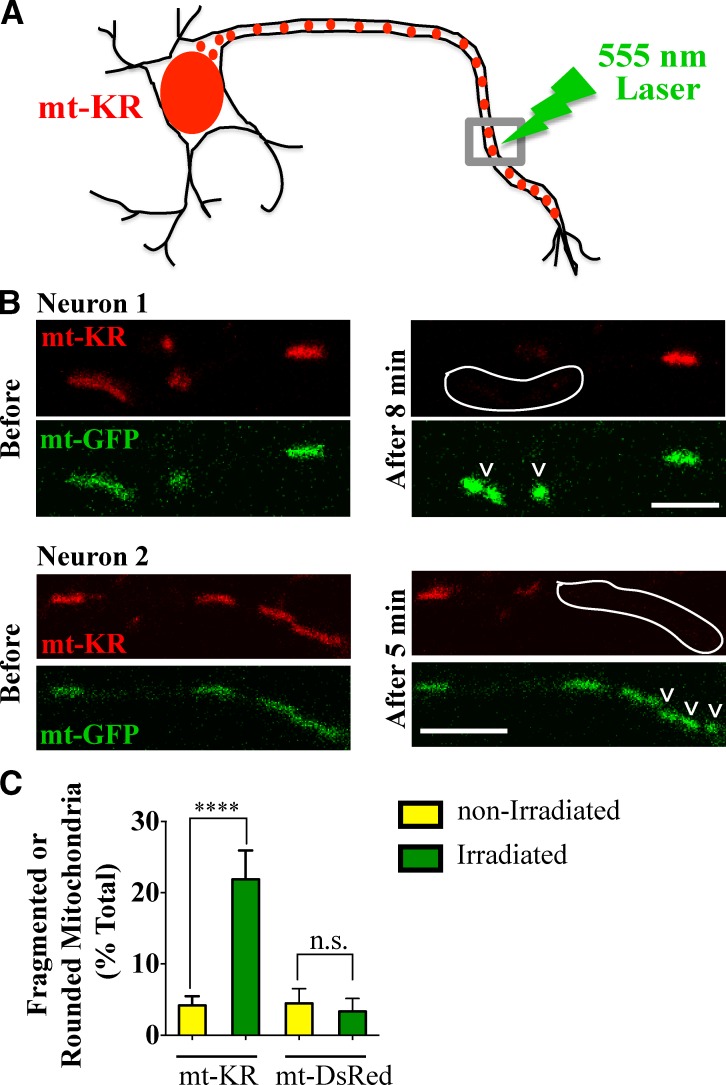
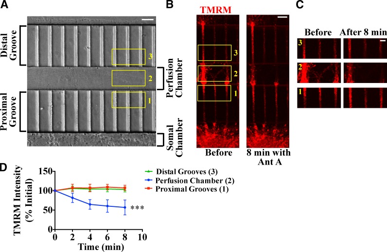
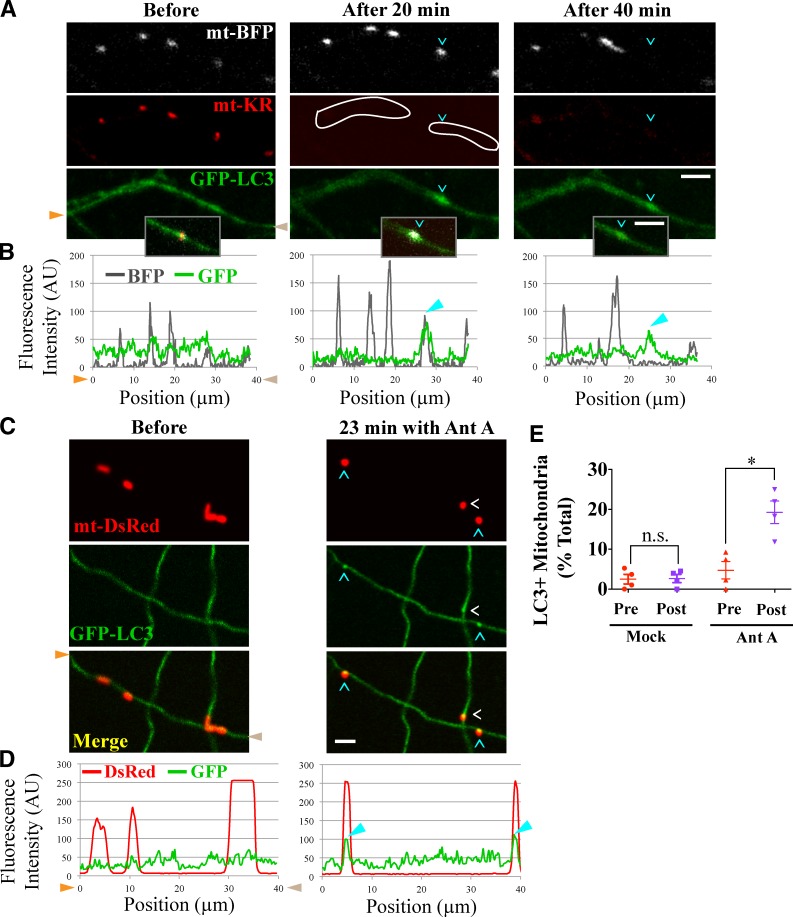
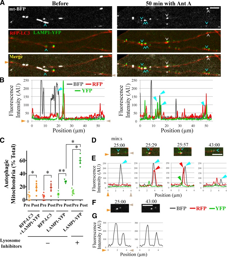
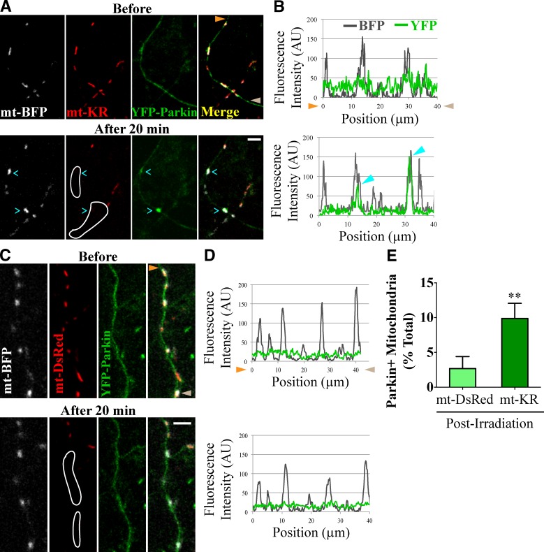
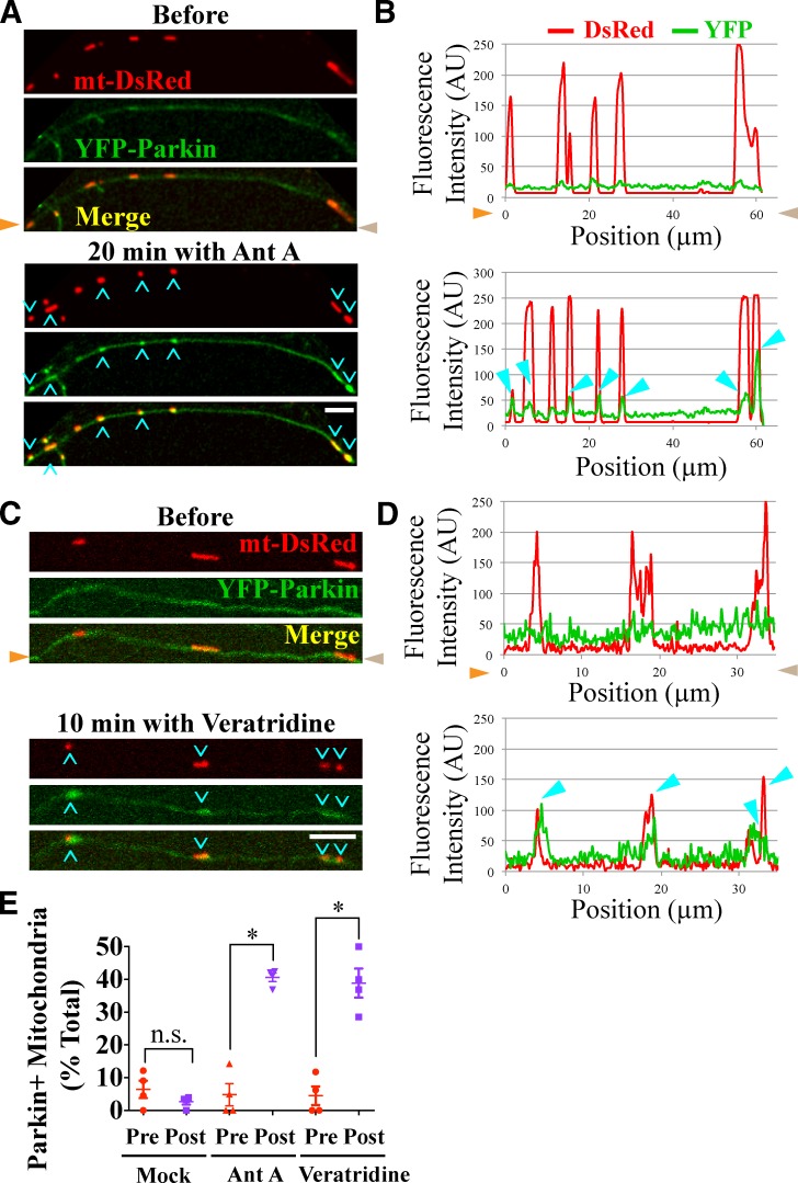
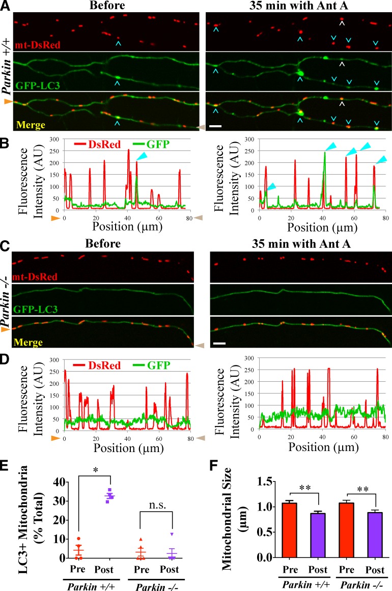
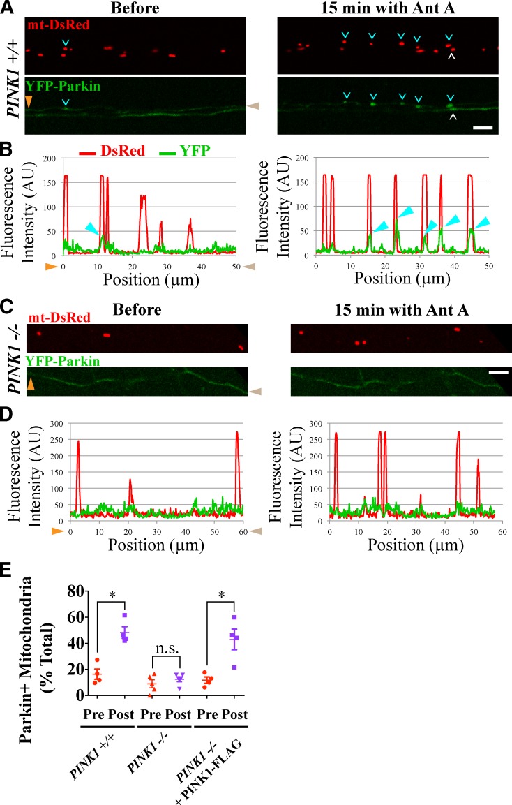
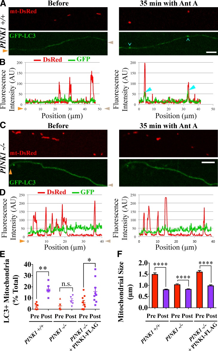
To minimize oxidative damage to the cell, malfunctioning mitochondria need to be removed by mitophagy. In neuronal axons, mitochondrial damage may occur in distal regions, far from the soma where most lysosomal degradation is thought to occur. In this paper, we report that PINK1 and Parkin, two Parkinson’s disease–associated proteins, mediate local mitophagy of dysfunctional mitochondria in neuronal axons. To reduce cytotoxicity and mimic physiological levels of mitochondrial damage, we selectively damaged a subset of mitochondria in hippocampal axons. Parkin was rapidly recruited to damaged mitochondria in axons followed by formation of LC3-positive autophagosomes and LAMP1-positive lysosomes. In PINK1−/− axons, damaged mitochondria did not accumulate Parkin and failed to be engulfed in autophagosomes. Similarly, initiation of mitophagy was blocked in Parkin−/− axons. Our findings demonstrate that the PINK1–Parkin-mediated pathway is required for local mitophagy in distal axons in response to focal damage. Local mitophagy likely provides rapid neuroprotection against oxidative stress without a requirement for retrograde transport to the soma.
Overview
The claims and statistics of this paper haven’t been checked yet.
Run the check — free, no account needed. It reads the open-access text and checks the sources it cites.
Integrity signals
Registry
Cites
Statistics
Figures
Verification
Claims the paper makes
The claims and statistics of this paper haven’t been checked yet.
Run the check — free, no account needed. It reads the open-access text and checks the sources it cites.
Claims other papers make about this one
Each is checked against this paper’s own text: does it say what the citing paper says it does?
The cited source says what the claim attributes to it.
In the citing paper
“Axonal autophagy is dependent on specialized enzymatic apparatuses, including the PINK1-PRKN complex for localized mitophagy [24], KIF1A/UNC-104 for autophagosome formation at the synapse [22,25], and the HTT-HAP1 complex and MAPK8IP1/JIP1 for retrograde transport of autophagosomes [23,26].”
From Autophagy and ALS: mechanistic insights and therapeutic implications (2022) · cites this paper as [22]
Self-check
Percentages, sums and reported differences re-computed from the paper’s own numbers.
The self-check needs the paper’s full text.
Citations
Cites
NIH Image to ImageJ: 25 years of image analysis
Schneider, C. A., Rasband, W. S., & Eliceiri, K. W. (2012) · Nature Methods · doi:10.1038/nmeth.2089 · Cited by 64,638 · cited 1×
“…of Parkin on mitochondria before and after 15–20 min of Antimycin A (or mock) treatment, plots of fluorescence intensity versus distance along the axon were generated for both DsRed (mitochondria) and YFP (Parkin) channels using the line scan function of ImageJ software (Schneider et al., 2012).”
Autophagy: Renovation of Cells and Tissues
Mizushima, N., & Komatsu, M. (2011) · Cell · doi:10.1016/j.cell.2011.10.026 · Cited by 6,615 · cited 1×
“GFPLC3 is a cytosolic protein, but upon induction of autophagy, it gets lipidated and incorporated into autophagosomes forming distinct puncta (Mizushima and Komatsu, 2011).”
BackgroundParkin is recruited selectively to impaired mitochondria and promotes their autophagy
Narendra, D., Tanaka, A., Suen, D.-F., & Youle, R. J. (2008) · The Journal of Cell Biology · doi:10.1083/jcb.200809125 · Cited by 4,026 · cited 4×
“Unlike many cell lines where the entire mitochondrial network can be cleared by mitophagy (Narendra et al., 2008, 2010; Geisler et al., 2010), neurons are strictly dependent on mitochondrial ATP production (Almeida et al., 2001, 2004).”
InfluentialChronic systemic pesticide exposure reproduces features of Parkinson's disease
Betarbet, R. S., Sherer, T., MacKenzie, G. M., Garcia‐Osuna, M., Panov, A. V., & Greenamyre, J. T. (2000) · Nature Neuroscience · doi:10.1038/81834 · Cited by 3,770 · cited 1×
“…2006; Navarro et al., 2009) and PDlike symptoms caused by mitochondrial drugs, such as Rotenone and MPTP (1methyl4phenyl1,2,3,6tetrahydro pyridine; Betarbet et al., 2000; Przedborski et al., 2004), were the first lines of evidence linking mitochondrial dysfunction to the pathology of PD.”
Youle, R. J., & Narendra, D. P. (2011) · Nature Reviews Molecular Cell Biology · doi:10.1038/nrm3028 · Cited by 3,273 · cited 1×
“The final step of mitophagy involves of autophagosome/ lysosome fusion and enzymatic degradation of the mitochon drion (Youle and Narendra, 2011).”
Wallace, D. C. (2005) · Annual Review of Genetics · doi:10.1146/annurev.genet.39.110304.095751 · Cited by 3,185 · cited 1×
“Mitochondria, particularly when dysfunctional, produce excessive reactive oxygen species (ROS; Wallace, 2005) and must be removed by quality control pathways.”
PINK1 Is Selectively Stabilized on Impaired Mitochondria to Activate Parkin
Narendra, D. P., Jin, S. M., Tanaka, A., Suen, D.-F., Gautier, C. A., Shen, J., Cookson, M. R., & Youle, R. J. (2010) · PLoS Biology · doi:10.1371/journal.pbio.1000298 · Cited by 2,944 · cited 5×
“Therefore, to rescue the recruitment of Parkin to depolarized mitochon dria in PINK1/ axons, we expressed PINK1FLAG at low mitochondrial depolarization (Narendra et al., 2010).”
BackgroundInfluentialPINK1/Parkin-mediated mitophagy is dependent on VDAC1 and p62/SQSTM1
Geisler, S., Holmström, K. M., Skujat, D., Fiesel, F. C., Rothfuss, O. C., Kahle, P. J., & Springer, W. (2010) · Nature Cell Biology · doi:10.1038/ncb2012 · Cited by 2,835 · cited 3×
“Unlike many cell lines where the entire mitochondrial network can be cleared by mitophagy (Narendra et al., 2008, 2010; Geisler et al., 2010), neurons are strictly dependent on mitochondrial ATP production (Almeida et al., 2001, 2004).”
Autophagosome formation: core machinery and adaptations
Xie, Z., & Klionsky, D. J. (2007) · Nature Cell Biology · doi:10.1038/ncb1007-1102 · Cited by 2,237 · cited 1×
“Degradation of autophagosomal cargoes requires fusion of au tophagosomes with lysosomes to form autolysosomes (Xie and Klionsky, 2007), and it was suggested that autophagosomes would have to translocate to neuronal soma to fuse with ly sosomes (Cai et al., 2012).”
Drosophila pink1 is required for mitochondrial function and interacts genetically with parkin
Clark, I. E., Dodson, M. W., Jiang, C., Cao, J. H., Huh, J. R., Seol, J. H., Yoo, S. J., Hay, B. A., & Guo, M. (2006) · Nature · doi:10.1038/nature04779 · Cited by 1,788 · cited 1×
“Genetic studies have indicated that PINK1 acts upstream of Parkin in the regulation of mitochondrial integrity (Clark et al., 2006; Park et al., 2006), and stabilization of PINK1 on the outer mitochondrial membrane is essential for Parkin re cruitment in nonneuronal cells (Geisler et al., 2010;…”
Cited by
Sorted by citations
Items 101–200 of 501
Autophagy Activator Drugs: A New Opportunity in Neuroprotection from Misfolded Protein Toxicity
Thellung, S., Corsaro, A., Nizzari, M., Barbieri, F., & Florio, T. (2019) · International Journal of Molecular Sciences · doi:10.3390/ijms20040901 · Cited by 118
Defending the mitochondria: The pathways of mitophagy and mitochondrial-derived vesicles
Roberts, R. F., Tang, M. Y. H., Fon, E. A., & Durcan, T. M. (2016) · The International Journal of Biochemistry & Cell Biology · doi:10.1016/j.biocel.2016.07.020 · Cited by 121
“Consistent with this notion, several groups have been unable to observe mitophagy in neurons (Ashrafi et al., 2014; Bingol et al., 2014; Rakovic et al., 2013; Van Laar et al., 2011).”
Regulation of long-distance transport of mitochondria along microtubules
Melkov, A., & Abdu, U. (2017) · Cellular and Molecular Life Sciences · doi:10.1007/s00018-017-2590-1 · Cited by 129
Regulation and Roles of Autophagy at Synapses
Nikoletopoulou, V., & Tavernarakis, N. (2018) · Trends in Cell Biology · doi:10.1016/j.tcb.2018.03.006 · Cited by 116
Ihenacho, U. K., Nolden, K. A., Harwig, M. C., Widlansky, M. E., & Hill, R. B. (2021) · Frontiers in Endocrinology · doi:10.3389/fendo.2021.660095 · Cited by 117
Cell Death and NeurodegenerationPreprint
Andreone, B. J., Larhammar, M., & Lewcock, J. W. (2019) · Cold Spring Harbor Perspectives in Biology · doi:10.1101/cshperspect.a036434 · Cited by 124
“Studies using cultured neurons from PINK1 and Parkin knockout mice suggested that these proteins normally mediate the removal of damaged mitochondria from neuronal axons via mitophagy, a specialized form of autophagy (Ashrafi et al. 2014).”
BackgroundNeuronal autophagy and intercellular regulation of homeostasis in the brain
Kulkarni, A., Chen, J., & Maday, S. (2018) · Current Opinion in Neurobiology · doi:10.1016/j.conb.2018.02.008 · Cited by 125
The multifaceted role of mitochondria in autism spectrum disorder
Khaliulin, I., Hamoudi, W., & Amal, H. (2024) · Molecular Psychiatry · doi:10.1038/s41380-024-02725-z · Cited by 128
Axonal transport and maturation of lysosomes
Ferguson, S. M. (2018) · Current Opinion in Neurobiology · doi:10.1016/j.conb.2018.02.020 · Cited by 111
Moore, A. S., & Holzbaur, E. L. F. (2018) · Current Opinion in Physiology · doi:10.1016/j.cophys.2018.03.003 · Cited by 120
Agujetas, V. R., Barbero‐Camps, E., de Dios, C., Podlesniy, P., Abadin, X., Morales, A., Marı́, M., Trullás, R., & Colell, A. (2021) · Molecular Neurodegeneration · doi:10.1186/s13024-021-00435-6 · Cited by 113
A novel PINK1- and PARK2-dependent protective neuroimmune pathway in lethal sepsis
Kang, R., Zeng, L., Xie, Y., Yan, Z., Zhou, B., Cao, L., Klionsky, D. J., Tracey, K. J., Li, J., Wang, H., et al. (2016) · Autophagy · doi:10.1080/15548627.2016.1239678 · Cited by 110
“Previous studies have suggested that loss of PINK1 or PARK2 results in a mitophagy deficiency-associated decrease of ATP synthesis and an increase of apoptosis in multiple cells, including neurons.(6,10,11) Consistently, the tissue levels of ATP were significantly reduced in brains and livers (Fig.”
BackgroundStructure-guided mutagenesis reveals a hierarchical mechanism of Parkin activation
Tang, M. Y. H., Vranas, M., Krahn, A. I., Pundlik, S., Trempe, J. F., & Fon, E. A. (2017) · Nature Communications · doi:10.1038/ncomms14697 · Cited by 111
Protein Turnover in Aging and Longevity
Basisty, N., Meyer, J. G., & Schilling, B. (2018) · PROTEOMICS · doi:10.1002/pmic.201700108 · Cited by 111
Mechanisms of PINK1, ubiquitin and Parkin interactions in mitochondrial quality control and beyond
Bayne, A. N., & Trempe, J. (2019) · Cellular and Molecular Life Sciences · doi:10.1007/s00018-019-03203-4 · Cited by 121
The Role of Impaired Mitochondrial Dynamics in MFN2-Mediated Pathology
Zaman, M., & Shutt, T. E. (2022) · Frontiers in Cell and Developmental Biology · doi:10.3389/fcell.2022.858286 · Cited by 116
UPRmt-mediated cytoprotection and organismal aging
Schulz, A. M., & Haynes, C. M. (2015) · Biochimica et Biophysica Acta (BBA) - Bioenergetics · doi:10.1016/j.bbabio.2015.03.008 · Cited by 113
Mitochondria: A Therapeutic Target for Parkinson’s Disease?
Luo, Y., Hoffer, A., Hoffer, B. J., & Qi, X. (2015) · International Journal of Molecular Sciences · doi:10.3390/ijms160920704 · Cited by 108
“The failure of PINK1/parkin-mediated mitophagic process leads to accumulation of damaged mitochondria, which results in an increase in ROS and cell death [141,142].”
BackgroundMitophagy and reactive oxygen species interplay in Parkinson’s disease
Xiao, B., Kuruvilla, J., & Tan, E. (2022) · npj Parkinson s Disease · doi:10.1038/s41531-022-00402-y · Cited by 117
The Endolysosomal System and Proteostasis: From Development to Degeneration
Winckler, B., Faúndez, V., Maday, S., Cai, Q., Almeida, C. G., & Zhang, H. (2018) · Journal of Neuroscience · doi:10.1523/jneurosci.1665-18.2018 · Cited by 111
“…2017; Nikoletopoulou et al., 2017; Ok- erlund et al., 2017; Nikoletopoulou and Tavernarakis, 2018), mitochondria (Cai et al., 2012; Maday et al., 2012; Ashrafi et al., 2014; Rangaraju et al., 2014; Wong and Holzbaur, 2014b; Xie et al., 2015; Ebrahimi-Fakhari et al., 2016), and pathogenic protein…”
BackgroundFerguson, S. M. (2018) · Neuroscience Letters · doi:10.1016/j.neulet.2018.04.005 · Cited by 107
“autophagosomes to form around acutely damaged axonal mitochondria (13).”
BackgroundQuality Control in Neurons: Mitophagy and Other Selective Autophagy Mechanisms
Evans, C. S., & Holzbaur, E. L. F. (2019) · Journal of Molecular Biology · doi:10.1016/j.jmb.2019.06.031 · Cited by 109
“(2012), local damage to axonal mitochondria using the mitochondrially-targeted photosensitizer mitoKillerRed was reported to occur within an hour [37].”
BackgroundInfluentialDyslipidemia impairs mitochondrial trafficking and function in sensory neurons
Rumora, A. E., Lentz, S. I., Hinder, L. M., Jackson, S. W., Valesano, A. L., Levinson, G. E., & Feldman, E. L. (2017) · The FASEB Journal · doi:10.1096/fj.201700206r · Cited by 106
“In addition, the maintained bidirectional mitochondrial movement across glucose and palmitate concentrations suggests that a preference for retrograde movement is not necessary to transport damagedmitochondria to the cell body for degradation (44, 59, 61, 62).”
BackgroundMitochondrial dysfunction and Parkinson disease: a Parkin–AMPK alliance in neuroprotection
Hang, L., Thundyil, J., & Lim, K. (2015) · Annals of the New York Academy of Sciences · doi:10.1111/nyas.12820 · Cited by 103
Autophagy core machinery: overcoming spatial barriers in neurons
Ariosa, A., & Klionsky, D. J. (2016) · Journal of Molecular Medicine · doi:10.1007/s00109-016-1461-9 · Cited by 108
“It was recently shown that, when called for, autophagy could be initiated in the edges of a neuron, without the need for long-distance transport of autophagosomes [83].”
BackgroundInfluentialMitophagy in neurodegenerative diseases
Rodolfo, C., Campello, S., & Cecconi, F. (2017) · Neurochemistry International · doi:10.1016/j.neuint.2017.08.004 · Cited by 104
“The mitophagy process acting through PINK1/Parkin seems to be important not only to protect neurons from the damage caused by dysfunctional mitochondria in the soma, but it has been proposed that this system could be very efficient in the removal of damaged organelles in distal axons of these cells (Ashrafi et al., 20…”
BackgroundGlucocorticoid impairs mitochondrial quality control in neurons
Choi, G. E., & Han, H. J. (2021) · Neurobiology of Disease · doi:10.1016/j.nbd.2021.105301 · Cited by 95
“Lysosomal degradation at distal axons in PD is more likely to occur than in soma [92].”
PINK1/Parkin-Dependent Mitochondrial Surveillance: From Pleiotropy to Parkinson's Disease
Mouton‐Liger, F., Jacoupy, M., Corvol, J., & Corti, O. (2017) · Frontiers in Molecular Neuroscience · doi:10.3389/fnmol.2017.00120 · Cited by 100
“In a more physiological paradigm based on the local depolarization of the mitochondrial network in hippocampal neurons, using the mitochondrion-targeted photosensitizer mt-KillerRed, and the use of microfluidic devices for the investigation of neuronal subcompartments, mitophagic events were observed preferentially in…”
Inhibition of autophagy curtails visual loss in a model of autosomal dominant optic atrophy
Zaninello, M., Palikaras, K., Naon, D., Iwata, K., Herkenne, S., Quintana-Cabrera, R., Semenzato, M., Grespi, F., Ross-Cisneros, F. N., Carelli, V., et al. (2020) · Nature Communications · doi:10.1038/s41467-020-17821-1 · Cited by 92
Dagda, R. K., & Banerjee, T. D. (2015) · Reviews in the Neurosciences · doi:10.1515/revneuro-2014-0085 · Cited by 96
“…of neurotrophic support, continuous synaptic input, efficient anterograde movement of mitochondria, and maintenance of functional mitochondria through the continuous activation of mitochondrial quality control pathways (Reichardt, 2006; Wang et al., 2011a,b; Schwarz, 2013; Ashrafi et al., 2014).”
Genes associated with Parkinson's disease: regulation of autophagy and beyond
Beilina, A. G., & Cookson, M. (2015) · Journal of Neurochemistry · doi:10.1111/jnc.13266 · Cited by 98
“While some studies have reported that neurons are susceptible to depolarization-induced parkin recruitment (Cai et al. 2012; Joselin et al. 2012; Ashrafi et al. 2014; Ye et al. 2015), others have not (Van Laar et al.”
BackgroundMitochondria in Neuronal Health: From Energy Metabolism to Parkinson's Disease
Mahadevan, H. M., Hashemiaghdam, A., Ashrafi, G., & Harbauer, A. B. (2021) · Advanced Biology · doi:10.1002/adbi.202100663 · Cited by 112
Self-citationPellegrino, M. W., & Haynes, C. M. (2015) · BMC Biology · doi:10.1186/s12915-015-0129-1 · Cited by 99
“This is also consistent with studies on axons of rat primary neurons showing that PINK1 and Parkin mediate the degradation of defective mitochondria via mitophagy [59].”
ResultSynaptosomal Mitochondrial Dysfunction in 5xFAD Mouse Model of Alzheimer's Disease
Wang, L., Guo, L., Lu, L., Sun, H., Shao, M., Beck, S. J., Lin, L., Ramachandran, J., Du, Y., & Du, H. (2016) · PLoS ONE · doi:10.1371/journal.pone.0150441 · Cited by 90
“Mitochondrial quality control through mitophagy is a critical pathway to regulate mitochondrial metabolism and maintain mitochondrial fitness [31, 44].”
BackgroundSomatic autophagy of axonal mitochondria in ischemic neurons
Zheng, Y., Zhang, X., Wu, X., Jiang, L., Ahsan, A., Ma, S., Xiao, Z., Han, F., Qin, Z., Hu, W., et al. (2019) · The Journal of Cell Biology · doi:10.1083/jcb.201804101 · Cited by 94
“Several studies also suggested that axonal mitochondrial mobility was arrested by stress and axonal mitophagy occurred locally (Wang et al., 2011; Lovas and Wang, 2013; Ashrafi et al., 2014; Hsieh et al., 2016).”
InfluentialCurrent perspective of mitochondrial biology in Parkinson's disease
Kaidery, N. A., & Thomas, B. (2018) · Neurochemistry International · doi:10.1016/j.neuint.2018.03.001 · Cited by 95
Mitochondrial NIX Promotes Tumor Survival in the Hypoxic Niche of Glioblastoma
Jung, J., Zhang, Y., Celiku, O., Zhang, W., Song, H., Williams, B. J., Giles, A. J., Rich, J. N., Abounader, R., Gilbert, M. R., et al. (2019) · Cancer Research · doi:10.1158/0008-5472.can-19-0198 · Cited by 97
Our (Mother’s) Mitochondria and Our Mind
Krämer, P., & Bressan, P. (2017) · Perspectives on Psychological Science · doi:10.1177/1745691617718356 · Cited by 94
Roles for neuronal and glial autophagy in synaptic pruning during development
Lieberman, O. J., McGuirt, A. F., Tang, G., & Sulzer, D. (2019) · Neurobiology of Disease · doi:10.1016/j.nbd.2018.04.017 · Cited by 87
“In neurons, this can occur to mitochondria in axons (Ashrafi et al., 2014; Berthet et al., 2014) and in the somatodendritic region (Cai et al.”
BackgroundShedding light on mitophagy in neurons: what is the evidence for PINK1/Parkin mitophagy in vivo?
Cummins, N., & Götz, J. (2017) · Cellular and Molecular Life Sciences · doi:10.1007/s00018-017-2692-9 · Cited by 88
“, used the fluorophore KillerRed, which causes ROS formation upon stimulation with green light, to increase ROS levels in individual mitochondria in the axon [43].”
Lie, P. P. Y., Yang, D.-S., Stavrides, P., Goulbourne, C. N., Zheng, P., Mohan, P. S., Cataldo, A. M., & Nixon, R. A. (2021) · Cell Reports · doi:10.1016/j.celrep.2021.109034 · Cited by 97
“, 2019), whether these are absolute requirements is still under debate (Ashrafi et al., 2014).”
BackgroundTrevisan, T., Pendin, D., Montagna, A., Bova, S., Ghelli, A. M., & Daga, A. (2018) · Cell Reports · doi:10.1016/j.celrep.2018.04.017 · Cited by 86
“Furthermore, the occurrence of local mitophagy in distal axons has also been reported (Ashrafi et al., 2014).”
BackgroundEmerging Mitochondrial Therapeutic Targets in Optic Neuropathies
Sanchez, M. I. G. L., Crowston, J. G., Mackey, D. A., & Trounce, I. A. (2016) · Pharmacology & Therapeutics · doi:10.1016/j.pharmthera.2016.06.004 · Cited by 87
Mitochondria in Multiple Sclerosis: Molecular Mechanisms of Pathogenesis
Patergnani, S., Fossati, V., Bonora, M., Giorgi, C., Marchi, S., Missiroli, S., Rusielewicz, T., Wieckowski, M. R., & Pinton, P. (2016) · International review of cytology · doi:10.1016/bs.ircmb.2016.08.003 · Cited by 86
“Recent findings have suggested a possible role of altered mitophagic processes in several human neurological diseases, such as PD and AD (Ashrafi et al., 2014; Ye et al., 2015; Palikaras and Tavernarakis, 2012).”
BackgroundMethylene Blue Reduces Acute Cerebral Ischemic Injury via the Induction of Mitophagy
Yao, D., He, Y., Zhao, T., Huang, X., Wu, K.-W., Liu, S.-H., Zhao, Y., Fan, M., Wu, L., & Zhu, L.-L. (2015) · Molecular Medicine · doi:10.2119/molmed.2015.00038 · Cited by 86
Sung, H., Tandarich, L. C., Nguyen, K. K., & Hollenbeck, P. J. (2016) · Journal of Neuroscience · doi:10.1523/jneurosci.0633-16.2016 · Cited by 85
“Since the PINK1/Parkin pathway is proposed not only to target damaged mitochondria for local turnover (Narendra et al., 2008, 2010; Matsuda et al., 2010; Ashrafi et al., 2014) but also to inhibit organelle fusion (Deng et al.”
BackgroundResultInfluentialParkin Regulation and Neurodegenerative Disorders
Zhang, C., Hang, L., Yao, T., & Lim, K. (2016) · Frontiers in Aging Neuroscience · doi:10.3389/fnagi.2015.00248 · Cited by 84
“…and Parkin, the effect of which leading to arrested motility suggests that PINK1/Parkin Frontiers in Aging Neuroscience | www.frontiersin.org 7 January 2016 | Volume 7 | Article 248 pathway may quarantine damaged mitochondria prior to their clearance (Wang et al., 2011b; Ashrafi et al., 2014).”
BackgroundImpaired Mitophagy Plays a Role in Denervation of Neuromuscular Junctions in ALS Mice
Rogers, R. S., Tungtur, S., Tanaka, T., Nadeau, L. L., Badawi, Y., Wang, H., Ni, H.-M., Ding, W.-X., & Nishimune, H. (2017) · Frontiers in Neuroscience · doi:10.3389/fnins.2017.00473 · Cited by 83
“Mitophagy mediated degradation of damaged mitochondria has been shown to occur locally in distal axons (Ashrafi et al., 2014).”
BackgroundResultSAMM50 acts with p62 in piecemeal basal- and OXPHOS-induced mitophagy of SAM and MICOS components
Abudu, Y. P., Shrestha, B. K., Zhang, W., Palara, A., Brenne, H. B., Larsen, K. B., Wolfson, D. L., Dumitriu, G., Øie, C. I., Ahluwalia, B. S., et al. (2021) · The Journal of Cell Biology · doi:10.1083/jcb.202009092 · Cited by 79
“PINK1 and E3-ubiquitin ligase Parkin, whose mutations are linked to autosomal recessive Parkinson’s disease, function in this pathway to eliminate damaged mitochondria to protect neuronal cells (Ashrafi et al., 2014; Youle and Narendra, 2011).”
BackgroundRosa, A. I., Fonseca, I., Nunes, M. J., Moreira, S., Rodrigues, E., Carvalho, A. N., Rodrigues, C. M. P., Gama, M. J., & Castro-Caldas, M. (2017) · Biochimica et Biophysica Acta (BBA) - Molecular Basis of Disease · doi:10.1016/j.bbadis.2017.06.004 · Cited by 82
Connecting mitochondrial dynamics and life-or-death events via Bcl-2 family proteins
Aouacheria, A., Baghdiguian, S., Lamb, H. M., Huska, J. D., Pineda, F. J., & Hardwick, J. M. (2017) · Neurochemistry International · doi:10.1016/j.neuint.2017.04.009 · Cited by 81
“2016), consistent with previous studies (Ashrafi et al., 2014; Wang and Schwarz, 2009).”
Mitochondrial Mechanisms of Neuromuscular Junction Degeneration with Aging
Anagnostou, M., & Hepple, R. T. (2020) · Cells · doi:10.3390/cells9010197 · Cited by 84
Multitasking guardian of mitochondrial quality: Parkin function and Parkinson’s disease
Kamienieva, I., Duszyński, J., & Szczepanowska, J. (2021) · Translational Neurodegeneration · doi:10.1186/s40035-020-00229-8 · Cited by 84
“This particular type of selective autophagy is designated to eliminate dysfunctional mitochondria and can be triggered by depolarization of the mitochondrial membrane (experimentally induced with Δψdissipating agents, e.g., H+ ionophores CCCP [carbonyl cyanide 3-chlorophenylhydrazone] or FCCP [carbonyl cyanide 4-trifl…”
BackgroundInfluentialSelective neuronal vulnerability in Parkinson's disease
González‐Rodríguez, P., Zampese, E., & Surmeier, D. J. (2020) · Progress in brain research · doi:10.1016/bs.pbr.2020.02.005 · Cited by 80
Reciprocity in ROS and autophagic signaling
Wible, D. J., & Bratton, S. B. (2017) · Current Opinion in Toxicology · doi:10.1016/j.cotox.2017.10.006 · Cited by 83
Devireddy, S., Liu, A., Lampe, T., & Hollenbeck, P. J. (2015) · Journal of Neuroscience · doi:10.1523/jneurosci.1198-15.2015 · Cited by 78
“, 2010), studies in neurons found that Parkin is targeted to only a small fraction of depolarized mitochondria after photodamage or drug treatment (Ashrafi et al., 2014).”
BackgroundThe Role of Mitophagy in Regulating Cell Death
Li, S., Zhang, J., Liu, C., Wang, Q., Yan, J., Hui, L., Jia, Q., Shan, H., Tao, L., & Zhang, M. (2021) · Oxidative Medicine and Cellular Longevity · doi:10.1155/2021/6617256 · Cited by 76
Gutnick, A., Banghart, M. R., West, E. R., & Schwarz, T. L. (2019) · Nature Cell Biology · doi:10.1038/s41556-019-0317-2 · Cited by 78
Self-citationThe Ubiquitin System in Alzheimer’s Disease
Harris, L. D., Jasem, S., & Licchesi, J. D. F. (2020) · Advances in experimental medicine and biology · doi:10.1007/978-3-030-38266-7_8 · Cited by 79
Exploring new pathways of neurodegeneration in ALS: The role of mitochondria quality control
Palomo, G. M., & Manfredi, G. (2014) · Brain Research · doi:10.1016/j.brainres.2014.09.065 · Cited by 81
Mitochondrial UQCRC1 mutations cause autosomal dominant parkinsonism with polyneuropathy
Lin, C., Tsai, P.-I., Lin, H., Hattori, N., Funayama, M., Jeon, B., Sato, K., Abe, K., Mukai, Y., Takahashi, Y., et al. (2020) · Brain · doi:10.1093/brain/awaa279 · Cited by 73
“Parkin and PINK1 are critical regulators of mitophagy (Burman et al., 2012; Ashrafi et al., 2014).”
BackgroundCompartment‐specific dynamics and functions of autophagy in neurons
Kulkarni, V. V., & Maday, S. (2017) · Developmental Neurobiology · doi:10.1002/dneu.22562 · Cited by 77
“Recent work has suggested that selective autophagy may be compartmentalized within the neuron (Cai et al., 2012; Sung et al., 2016; Dragich et al., 2016; Ashrafi et al., 2014).”
BackgroundInfluentialmiR-103a-3p regulates mitophagy in Parkinson’s disease through Parkin/Ambra1 signaling
Zhou, J., Zhao, Y., Li, Z., Zhu, M., Wang, Z., Li, Y., Xu, T., Feng, D., Zhang, S., Tang, F., et al. (2020) · Pharmacological Research · doi:10.1016/j.phrs.2020.105197 · Cited by 76
Key Targets for Multi-Target Ligands Designed to Combat Neurodegeneration
Ramsay, R. R., Májeková, M., Medina, M., & Valoti, M. (2016) · Frontiers in Neuroscience · doi:10.3389/fnins.2016.00375 · Cited by 76
“…of functional lysosomes in axons has been evidenced to contribute to mitophagy of damaged mitochondria, and the local PINK1–Parkin-mediated mitophagy pathway provides rapid neuroprotection against oxidative stress without a requirement for retrograde transport to the soma (Ashrafi et al., 2014).”
BackgroundAutophagy and proteostasis in the control of synapse aging and disease
Liang, Y., & Sigrist, S. J. (2017) · Current Opinion in Neurobiology · doi:10.1016/j.conb.2017.12.006 · Cited by 74
Light-Activated ROS Production Induces Synaptic Autophagy
Hoffmann, S., Orlando, M., Andrzejak, E., Bruns, C., Trimbuch, T., Rosenmund, C., Garner, C. C., & Ackermann, F. (2019) · Journal of Neuroscience · doi:10.1523/jneurosci.1317-18.2019 · Cited by 71
“…showing that elevated ROS levels around organelles, such as mitochondria, lead to the activation of autophagy (Yang and Yang, 2011; Wang et al., 2012; Ashrafi et al., 2014), we anticipated that a similar generation of ROS around SVs may also induce a presynaptic autophagy-based clearance program.”
BackgroundSmith, G. A., Lin, T.-H., Sheehan, A. E., van Naters, W. V. D. G., Neukomm, L. J., Graves, H. K., Bis‐Brewer, D. M., Zuchner, S. L., & Freeman, M. R. (2019) · Neuron · doi:10.1016/j.neuron.2019.04.017 · Cited by 71
“A High-Throughput In Vivo Genetic Screen for Modifiers of Axonal Mitochondria Mitochondrial maintenance processes present in non-polarized cells may occur in the axonal compartment of neurons (Amiri and Hollenbeck, 2008; Ashrafi and Schwarz, 2015; Ashrafi et al., 2014; Cartoni et al., 2016; Harbauer, 2017; Lee et al.,…”
SUMO ‐regulated mitochondrial function in Parkinson's disease
Souza, A. C. G., Prediger, R. D., & Cimarosti, H. (2016) · Journal of Neurochemistry · doi:10.1111/jnc.13599 · Cited by 78
“The loss of the mitochondrial transmembrane potential induces PINK1-dependent higher parkin levels in the mitochondria and subsequent proteasomal degradation of mitochondrial proteins and mitophagy (Narendra et al. 2008; Yu et al. 2011; Ashrafi et al. 2014; Pickrell and Youle 2015).”
BackgroundTubulin polyglutamylation is a general traffic-control mechanism in hippocampal neurons
Bodakuntla, S., Schnitzler, A., Villablanca, C., González-Billault, C. E., Bièche, I., Janke, C., & Magiera, M. M. (2020) · Journal of Cell Science · doi:10.1242/jcs.241802 · Cited by 73
Bastian, T. W., von Hohenberg, W. C., Georgieff, M., & Lanier, L. M. (2018) · Journal of Neuroscience · doi:10.1523/jneurosci.1504-18.2018 · Cited by 76
“Although mitophagy can occur locally in distal axons (Ashrafi et al., 2014), it likely occurs preferentially in the soma over the axons or dendrites (Cai et al., 2012; Ye et al., 2015; McWilliams et al., 2016), requiring retrograde transport for distal mitochondria.”
BackgroundIncreasing Nrf2 Activity as a Treatment Approach in Neuropsychiatry
Morris, G., Walker, A. J., Walder, K. R., Berk, M., Marx, W., Carvalho, A. F., Maes, M. H. J., & Puri, B. K. (2021) · Molecular Neurobiology · doi:10.1007/s12035-020-02212-w · Cited by 74
Mitostasis, Calcium and Free Radicals in Health, Aging and Neurodegeneration
Godoy, J. A., Ríos, J. A., Picón-Pagès, P., Herrera-Fernández, V., Swaby, B., Crepin, G., Vicente, R., Fernández‐Fernández, J. M., & Muñoz, F. J. (2021) · Biomolecules · doi:10.3390/biom11071012 · Cited by 73
Mitochondrial dysfunction in Parkinson disease: evidence in mutant PARK2 fibroblasts
Zanellati, M. C., Monti, V., Barzaghi, C., Reale, C., Nardocci, N., Albanese, A., Valente, E. M., Ghezzi, D., & Garavaglia, B. (2015) · Frontiers in Genetics · doi:10.3389/fgene.2015.00078 · Cited by 72
“In association with PINK1, Parkin acts in mitochondrial fission and fusion, mitochondrial transport and removal of damaged mitochondria through mitophagy process (Narendra et al., 2008; Wang et al., 2011; Yu et al., 2011; Ashrafi et al., 2014; Cook et al., 2014).”
BackgroundScott, L., Dawson, V. L., & Dawson, T. M. (2017) · Experimental Neurology · doi:10.1016/j.expneurol.2017.04.008 · Cited by 72
“Though studies have failed to see abnormal aggregation of damaged mitochondria in PINK1 KO and parkin KO mice, more recent work using microfluidics and high-resolution microscopy has verified parkindependent mitophagy in primary neuronal culture (Ashrafi et al., 2014).”
ResultAxonal autophagy: Mini-review for autophagy in the CNS
Stavoe, A. K., & Holzbaur, E. L. F. (2018) · Neuroscience Letters · doi:10.1016/j.neulet.2018.03.025 · Cited by 72
“For example, Schwarz and colleagues [2]demonstrated the engulfment of mitochondria by the local formation of an autophagosome along the axon, in a pathway that is both PINK1- and Parkin-dependent and is induced in response to localized formation of ROS.”
BackgroundBao, F., Zhou, L., Zhou, R., Huang, Q., Chen, J., Zeng, S., Wu, Y., Yang, L., Qian, S., Wang, M., et al. (2022) · Science Advances · doi:10.1126/sciadv.abk2376 · Cited by 70
Wen, H., Li, L., Zhan, L., Zuo, Y., Li, K., Qiu, M., Li, H., Sun, W., & Xu, E. (2021) · Cell Death and Disease · doi:10.1038/s41419-021-03900-8 · Cited by 69
“Notably, primary hippocampal neurons undergo PINK1/Parkin-mediated mitophagy in response to mitochondrial damage [18].”
BackgroundMitochondrial retrograde signaling regulates neuronal function
Cagin, U., Duncan, O. F., Gatt, A. P., Dionne, M., Sweeney, S. T., & Bateman, J. E. (2015) · Proceedings of the National Academy of Sciences · doi:10.1073/pnas.1505036112 · Cited by 66
“Furthermore, a recent study using KillerRed demonstrated that local mitochondrial damage results in mitophagy in axons (37).”
BackgroundZheng, Y., Zhang, X., & Chen, Z. (2019) · CNS Neuroscience & Therapeutics · doi:10.1111/cns.13122 · Cited by 72
Mandal, A., Wong, H.-T., Pinter, K. L., Mosqueda, N., Beirl, A. J., Lomash, R. M., Won, S., Kindt, K. S., & Drerup, C. M. (2020) · Journal of Neuroscience · doi:10.1523/jneurosci.1316-20.2020 · Cited by 72
“For example, treatment of cultured neurons with drugs such as rotenone, antimycin A, FCCP, and others cause a loss of mitochondrial matrix potential and can lead to enhanced retrograde mitochondrial motility (Miller and Sheetz, 2004; Ashrafi et al., 2014; Lin et al., 2017).”
InfluentialMechanics of mitochondrial motility in neurons
Barnhart, E. L. (2016) · Current Opinion in Cell Biology · doi:10.1016/j.ceb.2016.02.022 · Cited by 71
Subcellular proteomics of dopamine neurons in the mouse brain
Hobson, B. D., Choi, S. J., Mosharov, E. V., Soni, R. K., Sulzer, D., & Sims, P. A. (2022) · eLife · doi:10.7554/elife.70921 · Cited by 68
“, 2012), although other findings implicate local autophagic degradation of damaged mitochondria in axons (Ashrafi et al., 2014).”
Miro sculpts mitochondrial dynamics in neuronal health and disease
Devine, M. J., Birsa, N., & Kittler, J. T. (2015) · Neurobiology of Disease · doi:10.1016/j.nbd.2015.12.008 · Cited by 69
The Development of Neuronal Polarity: A Retrospective View
Banker, G. A. (2018) · Journal of Neuroscience · doi:10.1523/jneurosci.1372-16.2018 · Cited by 72
“Hippocampal cultures have also been useful for understanding the factors that make neurons susceptible to disease and in assays seeking to enhance neuronal survival or foster axonal regeneration (Ashrafi et al., 2014; Fang et al., 2016; Al-Ali et al., 2017).”
BackgroundGao, Y., Li, J., Li, J., Hu, C., Zhang, L., Yan, J., Li, L., & Zhang, L. (2020) · Biochemical Pharmacology · doi:10.1016/j.bcp.2020.113997 · Cited by 65
Parkin contributes to synaptic vesicle autophagy in Bassoon-deficient mice
Hoffmann-Conaway, S., Brockmann, M. M., Schneider, K., Annamneedi, A., Rahman, K. A., Bruns, C., Textoris‐Taube, K., Trimbuch, T., Smalla, K., Rosenmund, C., et al. (2020) · eLife · doi:10.7554/elife.56590 · Cited by 61
Role of Impaired Mitochondrial Dynamics Processes in the Pathogenesis of Alzheimer’s Disease
Blagov, A. V., Grechko, A. V., Nikiforov, N. G., Borisov, E. E., Sadykhov, N. K., & Orekhov, A. N. (2022) · International Journal of Molecular Sciences · doi:10.3390/ijms23136954 · Cited by 70
“For this, autophagosomes fuse with late endosomes and form amphysomes together with dynein–snapin motors for AV transport back into the soma [44].”
BackgroundMitochondrial plasticity and synaptic plasticity crosstalk; in health and Alzheimer's disease
Sayehmiri, F., Motamedi, F., Batool, Z., Naderi, N., Shaerzadeh, F., Zoghi, A., Rezaei, O., Khodagholi, F., & Pourbadie, H. G. (2024) · CNS Neuroscience & Therapeutics · doi:10.1111/cns.14897 · Cited by 68
Evidence that phosphorylated ubiquitin signaling is involved in the etiology of Parkinson’s disease
Shiba‐Fukushima, K., Ishikawa, K., Inoshita, T., Izawa, N., Takanashi, M., Sato, S., Onodera, O., Akamatsu, W., Okano, H., Imai, Y., et al. (2017) · Human Molecular Genetics · doi:10.1093/hmg/ddx201 · Cited by 64
“Whether efficient mitophagy occurs in the axonal processes of neurons remains controversial (40,41).”
AKT signalling selectively regulates PINK1 mitophagy in SHSY5Y cells and human iPSC-derived neurons
Soutar, M. P. M., Kempthorne, L., Miyakawa, S., Annuario, E., Melandri, D., Harley, J., O’Sullivan, G. A., Wray, S., Hancock, D. C., Cookson, M. R., et al. (2018) · Scientific Reports · doi:10.1038/s41598-018-26949-6 · Cited by 65
Activation mechanisms of the E3 ubiquitin ligase parkin
Panicker, N., Dawson, V. L., & Dawson, T. M. (2017) · Biochemical Journal · doi:10.1042/bcj20170476 · Cited by 64
Mitochondrial fission is an acute and adaptive response in injured motor neurons
Kiryu‐Seo, S., Tamada, H., Kato, Y., Yasuda, K., Ishihara, N., Nomura, M., Mihara, K., & Kiyama, H. (2016) · Scientific Reports · doi:10.1038/srep28331 · Cited by 64
Igarashi, R., Yamashita, S., Yamashita, T., Inoue, K., Fukuda, T., Fukuchi, T., & Kanki, T. (2020) · Scientific Reports · doi:10.1038/s41598-020-58315-w · Cited by 64
Lent, J. V., Verstraelen, P., Asselbergh, B., Adriaenssens, E., Mateiu, L. M., Verbist, C., Winter, V. D., Eggermont, K., Bosch, L. V. D., Vos, W. H. D., et al. (2021) · Brain · doi:10.1093/brain/awab226 · Cited by 60
“Similarly, mitophagy of dysfunctional mitochondria along the neurite also occurs in the axon itself.(41) It thus appears that local mitochondrial deficits can lead directly to mitochondrial stalling.”
BackgroundEmerging Concepts and Functions of Autophagy as a Regulator of Synaptic Components and Plasticity
Liang, Y. (2019) · Cells · doi:10.3390/cells8010034 · Cited by 59
Rojas-Charry, L., Nardi, L., Methner, A., & Schmeißer, M. J. (2020) · Journal of Molecular Medicine · doi:10.1007/s00109-020-02018-2 · Cited by 62
Lee, S., Zhang, C., & Liu, X. (2014) · Journal of Biological Chemistry · doi:10.1074/jbc.m114.606798 · Cited by 58
Regulation and roles of mitophagy at synapses
Palikaras, K., & Tavernarakis, N. (2020) · Mechanisms of Ageing and Development · doi:10.1016/j.mad.2020.111216 · Cited by 60
Son, T., Lee, D., Lee, C., Moon, G., Ha, G. E., Lee, H., Kwak, H., Cheong, E., & Kim, D. (2019) · ACS Nano · doi:10.1021/acsnano.8b08178 · Cited by 57
Epigenetic Treatment of Neurodegenerative Disorders: Alzheimer and Parkinson Diseases
Irwin, M. H., Moos, W. H., Faller, D. V., Steliou, K., & Pinkert, C. A. (2016) · Drug Development Research · doi:10.1002/ddr.21294 · Cited by 57
“…al., 2015] thereby affording a new and actionable therapeutic target in the treatment of AD and PD [Narendra and Youle; 2011; Lazarou et al., 2012; Ashrafi et al., 2014; Hattori et al., 2014; McLelland et al., 2014; Tufi et al., 2014; Frake et al., 2015; Kazlauskaite and Muqit, 2015; Kroemer,…”
Background
Metadata
- Authors
- Ashrafi, G., Schlehe, J. S., LaVoie, M. J., & Schwarz, T. L.
- Affiliations
- Boston Children's Hospital (US)
- Brigham and Women's Hospital (US)
- Corresponding author
- Schwarz, Thomas L.
- Venue
- Journal of Cell Biology
- Publisher
- Rockefeller University Press
- Published
- 2014-08-25
- Volume
- 206
- Issue
- 5
- Pages
- 655-670
- ISSN
- 0021-9525 (ISSN-L)
- 1540-8140
- PMC
- PMC4151150
- OpenAlex
- W1992264711
- Cited by
- 502
- References
- 65
- Citations by year
- 2026: 18 · 2025: 22 · 2024: 39 · 2023: 25 · 2022: 39 · 2021: 53 · 2020: 53 · 2019: 51 · 2018: 49 · 2017: 49 · 2016: 51 · 2015: 46 · 2014: 7
- FWCI
- 38.33
- Type
- journal article
- Language
- en
- Open access
- hybrid
- doi.org
- Licence
- cc-by-nc-sa
- Locations
- The Journal of Cell Biology (publishedVersion, cc-by-nc-sa)
- PubMed Central (publishedVersion, cc-by-nc-sa)
- PubMed (publishedVersion)
- Digital Access to Scholarship at Harvard (DASH) (Harvard University) (submittedVersion, cc-by)
- Funders
- National Institute of General Medical Sciences (GM069808, R01GM069808)
- National Institute of Neurological Disorders and Stroke (R21NS069949, R01NS065013, NS065013, NS069949)
- Eunice Kennedy Shriver National Institute of Child Health and Human Development (HD-01866, P30HD018655)
- Topics
- Life Sciences › Biochemistry, Genetics and Molecular Biology › Cell Biology › Autophagy in Disease and Therapy
- Health Sciences › Medicine › Clinical Neurology › Parkinson's Disease Mechanisms and Treatments
- Life Sciences › Biochemistry, Genetics and Molecular Biology › Molecular Biology › Ubiquitin and proteasome pathways
- Keywords
- mitophagy, PINK1, Parkin, axons, mitochondrial damage, Parkinson's disease, autophagosomes, lysosomes, oxidative stress, neuroprotection, hippocampal neurons, retrograde transport, mitochondrial quality control
- MeSH terms
- PTEN-Induced Putative Kinase, Animals, Antimycin A, Autophagy, Axons, Cells, Cultured, Female, Lysosomes, Mice, Inbred C57BL, Microtubule-Associated Proteins, Mitochondria, Phagosomes, Protein Kinases, Veratridine, Rats, Sprague-Dawley, Mice, Knockout, Rats, Long-Evans, Protein Transport, Ubiquitin-Protein Ligases, Mice, Rats, Membrane Potential, Mitochondrial, Mice, 129 Strain, Mitophagy
About these checks
Registry, retraction, Cites and statistics results are computed, not judged, with one exception: a printed reference that no index lists is read once by a model before it counts as a citation error. Claims are extracted by a model and kept only when their quote appears verbatim in the text; each cited source is then checked, with a model’s help, for whether it says what the claim relies on. Those verdicts can be wrong — read the claim’s quote and the cited source before relying on one.
Statistics and arithmetic are recomputed, not judged.
Confidence bands are shown only for fields calibrated on our annotated test set. None is calibrated yet, so no band is shown.
What counts as a claim ›How we measure confidence ›How we check this ›
Are you an author, or do you see a problem with this page? Request a review
