

The DOI matches its Crossref record; 107 of the 107 references it cites were found in a registry or an index.
Recent insights into the cellular biology of atherosclerosis
Open accessAbstract
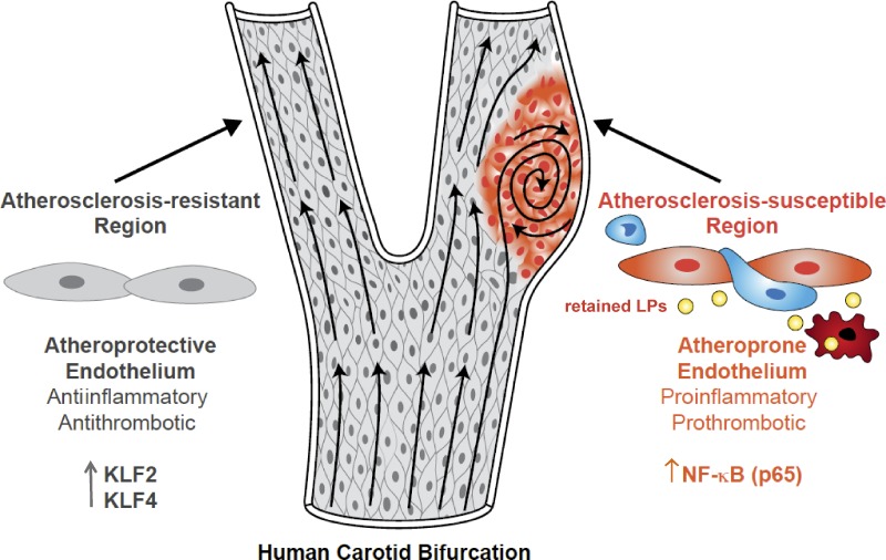
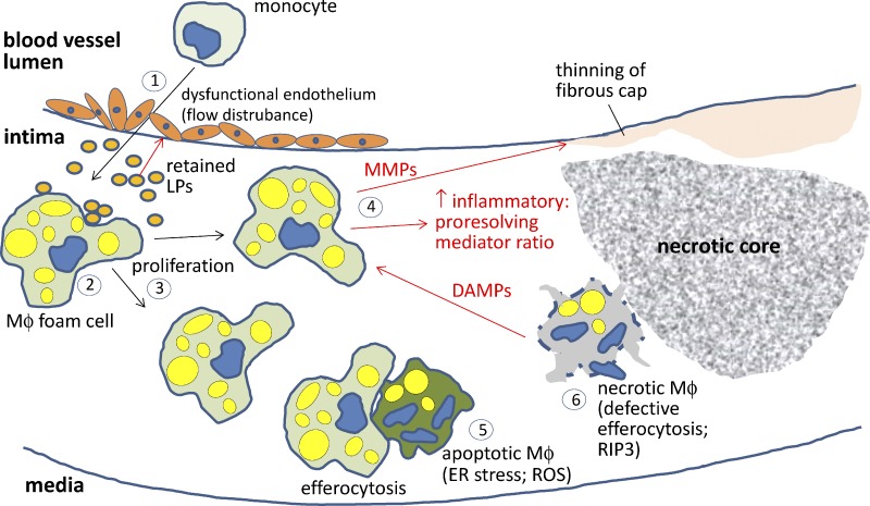
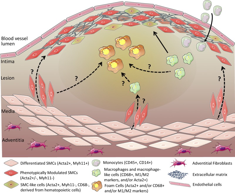
Atherosclerosis occurs in the subendothelial space (intima) of medium-sized arteries at regions of disturbed blood flow and is triggered by an interplay between endothelial dysfunction and subendothelial lipoprotein retention. Over time, this process stimulates a nonresolving inflammatory response that can cause intimal destruction, arterial thrombosis, and end-organ ischemia. Recent advances highlight important cell biological atherogenic processes, including mechanotransduction and inflammatory processes in endothelial cells, origins and contributions of lesional macrophages, and origins and phenotypic switching of lesional smooth muscle cells. These advances illustrate how in-depth mechanistic knowledge of the cellular pathobiology of atherosclerosis can lead to new ideas for therapy.
Overview
The claims and statistics of this paper haven’t been checked yet.
Run the check — free, no account needed. It reads the open-access text and checks the sources it cites.
Integrity signals
Registry
Cites
Statistics
Figures
Verification
Claims the paper makes
The claims and statistics of this paper haven’t been checked yet.
Run the check — free, no account needed. It reads the open-access text and checks the sources it cites.
Claims other papers make about this one
Each is checked against this paper’s own text: does it say what the citing paper says it does?
The check wasn’t confident either way, so it gives no verdict — never disputed.
In the citing paper
“Adding growth factors such as VEGF and PDGF to PLCL vascular scaffolds can effectively promote vascular endothelialization, accelerate migration and aggregation of endothelial cells and smooth muscle cells, and promote vascular regeneration and reconstruction [161].”
From Application of PLCL as a biodegradable polymer in biomedical engineering (2026) · cites this paper as [161]
Experimental — accuracy not yet measuredIn the citing paper
“Atherosclerosis is an age‐associated vascular disease characterised by the formation of plaques in arterial vessels, which can lead to the development of various cardiovascular disorders including myocardial infarction, stroke and sudden cardiac death (Tabas et al, 2015).”
From The struggle of a good friend getting old: cellular senescence in viral responses and therapy (2021) · cites this paper as Tabas et al, 2015
In the citing paper
“Hypercholesterolemia, defined by elevated plasma low‐density lipoprotein (LDL) cholesterol, is a well‐established risk factor not only for cardiovascular disease (Borén et al. 2022; Steinberg 2013; Tabas et al. 2015; Boffa and Koschinsky 2019), but also for cognitive decline and dementia in later life (Kivipelto et al. 2001; Kivipelto and Solomon 2006; Livingston et al. 2024; Sparks et al. 2000, 2”
From LDL Cholesterol Modulates Astrocyte Metabolism, Lipid Handling, and Morphology: Evidence From In Vitro and In Vivo Models (2026) · cites this paper as Tabas et al. 2015
Self-check
Percentages, sums and reported differences re-computed from the paper’s own numbers.
The self-check needs the paper’s full text.
Citations
Cites
A role for mitochondria in nlrp3 inflammasome activation
Zhou, R., Yazdi, A. S., Menu, P., & Tschopp, J. (2011) · Nature · doi:10.1038/nature09663 · Cited by 5,602
Lusis, A. J. (2000) · Nature · doi:10.1038/35025203 · Cited by 4,880
NLRP3 inflammasomes are required for atherogenesis and activated by cholesterol crystals
Duewell, P., Kono, H., Rayner, K. J., Sirois, C. M., Vladimer, G., Bauernfeind, F. G., Abela, G. S., Franchi, L., Nuñez, G., Schnurr, M., et al. (2010) · Nature · doi:10.1038/nature08938 · Cited by 4,026
Glass, C. K., & Witztum, J. L. (2001) · Cell · doi:10.1016/s0092-8674(01)00238-0 · Cited by 3,094
Macrophages in the Pathogenesis of Atherosclerosis
Moore, K. J., & Tabas, I. (2011) · Cell · doi:10.1016/j.cell.2011.04.005 · Cited by 2,669
Self-citationIntegrating the mechanisms of apoptosis induced by endoplasmic reticulum stress
Tabas, I. A., & Ron, D. (2011) · Nature Cell Biology · doi:10.1038/ncb0311-184 · Cited by 2,655
Self-citationThe immune system in atherosclerosis
Hansson, G. K., & Hermansson, A. (2011) · Nature Immunology · doi:10.1038/ni.2001 · Cited by 2,389
Nathan, C., & Ding, A. (2010) · Cell · doi:10.1016/j.cell.2010.02.029 · Cited by 2,183
Cardiovascular Medicine at the Turn of the Millennium: Triumphs, Concerns, and Opportunities
Braunwald, E. (1997) · New England Journal of Medicine · doi:10.1056/nejm199711063371906 · Cited by 1,426
The Response-to-Retention Hypothesis of Early Atherogenesis
Williams, K. J., & Tabas, I. (1995) · Arteriosclerosis, Thrombosis, and Vascular Biology · doi:10.1161/01.atv.15.5.551 · Cited by 1,413
Self-citation
Cited by
Sorted by citations
Endothelial Cell Dysfunction and the Pathobiology of Atherosclerosis
Gimbrone, M. A., & García‐Cardeña, G. (2016) · Circulation Research · doi:10.1161/circresaha.115.306301 · Cited by 3,694
Self-citationLibby, P., Buring, J. E., Badimon, L., Hansson, G. K., Deanfield, J., Bittencourt, M. S., Tokgözoğlu, L., & Lewis, E. F. (2019) · Nature Reviews Disease Primers · doi:10.1038/s41572-019-0106-z · Cited by 3,211
Vascular smooth muscle cells in atherosclerosis
Bennett, M. R., Sinha, S., & Owens, G. K. (2016) · Circulation Research · doi:10.1161/circresaha.115.306361 · Cited by 2,179
Self-citationReactive Oxygen Species in Metabolic and Inflammatory Signaling
Forrester, S. J., Kikuchi, D. S., Hernandes, M. S., Xu, Q., & Griendling, K. K. (2018) · Circulation Research · doi:10.1161/circresaha.117.311401 · Cited by 2,081
Dietary fats and cardiovascular disease: a presidential advisory from the American Heart Association
Sacks, F. M., Lichtenstein, A. H., Wu, J. H. Y., Appel, L. J., Creager, M. A., Kris-Etherton, P. M., Miller, M., Rimm, E. B., Rudel, L. L., Robinson, J. G., et al. (2017) · Circulation · doi:10.1161/cir.0000000000000510 · Cited by 1,386
Cellular Senescence: Aging, Cancer, and Injury
Calcinotto, A., Kohli, J., Zagato, E., Pellegrini, L., Demaria, M., & Alimonti, A. (2019) · Physiological Reviews · doi:10.1152/physrev.00020.2018 · Cited by 1,270
“Many vascular diseases arise because of this sequence of events including myocardial infarction, stroke, unstable angina, and sudden cardiac death (63, 309).”
BackgroundSenescent cells: an emerging target for diseases of ageing
Childs, B. G., Gluscevic, M., Baker, D. J., Laberge, R.-M., Marquess, D., Dananberg, J., & van Deursen, J. M. (2017) · Nature Reviews Drug Discovery · doi:10.1038/nrd.2017.116 · Cited by 1,284
Senescent intimal foam cells are deleterious at all stages of atherosclerosis
Childs, B. G., Baker, D. J., Wijshake, T., Conover, C. A., Campisi, J., & van Deursen, J. M. (2016) · Science · doi:10.1126/science.aaf6659 · Cited by 1,195
Clinical Update: Cardiovascular Disease in Diabetes Mellitus
Wang, C. C. L., Hess, C. N., Hiatt, W. R., & Goldfine, A. B. (2016) · Circulation · doi:10.1161/circulationaha.116.022194 · Cited by 1,222
Xu, S., Ilyas, I., Little, P. J., Li, H., Kamato, D., Zheng, X., Luo, S., Li, Z., Liu, P., Han, J., et al. (2021) · Pharmacological Reviews · doi:10.1124/pharmrev.120.000096 · Cited by 1,205
“Many studies have demonstrated that abnormal lipid metabolism, inflammatory responses, and hemodynamic alterations are critical players in the occurrence of endothelial dysfunction and atherosclerosis (Tabas et al., 2015).”
BackgroundResearch Progress on the Relationship between Atherosclerosis and Inflammation
Zhu, Y., Xian, X., Wang, Z., Bi, Y., Chen, Q., Han, X., Tang, D., & Chen, R. (2018) · Biomolecules · doi:10.3390/biom8030080 · Cited by 900
“In early-stage atherosclerosis, the causes of atherosclerosis are endothelial injury, abnormal lipid metabolism, and hemodynamic damage; and the atherogenic process is considered to be accompanied by flow-mediated inflammatory changes in endothelial cells (ECs) [5].”
BackgroundInflammation and plaque vulnerability
Hansson, G. K., Libby, P., & Tabas, I. A. (2015) · Journal of Internal Medicine · doi:10.1111/joim.12406 · Cited by 809
Self-citationOrganotypic vasculature: From descriptive heterogeneity to functional pathophysiology
Augustin, H. G., & Koh, G. Y. (2017) · Science · doi:10.1126/science.aal2379 · Cited by 790
“Yet growing evidence suggests that ECs play a pivotal role in the pathogenesis of atherosclerosis (92, 93).”
BackgroundThe Role of Inflammation in Cardiovascular Disease
Henein, M. Y., Vancheri, S., Longo, G., & Vancheri, F. (2022) · International Journal of Molecular Sciences · doi:10.3390/ijms232112906 · Cited by 802
“These changes lead to a subendothelial accumulation of cholesterol-containing lipoproteins which triggers a low-grade inflammatory response (Figure 1) [44,45].”
BackgroundLiver X receptors in lipid signalling and membrane homeostasis
Wang, B., & Tontonoz, P. (2018) · Nature Reviews Endocrinology · doi:10.1038/s41574-018-0037-x · Cited by 722
Therapeutic targeting of the angiopoietin–TIE pathway
Saharinen, P., Eklund, L., & Alitalo, K. (2017) · Nature Reviews Drug Discovery · doi:10.1038/nrd.2016.278 · Cited by 620
Cell Type-Specific Roles of NF-κB Linking Inflammation and Thrombosis
Mussbacher, M., Salzmann, M., Brostjan, C., Hoesel, B., Schoergenhofer, C., Datler, H., Hohensinner, P., Basílio, J., Petzelbauer, P., Assinger, A., et al. (2019) · Frontiers in Immunology · doi:10.3389/fimmu.2019.00085 · Cited by 596
“An important aspect is that SMC-derived macrophage-like cells are apparently less efficient in phagocytosis of deposits and apoptotic cells within the plaque as compared to “real” macrophages, which exacerbates necrotic core formation rendering the plaque unstable and prone for rupture (338, 339).”
BackgroundEndothelial-to-mesenchymal transition drives atherosclerosis progression
Chen, P., Qin, L., Baeyens, N., Li, G., Afolabi, T., Budatha, M., Tellides, G., Schwartz, M. A., & Simons, M. (2015) · Journal of Clinical Investigation · doi:10.1172/jci82719 · Cited by 592
“The two key factors regulating endothelial FGFR1 expression, disturbed shear stress and vessel wall inflammation, are also well-known atherosclerosis risk factors (1, 5).”
BackgroundEelen, G., de Zeeuw, P., Treps, L., Harjes, U., Wong, B. W., & Carmeliet, P. (2017) · Physiological Reviews · doi:10.1152/physrev.00001.2017 · Cited by 614
Animal models of atherosclerosis
Emini Veseli, B., Perrotta, P., De Meyer, G. R. A., Roth, L., Van der Donckt, C., Martinet, W., & De Meyer, G. R. Y. (2017) · European Journal of Pharmacology · doi:10.1016/j.ejphar.2017.05.010 · Cited by 548
Brozovich, F. V., Nicholson, C., Degen, C. V., Gao, Y. Z., Aggarwal, M., & Morgan, K. G. (2016) · Pharmacological Reviews · doi:10.1124/pr.115.010652 · Cited by 568
“Recent Direct Confirmation of Changes in Vascular Tone/Resistance Related to Changes in Systemic Vascular Resistance and Blood Pressure and the Importance of Vascular Smooth Muscle Contraction in both Normal Physiology and Pathophysiology—Hypertension.”
BackgroundInfluentialPhospholipid Remodeling in Physiology and Disease
Wang, B., & Tontonoz, P. (2018) · Annual Review of Physiology · doi:10.1146/annurev-physiol-020518-114444 · Cited by 558
Smooth muscle cell fate and plasticity in atherosclerosis
Allahverdian, S., Chaâbane, C., Boukais, K., Francis, G. A., & Bochaton‐Piallat, M. (2018) · Cardiovascular Research · doi:10.1093/cvr/cvy022 · Cited by 513
Cerebral Vascular Disease and Neurovascular Injury in Ischemic Stroke
Hu, X., Silva, T. M. D., Chen, J., & Faraci, F. M. (2017) · Circulation Research · doi:10.1161/circresaha.116.308427 · Cited by 456
“of monocytes, and other inflammatory cells, along with phenotypic switching of vascular muscle.(15) Atherosclerosis is primarily a disease of large arteries,(97) but can extend into smaller vessels with the addition of hypertension,(98) and perhaps other”
BackgroundThe molecular basis of endothelial cell plasticity
Dejana, E., Hirschi, K. K., & Simons, M. (2017) · Nature Communications · doi:10.1038/ncomms14361 · Cited by 451
Wang, J., Wang, L., Lou, G., Zeng, H., Ju, H., Huang, Q., Peng, W., & Yang, X.-B. (2019) · Pharmaceutical Biology · doi:10.1080/13880209.2019.1577466 · Cited by 427
Wang, Y., Zhang, K., Li, T., Maruf, A., Qin, X., Luo, L., Zhong, Y., Qiu, J., McGinty, S., Pontrelli, G., et al. (2020) · Theranostics · doi:10.7150/thno.47841 · Cited by 412
“, VCAM-1) and secrete chemokines to recruit circulating monocyte macrophages and promote them to traverse the subendothelial space and migrate into the intimal layer [55,56].”
BackgroundNakahara, T., Dweck, M. R., Narula, N., Pisapia, D. J., Narula, J., & Strausś, H. W. (2017) · JACC. Cardiovascular imaging · doi:10.1016/j.jcmg.2017.03.005 · Cited by 410
Tcheandjieu, C., Zhu, X., Hilliard, A. T., Clarke, S. L., Napolioni, V., Ma, S., Lee, K. M., Fang, H., Chen, F., Lu, Y., et al. (2022) · Nature Medicine · doi:10.1038/s41591-022-01891-3 · Cited by 363
SR-B1 drives endothelial cell LDL transcytosis via DOCK4 to promote atherosclerosis
Huang, L., Chambliss, K. L., Gao, X., Yuhanna, I. S., Behling‐Kelly, E. L., Bergaya, S., Ahmed, M. H., Michaely, P., Luby‐Phelps, K., Darehshouri, A., et al. (2019) · Nature · doi:10.1038/s41586-019-1140-4 · Cited by 360
Failure of physiological transformation and spiral artery atherosis: their roles in preeclampsia
Staff, A. C., Fjeldstad, H. E., Fosheim, I. K., Moe, K., Turowski, G., Johnsen, G. M., Alnaes-Katjavivi, P., & Sugulle, M. (2022) · American Journal of Obstetrics and Gynecology · doi:10.1016/j.ajog.2020.09.026 · Cited by 354
“Foam cells themselves are products of inflammatory stress, typically of the intima, and are characteristic of early stages of atherosclerosis.(74)”
BackgroundEndothelial TGF-β signalling drives vascular inflammation and atherosclerosis
Chen, P., Qin, L., Li, G., Wang, Z., Dahlman, J. E., Malagon-Lopez, J., Gujja, S., Cilfone, N. A., Kauffman, K. J., Sun, L., et al. (2019) · Nature Metabolism · doi:10.1038/s42255-019-0102-3 · Cited by 348
Inflamm-ageing: the role of inflammation in age-dependent cardiovascular disease
Liberale, L., Montecucco, F., Tardif, J., Libby, P., & Camici, G. G. (2020) · European Heart Journal · doi:10.1093/eurheartj/ehz961 · Cited by 363
“Cells within atherosclerotic plaques often show features of senescence.(8,90) Cytokines and inflammatory cells participate in atherosclerosis at all stages, able to exert either pro- or anti-atherogenic effects.”
BackgroundThe Contribution of Homocysteine Metabolism Disruption to Endothelial Dysfunction: State-of-the-Art
Esse, R., Barroso, M., de Almeida, I. T., & Castro, R. (2019) · International Journal of Molecular Sciences · doi:10.3390/ijms20040867 · Cited by 352
“and results in a chronic condition in which arteries harden through the build-up of plaques in the vessel wall [3].”
BackgroundWei, X., Man, Y. M., Dehaini, D., Su, Y., Kroll, A. V., Zhou, J., Gao, W., Fang, R. H., Chien, S., & Zhang, L. (2017) · ACS Nano · doi:10.1021/acsnano.7b07720 · Cited by 336
Leukocytes Link Local and Systemic Inflammation in Ischemic Cardiovascular Disease
Libby, P., Nahrendorf, M., & Świrski, F. K. (2016) · Journal of the American College of Cardiology · doi:10.1016/j.jacc.2015.12.048 · Cited by 335
“(5,29) popularized the concept of defective efferocytosis of phagocytes within atheromatous plaques.”
BackgroundThe role of cytokines in the development of atherosclerosis
Fatkhullina, A. R., Peshkova, I. O., & Koltsova, E. K. (2016) · Biochemistry (Moscow) · doi:10.1134/s0006297916110134 · Cited by 338
“11 2016 Later stages of atherosclerosis are characterized by so called unresolved inflammation that is maintained by various factors including increased levels of oxLDLs and high blood pressure [14].”
BackgroundSoft Drusen in Age-Related Macular Degeneration: Biology and Targeting Via the Oil Spill Strategies
Curcio, C. A. (2018) · Investigative Ophthalmology & Visual Science · doi:10.1167/iovs.18-24882 · Cited by 322
“Atherosclerosis research may be a source of ideas.(237,238)”
BackgroundKramann, R., Goettsch, C., Wongboonsin, J., Iwata, H., Schneider, R. K., Kuppe, C., Kaesler, N., Chang-Panesso, M., Machado, F. G., Gratwohl, S., et al. (2016) · Cell stem cell · doi:10.1016/j.stem.2016.08.001 · Cited by 308
“A classical hypothesis holds that in response to injury, terminally differentiated vSMCs undergo a phenotypic switch, downregulating contractile proteins and proliferating and migrating into the neointima (Tabas et al., 2015).”
BackgroundRen, J., Fu, L., Nile, S. H., Zhang, J., & Kai, G. (2019) · Frontiers in Pharmacology · doi:10.3389/fphar.2019.00753 · Cited by 310
Li, N., Zhou, H., & Tang, Q. (2017) · Disease Markers · doi:10.1155/2017/7089493 · Cited by 293
“Atherosclerosis usually occurs in the intima of medium-sized arteries at regions of disturbed blood flow, which is triggered by an interaction between endothelial dysfunction and subendothelial retention of apolipoprotein B- (apoB-) containing lipoproteins (LPS) in focal areas of arteries [30].”
BackgroundAkt Signaling in Macrophage Polarization, Survival, and Atherosclerosis
Linton, M. F., Moslehi, J. J., & Babaev, V. R. (2019) · International Journal of Molecular Sciences · doi:10.3390/ijms20112703 · Cited by 291
“Macrophage apoptosis significantly influences atherosclerosis formation at every stage of disease including early lesions, plaque progression, and plaque stability [67,68].”
BackgroundHypertension, Neurovascular Dysfunction, and Cognitive Impairment
Santisteban, M. M., Iadecola, C., & Carnevale, D. (2022) · Hypertension · doi:10.1161/hypertensionaha.122.18085 · Cited by 317
Macrophages in age-related chronic inflammatory diseases
Oishi, Y., & Manabe, I. (2016) · npj Aging and Mechanisms of Disease · doi:10.1038/npjamd.2016.18 · Cited by 262
“This necrotic debris also promotes inflammation and destabilization of the plaque, which can lead to plaque rupture.(63) It is currently unclear whether macrophage apoptosis is accelerated by aging, but peritoneal macrophages from aged mice are more susceptible to endoplasmic reticulum stress-induced apoptosis.”
BackgroundHeart in space: effect of the extraterrestrial environment on the cardiovascular system
Hughson, R. L., Helm, A., & Durante, M. (2018) · Nature Reviews Cardiology · doi:10.1038/nrcardio.2017.157 · Cited by 257
Macrophage Inflammation, Erythrophagocytosis, and Accelerated Atherosclerosis in Jak2 V617F Mice
Wang, W., Liu, W., Fidler, T., Wang, Y., Tang, Y., Woods, B., Welch, C., Cai, B., Silvestre-Roig, C., Ai, D., et al. (2018) · Circulation Research · doi:10.1161/circresaha.118.313283 · Cited by 262
Self-citationRecent progress regarding kaempferol for the treatment of various diseases (Review)
Ren, J., Lu, Y., Qian, Y., Chen, B., Wu, T., & Ji, G. (2019) · Experimental and Therapeutic Medicine · doi:10.3892/etm.2019.7886 · Cited by 272
“Endothelial cells, macrophages and smooth muscle cells are the major cell types involved in atherosclerosis (129).”
BackgroundDuan, H., Zhang, Q., Liu, J., Li, R., Wang, D., Peng, W., & Wu, C. (2021) · Pharmacological Research · doi:10.1016/j.phrs.2021.105599 · Cited by 262
Perivascular adipose tissue (PVAT) in atherosclerosis: a double-edged sword
Qi, X.-Y., Qu, S.-L., Xiong, W., Rom, O., Chang, L., & Jiang, Z. (2018) · Cardiovascular Diabetology · doi:10.1186/s12933-018-0777-x · Cited by 239
Targeting Foam Cell Formation in Atherosclerosis: Therapeutic Potential of Natural Products
Wang, D., Yang, Y., Lei, Y., Tzvetkov, N. T., Liu, X., Yeung, A. W. K., Xu, S., & Atanasov, A. G. (2019) · Pharmacological Reviews · doi:10.1124/pr.118.017178 · Cited by 223
“Normally, the necrotic core within the plaque is covered by a “protective” barrier—the fibrous cap, which serves as an “insulator” between platelets in circulating blood and prothrombotic substances within the lesion (Tabas et al., 2015).”
BackgroundInfluentialMacrophage Apoptosis and Efferocytosis in the Pathogenesis of Atherosclerosis
Linton, M. F., Babaev, V. R., Huang, J., Linton, E. F., Tao, H., & Yancey, P. G. (2016) · Circulation Journal · doi:10.1253/circj.cj-16-0924 · Cited by 235
Anti-inflammatory and immunoregulatory effects of icariin and icaritin
Bi, Z., Zhang, W. Y., & Yan, X. (2022) · Biomedicine & Pharmacotherapy · doi:10.1016/j.biopha.2022.113180 · Cited by 226
Integrin beta3 regulates clonality and fate of smooth muscle-derived atherosclerotic plaque cells
Misra, A., Feng, Z., Chandran, R. R., Kabir, I., Rotllan, N., Aryal, B., Sheikh, A. Q., Ding, L., Qin, L., Fernández-Hernando, C., et al. (2018) · Nature Communications · doi:10.1038/s41467-018-04447-7 · Cited by 207
Angiopoietin–Tie signalling in the cardiovascular and lymphatic systems
Eklund, L., Kangas, J., & Saharinen, P. (2016) · Clinical Science · doi:10.1042/cs20160129 · Cited by 210
Canfrán‐Duque, A., Rotllán, N., Zhang, X., Fernández‐Fuertes, M., Ramírez, C. M., Araldi, E., Daimiel, L., Busto, R., Fernandez-Hernando, C. F., & Suárez, Y. (2017) · EMBO Molecular Medicine · doi:10.15252/emmm.201607492 · Cited by 207
“As introduced above, in addition to macrophages apoptosis, the ability of phagocytes to clear apoptotic cells contributes to lesional necrosis, an important feature of advanced and unstable plaques (Tabas, 2010; Tabas et al , 2015).”
Non-HDL-cholesterol in dyslipidemia: Review of the state-of-the-art literature and outlook
Raja, V., Aguíar, C., Al-Sayed, N., Chibber, Y. S., ElBadawi, H., Владиславович, Е. М., Hermans, M. P., Pandey, R. C., Ray, K. K., Tokgözoğlu, L. S., et al. (2023) · Atherosclerosis · doi:10.1016/j.atherosclerosis.2023.117312 · Cited by 221
Senescent cells: a therapeutic target for cardiovascular disease
Childs, B. G., Li, H., & van Deursen, J. M. (2018) · Journal of Clinical Investigation · doi:10.1172/jci95146 · Cited by 192
“These foam cells secrete cytokines and chemokines such as IL-1α, TNF-α, and MCP-1 to drive further immune cell recruitment and form an atherosclerotic plaque (118, 119).”
BackgroundEpigenetics of Atherosclerosis: Emerging Mechanisms and Methods
Khyzha, N., Alizada, A., Wilson, M. D., & Fish, J. E. (2017) · Trends in Molecular Medicine · doi:10.1016/j.molmed.2017.02.004 · Cited by 196
Neurovascular Dysfunction in Vascular Dementia, Alzheimer's and Atherosclerosis
Shabir, O., Berwick, J., & Francis, S. E. (2018) · BMC Neuroscience · doi:10.1186/s12868-018-0465-5 · Cited by 191
Cao, H., Jia, Q., Li, Y., Chen, C., Xing, S., & Shen, D. (2019) · International Journal of Molecular Sciences · doi:10.3390/ijms20236093 · Cited by 189
“As a major component of AS lesions, foam cells play an important role in the development of AS [26].”
BackgroundResultTargeting smooth muscle cell phenotypic switching in vascular disease
Chakraborty, R., Chatterjee, P., Dave, J. M., Ostriker, A. C., Greif, D. M., Rzucidlo, E. M., & Martin, K. A. (2021) · JVS Vascular Science · doi:10.1016/j.jvssci.2021.04.001 · Cited by 182
The gut microbiota as a novel regulator of cardiovascular function and disease
Battson, M. L., Lee, D. M., Weir, T. L., & Gentile, C. L. (2017) · The Journal of Nutritional Biochemistry · doi:10.1016/j.jnutbio.2017.12.010 · Cited by 191
Ebnet, K. (2017) · Physiological Reviews · doi:10.1152/physrev.00004.2017 · Cited by 179
“Atherosclerosis is initiated by the formation of an atherosclerotic lesion caused by the retention of lipopoteins in the subendothelial space, which triggers a local inflammatory response characterized by the recruitment of various leukocyte subsets and the accumulation of extracellular material (212).”
BackgroundMicrovascular Dysfunction and Cognitive Impairment
Silva, T. M. D., & Faraci, F. M. (2016) · Cellular and Molecular Neurobiology · doi:10.1007/s10571-015-0308-1 · Cited by 173
“Immune-related molecular and cellular determinants of atherosclerosis (predominantly a disease of larger arteries) have been studied for many years (Tabas et al. 2015).”
BackgroundEndothelial Cell Metabolism in Atherosclerosis
Theodorou, K., & Boon, R. A. (2018) · Frontiers in Cell and Developmental Biology · doi:10.3389/fcell.2018.00082 · Cited by 167
“Alternatively, superficial intimal erosions, as a consequence of EC apoptosis and detachment, may also trigger an atherothrombotic event and its clinical sequelae (Quillard et al., 2015; Tabas et al., 2015).”
BackgroundEndothelial Transcytosis of Lipoproteins in Atherosclerosis
Zhang, X., Sessa, W. C., & Fernandez-Hernando, C. F. (2018) · Frontiers in Cardiovascular Medicine · doi:10.3389/fcvm.2018.00130 · Cited by 158
“This promotes the formation of macrophage foam cells within atherosclerotic lesions (13).”
BackgroundThe Key Role of DNA Methylation and Histone Acetylation in Epigenetics of Atherosclerosis
Lee, H., Oh, S., Ro, D. H., Yoo, H., & Kwon, Y. (2020) · Journal of Lipid and Atherosclerosis · doi:10.12997/jla.2020.9.3.419 · Cited by 168
The Dual Role of Myeloperoxidase in Immune Response
Arnhold, J. (2020) · International Journal of Molecular Sciences · doi:10.3390/ijms21218057 · Cited by 172
Simonetto, M., Infante, M., Sacco, R. L., Rundek, T., & Della‐Morte, D. (2019) · Nutrients · doi:10.3390/nu11102279 · Cited by 164
“Atherosclerosis begins with inflammatory changes within the endothelium caused by low-density lipoproteins (LDLs) being deposited on endothelial cells [41,42].”
BackgroundNon-alcoholic fatty liver disease and cardiovascular disease: assessing the evidence for causality
Brouwers, M. C. G. J., Simons, N., Stehouwer, C. D. A., & Isaacs, A. J. (2019) · Diabetologia · doi:10.1007/s00125-019-05024-3 · Cited by 154
Elmarasi, M., Elmakaty, I., Elsayed, B., Elsayed, A., Zein, J. A., Boudaka, A., & Eid, A. H. (2024) · Journal of Cellular Physiology · doi:10.1002/jcp.31200 · Cited by 159
“It is a chronic progressive inflammatory disease where lesions develop in the wall of large ‐ and medium ‐ sized arteries (Tabas et al., 2015).”
BackgroundNovel Insights Into the NLRP3 Inflammasome in Atherosclerosis
Jin, Y. H., & Fu, J. (2019) · Journal of the American Heart Association · doi:10.1161/jaha.119.012219 · Cited by 153
“A therosclerosis underlies the leading cause of death in industrialized countries.(1) Atherosclerosis is a maladap-”
BackgroundInfluentialThe role of smooth muscle cells in plaque stability: Therapeutic targeting potential
Harman, J. J., & Jørgensen, H. F. (2019) · British Journal of Pharmacology · doi:10.1111/bph.14779 · Cited by 147
“Thinning of the fibrous cap in advanced plaques increases the risk of rupture, which triggers thrombus formation and subsequent clinical complications including heart attack and stroke (Libby et al., 2011; Tabas et al., 2015).”
BackgroundThe Molecular Basis of Predicting Atherosclerotic Cardiovascular Disease Risk
Nayor, M. G., Brown, K., & Vasan, R. S. (2021) · Circulation Research · doi:10.1161/circresaha.120.315890 · Cited by 149
Cheng, H., Zhong, W., Wang, L., Zhang, Q. H., Ma, X., Wang, Y., Wang, S., He, C., Wei, Q., & Fu, C. (2023) · Biomedicine & Pharmacotherapy · doi:10.1016/j.biopha.2022.114198 · Cited by 157
PVAT and Its Relation to Brown, Beige, and White Adipose Tissue in Development and Function
Hildebrand, S., Stümer, J., & Pfeifer, A. (2018) · Frontiers in Physiology · doi:10.3389/fphys.2018.00070 · Cited by 143
“The retained particles are then modified in various ways (e.g., oxidation and glycation), which turns them into auto-antigens, inducing a low-grade inflammation (Rader and Daugherty, 2008; Tabas et al., 2015).”
BackgroundInfluentialMacrophage metabolism in atherosclerosis
Bories, G. F. P., & Leitinger, N. (2017) · FEBS Letters · doi:10.1002/1873-3468.12786 · Cited by 145
The impact of epigenetics on cardiovascular disease
Prasher, D., Greenway, S. C., & Singh, R. B. (2019) · Biochemistry and Cell Biology · doi:10.1139/bcb-2019-0045 · Cited by 143
Glycocalyx in Atherosclerosis-Relevant Endothelium Function and as a Therapeutic Target
Mitra, R., O’Neil, G., Harding, I. C., Cheng, M. J., Mensah, S. A., & Ebong, E. E. (2017) · Current Atherosclerosis Reports · doi:10.1007/s11883-017-0691-9 · Cited by 135
“The fibrous cap, which is in constant dynamic equilibrium, provides a protective barrier between platelets in the blood stream and pro-thrombotic plaque contents [15].”
BackgroundSARS-CoV-2 Mediated Endothelial Dysfunction: The Potential Role of Chronic Oxidative Stress
Chang, R. P., Mamun, A., Dominic, A., & Le, N. (2021) · Frontiers in Physiology · doi:10.3389/fphys.2020.605908 · Cited by 136
Kim, H. (2017) · Scientific Reports · doi:10.1038/s41598-017-07381-8 · Cited by 126
Zhang, X., McDonald, J. G., Aryal, B. K., Canfrán‐Duque, A., Goldberg, E. L., Araldi, E., Ding, W., Fan, Y., Thompson, B. M., Singh, A. K., et al. (2021) · Proceedings of the National Academy of Sciences · doi:10.1073/pnas.2107682118 · Cited by 136
He, L., Zhou, Q., Huang, Z., Xu, J., Zhou, H., Lv, D., Lu, L., Huang, S., Tang, M., Zhong, J., et al. (2018) · Journal of Cellular Physiology · doi:10.1002/jcp.27527 · Cited by 137
Freemerman, A. J., Zhao, L., Pingili, A. K., Teng, B., Cozzo, A. J., Fuller, A. M., Johnson, A., Milner, J. J., Lim, M. F., Galanko, J. A., et al. (2019) · The Journal of Immunology · doi:10.4049/jimmunol.1800002 · Cited by 132
“(LDL) particles, efferocytosis, and control of the immune milieu (32, 33).”
BackgroundCanfrán‐Duque, A., Rotllán, N., Zhang, X., Andrés‐Blasco, I., Thompson, B. M., Sun, J., Price, N. L., Fernández‐Fuertes, M., Fowler, J. W., Gómez‐Coronado, D., et al. (2022) · Circulation · doi:10.1161/circulationaha.122.059062 · Cited by 130
Vascular Cells in Blood Vessel Wall Development and Disease
Mazurek, R., Dave, J. M., Chandran, R. R., Misra, A., Sheikh, A. Q., & Greif, D. M. (2016) · Advances in pharmacology · doi:10.1016/bs.apha.2016.08.001 · Cited by 136
Cardiovascular effects of air pollution: current evidence from animal and human studies
Lederer, A. M., Fredriksen, P. M., Nkeh-Chungag, B. N., Everson, F., Strijdom, H., Boever, P. D., & Goswami, N. (2021) · American Journal of Physiology-Heart and Circulatory Physiology · doi:10.1152/ajpheart.00706.2020 · Cited by 130
Epigenetic Control of Smooth Muscle Cell Identity and Lineage Memory
Gomez, D. A., Swiatlowska, P., & Owens, G. K. (2015) · Arteriosclerosis Thrombosis and Vascular Biology · doi:10.1161/atvbaha.115.305044 · Cited by 131
Self-citationGalectin‐3 Is a Potential Mediator for Atherosclerosis
Gao, Z., Liu, Z., Wang, R., Zheng, Y., Li, H., & Yang, L. M. (2020) · Journal of Immunology Research · doi:10.1155/2020/5284728 · Cited by 123
“…in fl ammation, sustained activation of ECs by in fl ammatory stimuli causes alterations in normal endothelial function, resulting in endothelial dysfunction which has been considered to be the basis and initial step of atherosclerosis, the most common cause of cardiovascular diseases [34, 35].”
Atheroprotective Effects and Molecular Targets of Tanshinones Derived From Herbal Medicine Danshen
Fang, J., Little, P. J., & Xu, S. (2017) · Medicinal Research Reviews · doi:10.1002/med.21438 · Cited by 122
Endothelial cells, endoplasmic reticulum stress and oxysterols
Luchetti, F., Crinelli, R., Cesarini, E., Canonico, B., Guidi, L., Zerbinati, C., Sario, G. D., Zamai, L., Magnani, M., Papa, S., et al. (2017) · Redox Biology · doi:10.1016/j.redox.2017.07.014 · Cited by 125
“contrast, unbranched arteries that are exposed to uniform laminar shear stress largely do not develop lesions [2].”
BackgroundEndothelial to Mesenchymal Transition in Health and Disease
Xu, Y., & Kovacic, J. C. (2022) · Annual Review of Physiology · doi:10.1146/annurev-physiol-032222-080806 · Cited by 125
Endothelial-to-Mesenchymal Transition, Vascular Inflammation, and Atherosclerosis
Chen, P., Schwartz, M. A., & Simons, M. (2020) · Frontiers in Cardiovascular Medicine · doi:10.3389/fcvm.2020.00053 · Cited by 119
“The process is initiated by vascular injury–induced primarily by hyperlipidemia, albeit other factors, such as tobacco, hypercysteinemia, diabetes, and hypertension, also play a role (1, 2).”
BackgroundVulnerable Plaque, Characteristics, Detection, and Potential Therapies
Hafiane, A. (2019) · Journal of Cardiovascular Development and Disease · doi:10.3390/jcdd6030026 · Cited by 130
“Incidences of this disease are continuing to rise despite recent advances in primary prevention and therapeutic technology [1,3,4].”
BackgroundLecce, L., Xu, Y., V’Gangula, B., Chandel, N., Pothula, V., Caudrillier, A., Santini, M. P., d’Escamard, V., Ceholski, D. K., Gorski, P. A., et al. (2021) · Journal of Clinical Investigation · doi:10.1172/jci131178 · Cited by 118
“Atherosclerosis is the most common cause of cardiovascular disease and is a chronic process involving cellular, metabolic, and inflammatory events (45, 46).”
BackgroundZhang, L., Cheng, H., Yue, Y., Li, S., Zhang, D., & He, R. (2017) · Cardiovascular Pathology · doi:10.1016/j.carpath.2017.11.004 · Cited by 115
Extracellular MicroRNA-92a Mediates Endothelial Cell–Macrophage Communication
Chang, Y., Li, Y.-S. J., Wu, C., Wang, K.-C., Huang, T.-C., Chen, Z., & Chien, S. (2019) · Arteriosclerosis Thrombosis and Vascular Biology · doi:10.1161/atvbaha.119.312707 · Cited by 116
“Our current study has demonstrated that (1) miR-92a is transported from atherogenic ECs to macrophages through extracellular vesicles (EVs); (2) the pathophysiologic macrophage phenotypes, such as inflammatory responses, LDL uptake, and impaired migration ability, are induced by intravesicular miR-92a; and (3) that ma…”
BackgroundEndothelial protein kinase MAP4K4 promotes vascular inflammation and atherosclerosis
Flach, R. J. R., Skoura, A., Matevossian, A., Danai, L. V., Zheng, W., Cortes, C., Bhattacharya, S. K., Aouadi, M., Hagan, N., Yawe, J. C., et al. (2015) · Nature Communications · doi:10.1038/ncomms9995 · Cited by 114
Che, J., Liang, B., Zhang, Y., Wang, Y., Tang, J., & Shi, G. (2017) · Cardiovascular Pathology · doi:10.1016/j.carpath.2017.08.001 · Cited by 112
The Protective Effect of Antioxidants Consumption on Diabetes and Vascular Complications
Dal-Ros, S., & Sigrist, S. (2016) · Diseases · doi:10.3390/diseases4030024 · Cited by 117
“These disorders enable endothelial dysfunction as an early step in pathologies such as atherosclerosis and heart failure [181–186] and aging [187], as well as metabolic syndrome [188,189] and diabetes [190,191].”
Background
Metadata
- Authors
- Tabas, I., García-Cardeña, G., & Owens, G. K.
- Affiliations
- Columbia University Irving Medical Center (US)
- Program in Human Biology and Translational Medicine, Harvard Medical School, Boston, MA 02115 4
- University of Virginia (US)
- Corresponding author
- Tabas, Ira
- Venue
- Journal of Cell Biology
- Publisher
- Rockefeller University Press
- Published
- 2015-04-13
- Volume
- 209
- Issue
- 1
- Pages
- 13-22
- ISSN
- 0021-9525 (ISSN-L)
- 1540-8140
- PMC
- PMC4395483
- OpenAlex
- W1949072443
- Cited by
- 1,016
- References
- 107
- Citations by year
- 2026: 45 · 2025: 68 · 2024: 70 · 2023: 96 · 2022: 107 · 2021: 94 · 2020: 96 · 2019: 124 · 2018: 108 · 2017: 103 · 2016: 78 · 2015: 23
- FWCI
- 9.82
- Type
- journal article
- Language
- en
- Open access
- bronze
- doi.org
- Locations
- The Journal of Cell Biology (publishedVersion)
- Europe PMC (PubMed Central) (publishedVersion, cc-by-nc-sa)
- PubMed Central (publishedVersion, cc-by-nc-sa)
- PubMed (publishedVersion)
- Digital Access to Scholarship at Harvard (DASH) (Harvard University) (submittedVersion, cc-by)
- Funders
- National Institutes of Health
- National Institute on Aging (AG032443, R01AG032443)
- National Heart, Lung, and Blood Institute (R44HL118826, HL057353, HL087867, HL107497, HL075662, HL121008, HL118826, R01HL087867, R01HL057353, R01HL075662, R37HL057353, R01HL121008, R01HL107497, R41HL118826)
- Topics
- Life Sciences › Immunology and Microbiology › Immunology › Atherosclerosis and Cardiovascular Diseases
- Life Sciences › Biochemistry, Genetics and Molecular Biology › Biochemistry › Cholesterol and Lipid Metabolism
- Health Sciences › Medicine › Physiology (medical) › Nitric Oxide and Endothelin Effects
- Keywords
- atherosclerosis, endothelial dysfunction, disturbed blood flow, nonresolving inflammation, arterial thrombosis, end-organ ischemia, mechanotransduction, endothelial inflammation, macrophage origin, smooth muscle cell phenotypic switching, atherosclerosis treatment
- MeSH terms
- Animals, Endothelium, Vascular, Humans, Macrophages, Muscle, Smooth, Vascular, Myocytes, Smooth Muscle, Mechanotransduction, Cellular, Endothelial Cells, Atherosclerosis
About these checks
Registry, retraction, Cites and statistics results are computed, not judged, with one exception: a printed reference that no index lists is read once by a model before it counts as a citation error. Claims are extracted by a model and kept only when their quote appears verbatim in the text; each cited source is then checked, with a model’s help, for whether it says what the claim relies on. Those verdicts can be wrong — read the claim’s quote and the cited source before relying on one.
Statistics and arithmetic are recomputed, not judged.
Confidence bands are shown only for fields calibrated on our annotated test set. None is calibrated yet, so no band is shown.
What counts as a claim ›How we measure confidence ›How we check this ›
Are you an author, or do you see a problem with this page? Request a review
