

The DOI matches its Crossref record; 107 of the 107 references it cites were found in a registry or an index.
Recent insights into the cellular biology of atherosclerosis
Open accessAbstract
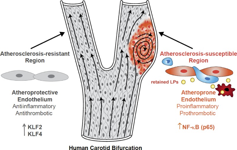
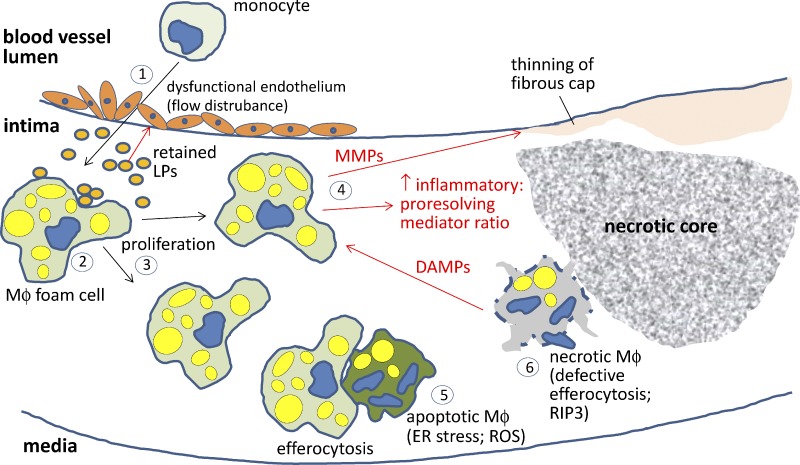
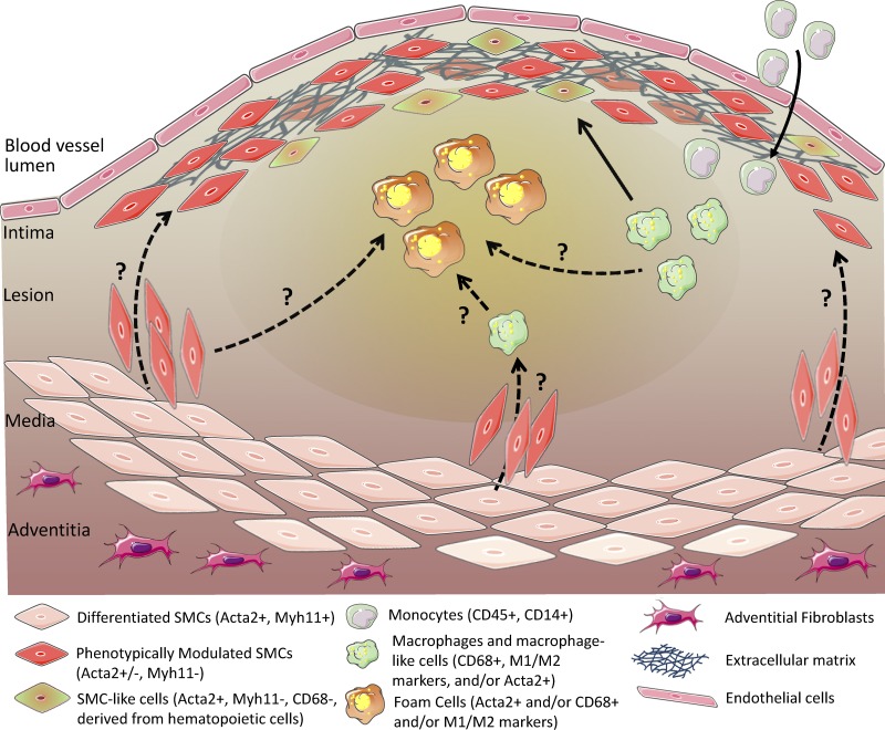
Atherosclerosis occurs in the subendothelial space (intima) of medium-sized arteries at regions of disturbed blood flow and is triggered by an interplay between endothelial dysfunction and subendothelial lipoprotein retention. Over time, this process stimulates a nonresolving inflammatory response that can cause intimal destruction, arterial thrombosis, and end-organ ischemia. Recent advances highlight important cell biological atherogenic processes, including mechanotransduction and inflammatory processes in endothelial cells, origins and contributions of lesional macrophages, and origins and phenotypic switching of lesional smooth muscle cells. These advances illustrate how in-depth mechanistic knowledge of the cellular pathobiology of atherosclerosis can lead to new ideas for therapy.
Overview
The claims and statistics of this paper haven’t been checked yet.
Run the check — free, no account needed. It reads the open-access text and checks the sources it cites.
Integrity signals
Registry
Cites
Statistics
Figures
Verification
Claims the paper makes
The claims and statistics of this paper haven’t been checked yet.
Run the check — free, no account needed. It reads the open-access text and checks the sources it cites.
Claims other papers make about this one
Each is checked against this paper’s own text: does it say what the citing paper says it does?
The check wasn’t confident either way, so it gives no verdict — never disputed.
In the citing paper
“Adding growth factors such as VEGF and PDGF to PLCL vascular scaffolds can effectively promote vascular endothelialization, accelerate migration and aggregation of endothelial cells and smooth muscle cells, and promote vascular regeneration and reconstruction [161].”
From Application of PLCL as a biodegradable polymer in biomedical engineering (2026) · cites this paper as [161]
Experimental — accuracy not yet measuredIn the citing paper
“Atherosclerosis is an age‐associated vascular disease characterised by the formation of plaques in arterial vessels, which can lead to the development of various cardiovascular disorders including myocardial infarction, stroke and sudden cardiac death (Tabas et al, 2015).”
From The struggle of a good friend getting old: cellular senescence in viral responses and therapy (2021) · cites this paper as Tabas et al, 2015
In the citing paper
“Hypercholesterolemia, defined by elevated plasma low‐density lipoprotein (LDL) cholesterol, is a well‐established risk factor not only for cardiovascular disease (Borén et al. 2022; Steinberg 2013; Tabas et al. 2015; Boffa and Koschinsky 2019), but also for cognitive decline and dementia in later life (Kivipelto et al. 2001; Kivipelto and Solomon 2006; Livingston et al. 2024; Sparks et al. 2000, 2”
From LDL Cholesterol Modulates Astrocyte Metabolism, Lipid Handling, and Morphology: Evidence From In Vitro and In Vivo Models (2026) · cites this paper as Tabas et al. 2015
Self-check
Percentages, sums and reported differences re-computed from the paper’s own numbers.
The self-check needs the paper’s full text.
Citations
Cites
A role for mitochondria in nlrp3 inflammasome activation
Zhou, R., Yazdi, A. S., Menu, P., & Tschopp, J. (2011) · Nature · doi:10.1038/nature09663 · Cited by 5,602
Lusis, A. J. (2000) · Nature · doi:10.1038/35025203 · Cited by 4,880
NLRP3 inflammasomes are required for atherogenesis and activated by cholesterol crystals
Duewell, P., Kono, H., Rayner, K. J., Sirois, C. M., Vladimer, G., Bauernfeind, F. G., Abela, G. S., Franchi, L., Nuñez, G., Schnurr, M., et al. (2010) · Nature · doi:10.1038/nature08938 · Cited by 4,026
Glass, C. K., & Witztum, J. L. (2001) · Cell · doi:10.1016/s0092-8674(01)00238-0 · Cited by 3,094
Macrophages in the Pathogenesis of Atherosclerosis
Moore, K. J., & Tabas, I. (2011) · Cell · doi:10.1016/j.cell.2011.04.005 · Cited by 2,669
Self-citationIntegrating the mechanisms of apoptosis induced by endoplasmic reticulum stress
Tabas, I. A., & Ron, D. (2011) · Nature Cell Biology · doi:10.1038/ncb0311-184 · Cited by 2,655
Self-citationThe immune system in atherosclerosis
Hansson, G. K., & Hermansson, A. (2011) · Nature Immunology · doi:10.1038/ni.2001 · Cited by 2,389
Nathan, C., & Ding, A. (2010) · Cell · doi:10.1016/j.cell.2010.02.029 · Cited by 2,183
Cardiovascular Medicine at the Turn of the Millennium: Triumphs, Concerns, and Opportunities
Braunwald, E. (1997) · New England Journal of Medicine · doi:10.1056/nejm199711063371906 · Cited by 1,426
The Response-to-Retention Hypothesis of Early Atherogenesis
Williams, K. J., & Tabas, I. (1995) · Arteriosclerosis, Thrombosis, and Vascular Biology · doi:10.1161/01.atv.15.5.551 · Cited by 1,413
Self-citation
Cited by
Sorted by citations
Items 301–400 of 1,000
Using Yoda-1 to mimic laminar flow in vitro: A tool to simplify drug testing
Davies, J. E., Lopresto, D., Apta, B. H. R., Lin, Z., Ma, W., & Harper, M. T. (2019) · Biochemical Pharmacology · doi:10.1016/j.bcp.2019.08.013 · Cited by 36
“to effectively mimic constant shear stress from laminar flow, the endothelial response to turbulent flow is often very different and pathologically-relevant [42].”
BackgroundLipid efflux mechanisms, relation to disease and potential therapeutic aspects
Castaño, D., Rattanasopa, C., Monteiro-Cardoso, V. F., Corlianò, M., Liu, Y., Zhong, A., Rusu, M., Liehn, E. A., & Singaraja, R. R. (2020) · Advanced Drug Delivery Reviews · doi:10.1016/j.addr.2020.04.013 · Cited by 37
Roche-Molina, M., Hardwick, B. S., Sánchez‐Ramos, C., Sanz‐Rosa, D., Gewert, D. R., Cruz, F. M., González-Guerra, A., Andrés, V., Palma, J. A., Ibáñez, B., et al. (2020) · Scientific Reports · doi:10.1038/s41598-020-68350-2 · Cited by 37
Roles and mechanisms of garlic and its extracts on atherosclerosis: A review
Li, M., Yun, W., Wang, G., Li, A., Gao, J., & He, Q. (2022) · Frontiers in Pharmacology · doi:10.3389/fphar.2022.954938 · Cited by 37
“The activation of the NF-κB signaling pathway stimulates the formation of the inflammatory process, which leads ECs to take on AS phenotypes in the carotid sinus (Tabas et al., 2015).”
BackgroundIncretin drugs as modulators of atherosclerosis
Gallego-Colon, E., Wojakowski, W., & Francuz, T. (2018) · Atherosclerosis · doi:10.1016/j.atherosclerosis.2018.09.011 · Cited by 35
Wang, J., Xu, P., Xie, X., Li, J., Wang, J., Wang, J., Hong, F., Li, J., Zhang, Y., Song, Y., et al. (2017) · Journal of the American Heart Association · doi:10.1161/jaha.117.006297 · Cited by 35
“Monocytes and macrophages are the predominant cellular components of atherosclerotic lesions.(2,3,7) Inflammation induces the recruitment of monocytes to the subendothelium, and they subsequently transmigrate through the vascular endothelial layer, internalize modified lipoproteins, and then invade the vessel wall to…”
BackgroundVascular smooth muscle cell contractile function and mechanotransduction
Ahmed, S., & Warren, D. T. (2018) · Vessel Plus · doi:10.20517/2574-1209.2018.51 · Cited by 37
“Atherosclerosis is an inf lammatory CVD that acts as the underlying cause of heart attack, stroke and cardiac death([87]).”
BackgroundJayawardena, R., Swarnamali, H., Lanerolle, P., & Ranasinghe, P. D. (2020) · Diabetes & Metabolic Syndrome Clinical Research & Reviews · doi:10.1016/j.dsx.2020.09.033 · Cited by 44
Rami, A. Z. A., Hamid, A. A., Anuar, N. N. M., Aminuddin, A., & Ugusman, A. (2022) · Mediators of Inflammation · doi:10.1155/2022/2734321 · Cited by 37
“The migrated monocytes transformed into macrophages that secrete proinflammatory cytokines such as MCP-1, TNF-α, IFN-γ, and IL-6, which further aggravate monocyte recruitment and low-density lipoprotein (LDL) oxidation [98].”
BackgroundAnnexin A protein family in atherosclerosis
Li, Y., Wang, Y., Huang, L., Zhao, Y.-Y., Chen, L.-H., & Zhang, C. (2022) · Clinica Chimica Acta · doi:10.1016/j.cca.2022.05.009 · Cited by 37
Mao, L., Yin, R., Yang, L., & Zhao, D. (2022) · Frontiers in Endocrinology · doi:10.3389/fendo.2022.983723 · Cited by 42
Transcription factors: key regulatory targets of vascular smooth muscle cell in atherosclerosis
Jiang, Y., & Qian, H. (2023) · Molecular Medicine · doi:10.1186/s10020-022-00586-2 · Cited by 36
Post-transcriptional down regulation of ICAM-1 in feto-placental endothelium in GDM
Díaz-Pérez, F., Hiden, U., Gauster, M., Lang, I., Kónya, V., Heinemann, Á., Loegl, J., Saffery, R., Desoyé, G., & Cvitic, S. (2016) · Cell Adhesion & Migration · doi:10.1080/19336918.2015.1127467 · Cited by 35
“lial functions include the formation of the endothelial barrier, leukocyte trafficking, regulation of vascular tone, blood coagulation, nutrient uptake and neovascularization.(1,2) Endothelial dysfunction is the inability of endothelial cells to perform their physiological functions which is paralleled by a change of…”
BackgroundInflammation in human carotid atheroma plaques
Goikuria, H., Vandenbroeck, K., & Alloza, I. (2018) · Cytokine & Growth Factor Reviews · doi:10.1016/j.cytogfr.2018.01.006 · Cited by 35
Erbilgin, A., Seldin, M. M., Wu, X., Mehrabian, M., Zhou, Z., Qi, H., Dabirian, K. S., Packard, R. R. S., Hsieh, W. Y., Bensinger, S. J., et al. (2018) · Arteriosclerosis Thrombosis and Vascular Biology · doi:10.1161/atvbaha.118.311266 · Cited by 32
Atheroma Niche‐Responsive Nanocarriers for Immunotherapeutic Delivery
Peters, E. B., Tsihlis, N. D., Karver, M. R., Chin, S. M., Musetti, B., Ledford, B. T., Bahnson, E. M., Stupp, S. I., & Kibbe, M. R. (2019) · Advanced Healthcare Materials · doi:10.1002/adhm.201801545 · Cited by 35
KLF4-Induced Connexin40 Expression Contributes to Arterial Endothelial Quiescence
Denis, J.-F., Diagbouga, M. R., Molica, F., Hautefort, A., Linnerz, T., Watanabe, M., Lemeille, S., Bertrand, J. Y., & Kwak, B. R. (2019) · Frontiers in Physiology · doi:10.3389/fphys.2019.00080 · Cited by 35
“, KLF2 and KLF4, are highly expressed in regions exposed to athero-protective flow (Tabas et al., 2015).”
BackgroundThe TICE Pathway: Mechanisms and Lipid-Lowering Therapies
Grefhorst, A., Verkade, H. J., & Groen, A. K. (2019) · Methodist DeBakey Cardiovascular Journal · doi:10.14797/mdcj-15-1-70 · Cited by 35
“(3) Excretion over the apical membrane into the intestinal lumen, a process that is mainly mediated by the ATP-binding cassettes G5/G8 (ABCG5/ G8) transporter.”
BackgroundPerivascular adipose tissue in cardiovascular diseases – an update
Grigoraş, A. (2019) · The Anatolian Journal of Cardiology · doi:10.14744/anatoljcardiol.2019.91380 · Cited by 38
“Among these, leptin, TNF-α, IL-6, and H2O2 have a dual action, vasoconstrictory and vasodilatory, their balance being perturbed in the pathological status (2, 10, 36, 46).”
BackgroundInfluentialWiciński, M., Górski, K. M., Walczak, M., Wódkiewicz, E., Słupski, M., Pawlak-Osińska, K., & Malinowski, B. (2019) · International Journal of Molecular Sciences · doi:10.3390/ijms20164052 · Cited by 35
“In bends and at branch points, blood flow dynamics are disturbed and endothelial cells in this area exhibit a pro-inflammatory and prothrombotic phenotype with reduced barrier function [56].”
BackgroundThe Angiopoietin ligands and Tie receptors: potential diagnostic biomarkers of vascular disease
Bilimoria, J., & Singh, H. (2019) · Journal of Receptors and Signal Transduction · doi:10.1080/10799893.2019.1652650 · Cited by 37
“Disturbed blood flow in the vasculature is another component that promotes endothelial inflammation and is associated with atherosclerosis [45].”
BackgroundCarballo-Perich, L., Puigoriol‐Illamola, D., Bashir, S., Terceño, M., Silva, Y., Gubern, C., & Serena, J. L. (2022) · International Journal of Molecular Sciences · doi:10.3390/ijms23095149 · Cited by 33
“Atherosclerosis is associated with age and the presence of diverse classical vascular risk factors (VRFs) including: hypertension, dyslipidaemia, diabetes, tobacco use, obesity, and a sedentary lifestyle [4].”
BackgroundYu, W., Ilyas, I., Hu, X., Xu, S., & Yu, H. (2022) · Frontiers in Immunology · doi:10.3389/fimmu.2022.1072007 · Cited by 34
“The conversion of macrophages to foam cells is consistently accompanied by anoverproduction of pro-inflammatory cytokines and chemokines (4), which deepens and aggravates atherosclerosis.”
BackgroundGong, F., Cheng, W., Wang, H., Gao, M.-M., Qin, J., Zhang, Y., Li, X., Zhu, X., Xia, H., & She, Z. (2018) · Journal of Lipid Research · doi:10.1194/jlr.m082651 · Cited by 34
Hou, Y., Lu, C., Zheng, C., Liu, W.-C., Yen, T., Chen, R., Lin, Y., Chao, C., & Lu, K. (2020) · International Journal of Molecular Sciences · doi:10.3390/ijms21072466 · Cited by 34
“The intimal calcification is initiated by the focal retention of apo B–containing lipoproteins in the subendothelial extracellular matrix [19].”
BackgroundWang, J., Li, P., Xu, X., Zhang, B., & Zhang, J. (2020) · Frontiers in Immunology · doi:10.3389/fimmu.2020.00907 · Cited by 34
“It has been reported that endothelial dysfunction is central to the progression of atherosclerosis (4).”
BackgroundRetractedRole of Angiopoietin-Tie axis in vascular and lymphatic systems and therapeutic interventions
Wang, R., Yang, M., Jiang, L., & Huang, M. (2022) · Pharmacological Research · doi:10.1016/j.phrs.2022.106331 · Cited by 35
Hu, X., Li, Y., Chen, Q., Wang, T., Ma, L., Zhang, W., Yu, R., Zhang, J., Wan, J., Yu, C., et al. (2023) · International Immunopharmacology · doi:10.1016/j.intimp.2023.110410 · Cited by 34
Zhang, Y., Wang, L., Zhang, J.-J., Xiong, X.-M., Zhang, D., Tang, X.-M., Luo, X., Ma, Q., & Peng, J. (2018) · Atherosclerosis · doi:10.1016/j.atherosclerosis.2018.04.031 · Cited by 36
Huang, K., Jo, H., Echesabal-Chen, J., & Stamatikos, A. (2021) · Metabolites · doi:10.3390/metabo11090640 · Cited by 34
“Historically, the consequence of intimal arterial macrophages becoming cholesterol-filled has traditionally been considered the prime reason for atherogenesis and atherosclerosis progression [8–10].”
BackgroundCai, C., Zhu, H., Ning, X., Lin, L., Yang, B., Chen, S., Wang, L., Lu, X., & Gu, D. (2019) · Atherosclerosis · doi:10.1016/j.atherosclerosis.2019.04.204 · Cited by 33
Different Approaches in Therapy Aiming to Stabilize an Unstable Atherosclerotic Plaque
Kowara, M., & Cudnoch‐Jędrzejewska, A. (2021) · International Journal of Molecular Sciences · doi:10.3390/ijms22094354 · Cited by 33
“These processes change the microenvironment of the arterial wall intima, which promotes subsequent alterations—foam cell generation, vascular smooth muscle cell migration, and their conversion from contractile into the synthetic phenotype, extracellular matrix remodeling, plaque growth, fibrous cap formation, and fina…”
BackgroundMechanisms of continual efferocytosis by macrophages and its role in mitigating atherosclerosis
Kumar, D., Pandit, R., & Yurdagul, A. (2023) · Immunometabolism · doi:10.1097/in9.0000000000000017 · Cited by 37
“These cytokines increase the expression of adhesion molecules on the surface of the endothelium and increase leukocyte–endothelial interactions [4].”
BackgroundGanguly, R., Sahu, S., Ohanyan, V. A., Haney, R. M., Chavez, R. J., Shah, S., Yalamanchili, S. P., & Raman, P. (2017) · Scientific Reports · doi:10.1038/srep45279 · Cited by 33
“This, in turn, may initiate release of a repertoire of growth factors and cytokines promoting VSMC activation, a key step responsible for initiation and progression of atherosclerotic lesions 41 .”
CRISPR-Cas9 Genome Editing for Treatment of Atherogenic Dyslipidemia
Chadwick, A. C., & Musunuru, K. (2017) · Arteriosclerosis Thrombosis and Vascular Biology · doi:10.1161/atvbaha.117.309326 · Cited by 33
“12 With atherosclerotic vascular disease being the leading cause of death in the United States and worldwide,(1) enormous effort has been focused on understanding the development and progression of disease, with an eye toward improving the prevention and treatment of disease.(2) In just the last decade, genotyping and…”
Combined PET/DCE-MRI in a Rabbit Model of Atherosclerosis
Calcagno, C., Lairez, O., Hawkins, J., Kerr, S. W., Dugas, M. S., Simpson, T., Epskamp, J., Robson, P. M., Eldib, M., Bander, I., et al. (2018) · JACC. Cardiovascular imaging · doi:10.1016/j.jcmg.2017.11.030 · Cited by 33
“The accumulation of oxidized lipids in the arterial wall precipitates an active inflammatory and immune response, accompanied by increased neovascularization and permeability, which is key in promoting plaque instability, thrombosis, and clinical events (2).”
BackgroundIncreased lymphocyte activation and atherosclerosis in CD47-deficient mice
Engelbertsen, D., Autio, A. K., Verwilligen, R. A. F., Depuydt, M. A. C., Newton, G. A., Rattik, S., Levinsohn, E. A., Saggu, G., Jarolı́m, P., Wang, H., et al. (2019) · Scientific Reports · doi:10.1038/s41598-019-46942-x · Cited by 32
Endothelial Dysfunction in Cardiovascular Diseases
Biswas, I., & Khan, G. A. (2020) · IntechOpen eBooks · doi:10.5772/intechopen.89365 · Cited by 33
Inflammatory Basis of Atherosclerosis: Modulation by Sex Hormones
Nasser, S. A., Afify, E. A., Kobeissy, F. H., Hamam, B., Eid, A. H., & El‐Mas, M. M. (2021) · Current Pharmaceutical Design · doi:10.2174/1381612827666210122142811 · Cited by 32
Olive Oil Consumption can Prevent Non-communicable Diseases and COVID-19: A Review
Majumder, D., Debnath, M., Sharma, K. N., Shekhawat, S. S., Prasad, G. B. K. S., Maiti, D., & Ramakrishna, S. (2021) · Current Pharmaceutical Biotechnology · doi:10.2174/1389201022666210412143553 · Cited by 32
Unexpected Role of MPO-Oxidized LDLs in Atherosclerosis: In between Inflammation and Its Resolution
Tangeten, C., Boudjeltia, K. Z., Delporte, C., Antwerpen, P. V., & Korpak, K. (2022) · Antioxidants · doi:10.3390/antiox11050874 · Cited by 31
“RvD1 enhanced efferocytosis, a critical cellular process of inflammation resolution that is defective in atherosclerosis [84,85].”
BackgroundWang, Y., Gao, H., Huang, X., Chen, Z., Kang, P., Zhou, Y., Qin, D., Zhang, W., & Liu, J. (2022) · Carbohydrate Polymers · doi:10.1016/j.carbpol.2022.119632 · Cited by 32
Sequelae and management of radiation vasculopathy in neurosurgical patients
Twitchell, S., Karsy, M. J., Guan, J., Couldwell, W. T., & Taussky, P. (2018) · Journal of neurosurgery · doi:10.3171/2017.12.jns172635 · Cited by 32
Karnewar, S., Karnewar, V., Shankman, L. S., & Owens, G. K. (2024) · JCI Insight · doi:10.1172/jci.insight.173863 · Cited by 36
“…in atherosclerosis, including macrophages (10), T cells (11), B cells (12), neutrophils (13), endothelial cells (EC) (9), and SMC (4, 14–16), can have beneficial or detrimental effects on lesional development or late-stage pathogenesis depending on the nature of their phenotypic transitions (17).”
BackgroundSelf-citationInteractions between nanoparticles and pathological changes of vascular in Alzheimer’s disease
Lei, T., Yang, Z., Li, H., Qin, M., & Gao, H. (2024) · Advanced Drug Delivery Reviews · doi:10.1016/j.addr.2024.115219 · Cited by 34
Cardiovascular Disease in Nonalcoholic Steatohepatitis: Screening and Management
Shroff, H., & VanWagner, L. B. (2020) · Current Hepatology Reports · doi:10.1007/s11901-020-00530-0 · Cited by 33
The angiopoietin receptor Tie2 is atheroprotective in arterial endothelium
Anisimov, A., Fang, S., Hemanthakumar, K. A., Örd, T., Avondt, K. V., Chèvre, R., Toropainen, A., Singha, P., Gilani, H., Nguyen, S. D., et al. (2023) · Nature Cardiovascular Research · doi:10.1038/s44161-023-00224-y · Cited by 33
“Endothelial cells are known to participate in the inflammation in atherosclerotic lesions via adhesion receptors and paracrine signaling 35,36 .”
Budatha, M., Zhang, J., Zhuang, Z. W., Yun, S., Dahlman, J. E., Anderson, D. G., & Schwartz, M. A. (2018) · Journal of the American Heart Association · doi:10.1161/jaha.117.007501 · Cited by 31
Rafi, U., Ahmad, S., Bokhari, S. S., Iqbal, M. A., Zia, A., Khan, M. A., & Roohi, N. (2021) · Mediators of Inflammation · doi:10.1155/2021/3425560 · Cited by 30
“The manifestation of atherosclerotic disorders is associated with monocytes/macrophages, endothelial cells, and vascular smooth muscle cells [63].”
Jin, Y., Lee, J. S., Min, S., Park, H., Kang, T. J., & Cho, S. (2016) · Advanced Functional Materials · doi:10.1002/adfm.201601430 · Cited by 30
Hemodynamic Control of Endothelial Cell Fates in Development
García‐Cardeña, G., & Slegtenhorst, B. R. (2016) · Annual Review of Cell and Developmental Biology · doi:10.1146/annurev-cellbio-100814-125610 · Cited by 30
Self-citationGanbaatar, B., Fukuda, D., Salim, H. M., Nishimoto, S., Tanaka, K., Higashikuni, Y., Hirata, Y., Yagi, S., Soeki, T., & Sata, M. (2018) · Atherosclerosis · doi:10.1016/j.atherosclerosis.2018.05.053 · Cited by 30
Wu, C., Zhou, Z., Wang, B., Ke, Z., Ge, Z., & Zhang, X. (2018) · Journal of Cellular Biochemistry · doi:10.1002/jcb.27469 · Cited by 30
“Endothelial dysfunction contributes a lot to atherosclerosis.(2) In addition, oxidized low‐density lipoprotein (ox‐LDL) can induce endothelial cells apoptosis for atherosclerosis.”
BackgroundThe function of long non-coding RNAs in vascular biology and disease
Kok, F. O., & Baker, A. H. (2019) · Vascular Pharmacology · doi:10.1016/j.vph.2018.06.004 · Cited by 30
Xu, Y., Wang, M., Xie, Y., Jiang, Y., Liu, M., Yu, S., Wang, B., & Liu, Q. (2019) · European Journal of Pharmacology · doi:10.1016/j.ejphar.2019.172451 · Cited by 30
Choi, B., Shin, M., Kim, E., Park, J., Lee, H., Kim, S. W., Song, J., & Chang, E. (2019) · Frontiers in Immunology · doi:10.3389/fimmu.2019.01701 · Cited by 32
“Endothelial dysfunction leads to increased permeability and subendothelial retention of modified low-density lipoprotein (LDL) particles, which trigger the subsequent recruitment of monocyte/macrophages into the subendothelial intima (1) and the initiation of plaque formation in the intima of blood vessels (2).”
BackgroundCell-specific and athero-protective roles for RIPK3 in a murine model of atherosclerosis
Colijn, S., Muthukumar, V., Xie, J., Gao, S., & Griffin, C. T. (2020) · Disease Models & Mechanisms · doi:10.1242/dmm.041962 · Cited by 29
“The cellular mechanisms that have been linked to atherosclerosis development are vast in number and include metabolic imbalances, inflammation, fibrosis, mechanotransduction and cell death (Lusis, 2000; Tabas et al., 2015).”
Collagenase‐Cleavable Peptide Amphiphile Micelles as a Novel Theranostic Strategy in Atherosclerosis
Chin, D. D., Poon, C. J., Trac, N. T., Wang, J., Cook, J., Joo, J., Jiang, Z., Maria, N. S. S., Jacobs, R. E., & Chung, E. J. (2020) · Advanced Therapeutics · doi:10.1002/adtp.201900196 · Cited by 29
Khoonrit, P., Mirdogan, A., Dehlinger, A., Mekboonsonglarp, W., Likhitwitayawuid, K., Priller, J., Böttcher, C., & Sritularak, B. (2020) · PLoS ONE · doi:10.1371/journal.pone.0238509 · Cited by 30
“Monocyte dysregulation, which is generally indicated by intense production of inflammatory mediators, has been observed in several inflammatory diseases, including sepsis [17], atherosclerosis [18], rheumatoid arthritis (RA) [19], and hepatic fibrosis [20].”
BackgroundCraparo, E. F., Cabibbo, M., Conigliaro, A., Barreca, M. M., Musumeci, T., Giammona, G., & Cavallaro, G. (2021) · Pharmaceutics · doi:10.3390/pharmaceutics13040503 · Cited by 32
“Cardiovascular diseases caused by atherosclerosis represent a serious problem for society, both in terms of lost lives and the economic investment linked to their management [1,2].”
BackgroundLiu, M., Zhang, Y., Ma, X., Zhang, B., Huang, Y., Zhao, J., Wang, S., Li, Y., Zhu, Y., Xiong, J., et al. (2022) · Oxidative Medicine and Cellular Longevity · doi:10.1155/2022/8006642 · Cited by 34
“Moreover, oxLDL triggers the transformation of macrophages into lipid-laden foam cells that critically a ff ected atherosclerosis ’ occurrence and progression [86].”
Atrial Fibrillation and Chronic Coronary Ischemia: A Challenging Vicious Circle
Oancea, A. F., Jigoranu, A. R., Morariu, P. C., Miftode, R.-Ş., Trandabat, B. A., Iov, D. E., Cojocaru, E., Costache, I. I., Baroi, L. G., Timofte, D. V., et al. (2023) · Life · doi:10.3390/life13061370 · Cited by 33
“In general, a reduction in luminal diameter of more than 70% is required to significantly restrict blood flow, generating the specific symptoms of chronic ischemia [51,52].”
BackgroundMatrix Metalloproteinase-13 in Atherosclerotic Plaque Is Increased by Influenza A Virus Infection
Lee, H. S., Noh, J. Y., Shin, O. S., Song, J. Y., Cheong, H. J., & Kim, W. J. (2019) · The Journal of Infectious Diseases · doi:10.1093/infdis/jiz580 · Cited by 29
Wang, S., Yuan, R., Liu, M., Zhang, Y., Jia, B., Ruan, J., Shen, J., Zhang, Y., Liu, M., & Wang, T. (2022) · Biomedicine & Pharmacotherapy · doi:10.1016/j.biopha.2022.113712 · Cited by 29
ABCA1 agonist peptides for the treatment of disease
Bielicki, J. K. (2015) · Current Opinion in Lipidology · doi:10.1097/mol.0000000000000258 · Cited by 29
Linking gut microbiota with human diseases
Vandana, U. K. (2020) · Bioinformation · doi:10.6026/97320630016196 · Cited by 32
Araujo, G., Valencia, L. M., Martin-Ozimek, A., Soto, Y., & Proctor, S. D. (2025) · Frontiers in Immunology · doi:10.3389/fimmu.2025.1485801 · Cited by 29
“…absence of a con fl uent luminal elastin sheet, and exposure of arterial proteoglycans, not only increase ApoB-containing lipoproteins delivery in the intima but also accelerate the subendothelial deposition of lipids and contribute to the onset and progression of atherosclerosis (40, 45, 47, 48).”
Ruiz, M., Coderre, L., Allen, B. G., & Rosiers, C. D. (2017) · Biochimica et Biophysica Acta (BBA) - Molecular Basis of Disease · doi:10.1016/j.bbadis.2017.07.015 · Cited by 28
Nanomaterials to Resolve Atherosclerosis
Peters, E. B., & Kibbe, M. R. (2020) · ACS Biomaterials Science & Engineering · doi:10.1021/acsbiomaterials.0c00281 · Cited by 28
Extension of Endocardium-Derived Vessels Generate Coronary Arteries in Neonates
Tang, J., Zhu, H., Tian, X., Wang, H., Liu, S., Liu, K., Zhao, H., He, L., Huang, X., Feng, Z., et al. (2022) · Circulation Research · doi:10.1161/circresaha.121.320335 · Cited by 28
Are apolipoprotein E fragments a promising new therapeutic target for Alzheimer’s disease?
Vecchio, F. L., Bisceglia, P., Imbimbo, B. P., Lozupone, M., Latino, R. R., Resta, E., Leone, M., Solfrizzi, V., Greco, A., Daniele, A., et al. (2022) · Therapeutic Advances in Chronic Disease · doi:10.1177/20406223221081605 · Cited by 28
“Mouse J20 line of hAPPFAD and overexpressing APOE 272-299 ApoE4 (1-271)1 (N-terminal) Decreases Aβ clearance and increases Aβ deposition In vivo/ In vitro Full-length ApoE3 and ApoE4 expressing mice were able to stimulate Aβ clearance Bien-Ly et al.”
BackgroundStudy of the activated macrophage transcriptome
Novoselov, V. V., Sazonova, M. A., Ivanova, E. A., & Orekhov, A. N. (2015) · Experimental and Molecular Pathology · doi:10.1016/j.yexmp.2015.09.014 · Cited by 29
Human cells involved in atherosclerosis have a sex
Franconi, F., Rosano, G. M. C., Basili, S., Montella, A., & Campesi, I. (2016) · International Journal of Cardiology · doi:10.1016/j.ijcard.2016.11.118 · Cited by 28
“1 Function of human ECs ECs have many functions [46], and their impairment is a risk factor for vascular diseases [47-49].”
BackgroundKim, H. J., Son, J. E., Kim, J. H., Lee, C. C., Yang, H. S., Yaghmoor, S., Ahmed, Y. M., Yousef, J. M., Abualnaja, K. O., Al‐Malki, A. L., et al. (2017) · Journal of Cellular Biochemistry · doi:10.1002/jcb.26138 · Cited by 28
“A is a chronic and progressive inflammatory disease of the cardiovascular system, and currently a major cause of death in the developed world through complications including coronary thrombosis, heart attack, hypertension, and stroke [Tabas et al., 2015; Norenberg et al., 2016].”
BackgroundLDL-C levels in older people: Cholesterol homeostasis and the free radical theory of ageing converge
Auley, M. T. M., & Mooney, K. M. (2017) · Medical Hypotheses · doi:10.1016/j.mehy.2017.05.013 · Cited by 29
Therapeutic strategy for atherosclerosis based on bone-vascular axis hypothesis
Kim, J., Lee, W. S., & Kim, J. (2019) · Pharmacology & Therapeutics · doi:10.1016/j.pharmthera.2019.107436 · Cited by 28
Oxysterols in Vascular Cells and Role in Atherosclerosis
Luquain‐Costaz, C., & Delton-v, I. (2023) · Advances in experimental medicine and biology · doi:10.1007/978-3-031-43883-7_11 · Cited by 27
“7-KC and 7β-OHC can also induce ROS production and decrease cellular antioxidants, therefore inducing a mitochondrial oxidative stress in the sub-endothelial space (Tabas et al. 2015; Ravi et al. 2021).”
BackgroundIndoxyl Sulfate-Induced Macrophage Toxicity and Therapeutic Strategies in Uremic Atherosclerosis
Wakamatsu, T., Yamamoto, S., Yoshida, S., & Narita, I. (2024) · Toxins · doi:10.3390/toxins16060254 · Cited by 29
“These monocytes differentiate into macrophages, proliferating and absorbing oxidized LDL in atherosclerotic lesions [52,53].”
BackgroundThe Pathological Mechanisms and Therapeutic Molecular Targets in Arteriovenous Fistula Dysfunction
Yan, R., Song, A., & Zhang, C. (2024) · International Journal of Molecular Sciences · doi:10.3390/ijms25179519 · Cited by 27
“…responsible for contractility; (ii) dedifferentiated VSMCs labeled with cytokeratins-8 and 18 (CK-8, CK-18) + [36,37], intercellular adhesion molecule-1 (ICAM-1) + [38], vascular cell adhesion molecule-1 (VCAM-1) + [39] acquire the ability to synthesize and secrete ECM, as well as proliferate.”
BackgroundLinc00299/miR-490-3p/AURKA axis regulates cell growth and migration in atherosclerosis
Liu, Y., Chen, Y., Tan, L., Zhao, H., & Xiao, N. (2019) · Heart and Vessels · doi:10.1007/s00380-019-01356-7 · Cited by 27
Li, C., Zhang, M., Dai, Y., & Xu, Z. (2020) · Experimental Physiology · doi:10.1113/ep088088 · Cited by 27
Recent progress in the detection and treatment of atherosclerosis by nanoparticles
Chen, L., Jiang, Z., Akakuru, O. U., Yang, L., Li, J., Ma, S., & Wu, A. (2020) · Materials Today Chemistry · doi:10.1016/j.mtchem.2020.100280 · Cited by 28
Microglia–neuron–vascular interactions in ischemia
Lénárt, N., Cserép, C., Császár, E., Pósfai, B., & Dénes, Á. (2023) · Glia · doi:10.1002/glia.24487 · Cited by 28
“Endothelial mechanisms also contribute to the throm-boresistance property of blood vessels and suppress proinflammatory gene expression, immune cell trafficking, and development of atherosclerosis (Tabas et al., 2015) (Wolf & Ley, 2019).”
BackgroundScavenger receptors and non-coding RNAs: relevance in atherogenesis
Dai, Y., Condorelli, G., & Mehta, J. L. (2015) · Cardiovascular Research · doi:10.1093/cvr/cvv236 · Cited by 27
Lü, J., Chen, X., Xu, X., Liu, J., Zhang, Z., Wang, M., Li, X., Chen, H., Zhao, D., Wang, J., et al. (2019) · Biomedicine & Pharmacotherapy · doi:10.1016/j.biopha.2019.108840 · Cited by 27
Liang, W., Wang, Q., Ma, H., Yan, W., & Yang, J. (2018) · Cellular Physiology and Biochemistry · doi:10.1159/000487569 · Cited by 26
Sun, S.-M., Xie, Z., Zhang, Y., Zhang, X., Zhou, C., Yin, J.-P., Yu, Y., Cui, S., Jiang, H., Li, T.-T., et al. (2020) · Acta Pharmacologica Sinica · doi:10.1038/s41401-020-0472-9 · Cited by 26
Xu, Y., Miao, C., Cui, J., & Bian, X. (2021) · Brazilian Journal of Medical and Biological Research · doi:10.1590/1414-431x20209386 · Cited by 26
“Endothelial injury and dysfunction are considered the base and the initial steps for atherosclerosis (3).”
BackgroundSong, L., Zhang, J., Lai, R., Li, Q., Ju, J., & Xu, H. (2021) · Frontiers in Pharmacology · doi:10.3389/fphar.2021.675999 · Cited by 27
Succinate: A Novel Mediator to Promote Atherosclerotic Lesion Progression
Zhang, S., Liang, Y., Lü, L., Chen, Y., Wu, P., & Wei, D. (2022) · DNA and Cell Biology · doi:10.1089/dna.2021.0345 · Cited by 27
“The dynamic interaction and crosstalk of these cells enhance the progression of atherosclerotic lesions and contribute to the instability (Tabas et al. , 2015; Gimbrone and Garcia-Cardena, 2016).”
Inhibition of macrophage-derived foam cells by Adipsin attenuates progression of atherosclerosis
Duan, Y., Zhang, X., Zhang, X., Lin, J. M., Shu, X., Man, W., Jiang, M., Zhang, Y., Wu, D., Zhao, Z., et al. (2022) · Biochimica et Biophysica Acta (BBA) - Molecular Basis of Disease · doi:10.1016/j.bbadis.2022.166533 · Cited by 26
Vascular homeostasis in atherosclerosis: A holistic overview
Xu, S., Lyu, Q., Ilyas, I., Tian, X.-Y., & Weng, J. (2022) · Frontiers in Immunology · doi:10.3389/fimmu.2022.976722 · Cited by 26
“The SES is a fertile “soil” (microenvironment or niche) for atherosclerosis development due to the intricate interplay among subendothelial low-density lipoprotein (LDL) retention, LDL transcytosis, VSMC proliferation and migration, endothelial dysfunction, foam cell formation, and necrotic core formation (21).”
BackgroundSu, C., Lu, Y., Wang, Z., Guo, J., Hou, Y., Wang, X., Qin, Z., Gao, J., Sun, Z., Dai, Y., et al. (2022) · Aging and Disease · doi:10.14336/ad.2022.1208 · Cited by 28
“secreted by macrophages and ECs can also enhance vascular smooth muscle proliferation and produce fibromuscular lesions [154].”
BackgroundMulti-photon microscopy in cardiovascular research.
Wu, Z., Rademakers, T., Kiessling, F., Vogt, M., Westein, E., Weber, C., Megens, R., & van Zandvoort, M. V. (2017) · Methods · doi:10.1016/j.ymeth.2017.04.013 · Cited by 19
“Monocyte-derived macrophages play a key role in both early and late stages of plaque growth, however, it is still not fully understood how lipid loading by macrophages affects their biology and promotes atherosclerosis [64].”
BackgroundBabaev, V. R., Huang, J., Ding, L., Zhang, Y., May, J. M., & Linton, M. F. (2018) · Frontiers in Immunology · doi:10.3389/fimmu.2018.00215 · Cited by 27
“These results are consistent with the current concepts that low numbers of circulating monocytes and high sensitivity of macrophages to pro-apoptotic stimuli are key drivers that suppress atherogenesis (66, 67).”
ResultSun, R., Fan, Y., Liang, X., Gong, T., Wang, Q., Liu, H., Shan, Z., & Lei, L. (2018) · BioMed Research International · doi:10.1155/2018/1010248 · Cited by 26
Role of the COP9 Signalosome (CSN) in Cardiovascular Diseases
Milic, J., Tian, Y., & Bernhagen, J. (2019) · Biomolecules · doi:10.3390/biom9060217 · Cited by 27
“Accordingly, atherosclerosis is the main underlying condition of acute myocardial infarction (AMI, heart attack, ischemic heart disease, IHD), ischemic stroke (large artery stroke), and peripheral artery disease (PAD) [64,76,77].”
BackgroundProtective Effect of Ganoderma (Lingzhi) on Cardiovascular System
Meng, J., & Yang, B. (2019) · Advances in experimental medicine and biology · doi:10.1007/978-981-32-9421-9_7 · Cited by 26
Cathelicidin Modulates Vascular Smooth Muscle Cell Phenotypic Switching through ROS/IL-6 Pathway
Dong, X., Wu, D., Zhang, Y., Jia, L., Pan, X., Sun, J., & Pan, L.-L. (2020) · Antioxidants · doi:10.3390/antiox9060491 · Cited by 25
Xu, L., Cheng, D., Huang, Z., Ding, S., Zhang, W., Tan, H., Shi, H., Chen, R., Zou, Y., Wang, T. C., et al. (2017) · Atherosclerosis · doi:10.1016/j.atherosclerosis.2017.05.024 · Cited by 25
Metadata
- Authors
- Tabas, I., García-Cardeña, G., & Owens, G. K.
- Affiliations
- Columbia University Irving Medical Center (US)
- Program in Human Biology and Translational Medicine, Harvard Medical School, Boston, MA 02115 4
- University of Virginia (US)
- Corresponding author
- Tabas, Ira
- Venue
- Journal of Cell Biology
- Publisher
- Rockefeller University Press
- Published
- 2015-04-13
- Volume
- 209
- Issue
- 1
- Pages
- 13-22
- ISSN
- 0021-9525 (ISSN-L)
- 1540-8140
- PMC
- PMC4395483
- OpenAlex
- W1949072443
- Cited by
- 1,016
- References
- 107
- Citations by year
- 2026: 45 · 2025: 68 · 2024: 70 · 2023: 96 · 2022: 107 · 2021: 94 · 2020: 96 · 2019: 124 · 2018: 108 · 2017: 103 · 2016: 78 · 2015: 23
- FWCI
- 9.82
- Type
- journal article
- Language
- en
- Open access
- bronze
- doi.org
- Locations
- The Journal of Cell Biology (publishedVersion)
- Europe PMC (PubMed Central) (publishedVersion, cc-by-nc-sa)
- PubMed Central (publishedVersion, cc-by-nc-sa)
- PubMed (publishedVersion)
- Digital Access to Scholarship at Harvard (DASH) (Harvard University) (submittedVersion, cc-by)
- Funders
- National Institutes of Health
- National Institute on Aging (AG032443, R01AG032443)
- National Heart, Lung, and Blood Institute (R44HL118826, HL057353, HL087867, HL107497, HL075662, HL121008, HL118826, R01HL087867, R01HL057353, R01HL075662, R37HL057353, R01HL121008, R01HL107497, R41HL118826)
- Topics
- Life Sciences › Immunology and Microbiology › Immunology › Atherosclerosis and Cardiovascular Diseases
- Life Sciences › Biochemistry, Genetics and Molecular Biology › Biochemistry › Cholesterol and Lipid Metabolism
- Health Sciences › Medicine › Physiology (medical) › Nitric Oxide and Endothelin Effects
- Keywords
- atherosclerosis, endothelial dysfunction, disturbed blood flow, nonresolving inflammation, arterial thrombosis, end-organ ischemia, mechanotransduction, endothelial inflammation, macrophage origin, smooth muscle cell phenotypic switching, atherosclerosis treatment
- MeSH terms
- Animals, Endothelium, Vascular, Humans, Macrophages, Muscle, Smooth, Vascular, Myocytes, Smooth Muscle, Mechanotransduction, Cellular, Endothelial Cells, Atherosclerosis
About these checks
Registry, retraction, Cites and statistics results are computed, not judged, with one exception: a printed reference that no index lists is read once by a model before it counts as a citation error. Claims are extracted by a model and kept only when their quote appears verbatim in the text; each cited source is then checked, with a model’s help, for whether it says what the claim relies on. Those verdicts can be wrong — read the claim’s quote and the cited source before relying on one.
Statistics and arithmetic are recomputed, not judged.
Confidence bands are shown only for fields calibrated on our annotated test set. None is calibrated yet, so no band is shown.
What counts as a claim ›How we measure confidence ›How we check this ›
Are you an author, or do you see a problem with this page? Request a review
